

FIGURE 1. Habitus of male representatives of the group Libanochlites. 1. Libanochlites eocenicus sp. nov. HT: Dip 00606. 2. L. neocomicus Brundin, 1976.
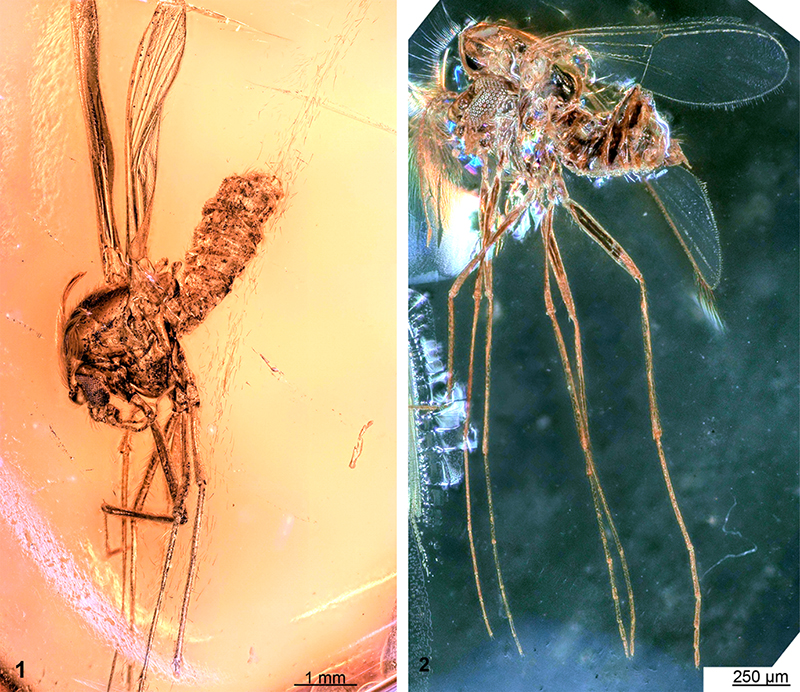
FIGURE 2. Tibial spurs of third thoracic appendages of male representatives of the group Libanochlites. 1. Libanochlites eocenicus sp. nov. HT: Dip 00606. 2. L. neocomicus Brundin, 1976.
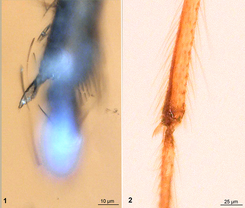
FIGURE 3. Mouthparts of male representatives of Chironomidae. 1. L. eocenicus sp. nov. HT: Dip 00606. 2. L. neocomicus Brundin, 1976. 3-4. Afrochlus harrisoni Freeman, 1964. 3. Mouthparts of A. harrisoni. 4. Head of A. harrisoni. Abbreviations: cly = clypeus; md = mandible; mp = maxilar palpi; lac = lacinia, lab = labrum.
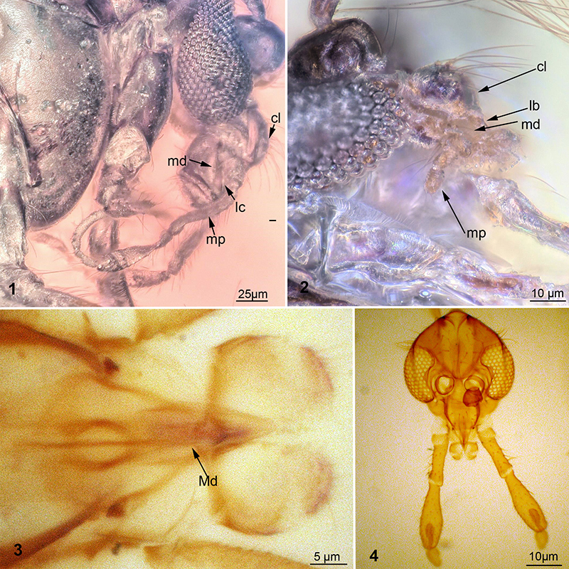
FIGURE 4. Wing of Libanochlites eocenicus sp. nov. HT: Dip 00606. 1. Marked wing. 2. unmarked wing. Abbreviations: mcu = medial-cubital vein; r1 = radial vein 1; r4+5 = radialvein 4+5; fcu = fork of the cubital vein.
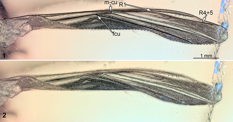
FIGURE 5. Hypopygium of male representative of Libanochlites eocenicus sp. nov. HT: Dip 00606. 1. Hypopygium, ventral view. 2. Gonocoxite, close-up view. 3. Hypopygium, line drawing, ventral view. 4. Hypopygium, line drawing dorsal view. Abbreviations: gc-gonocoxite, gs- gonostylus, t9-tergite 9, w- warts of Gonocoxite.
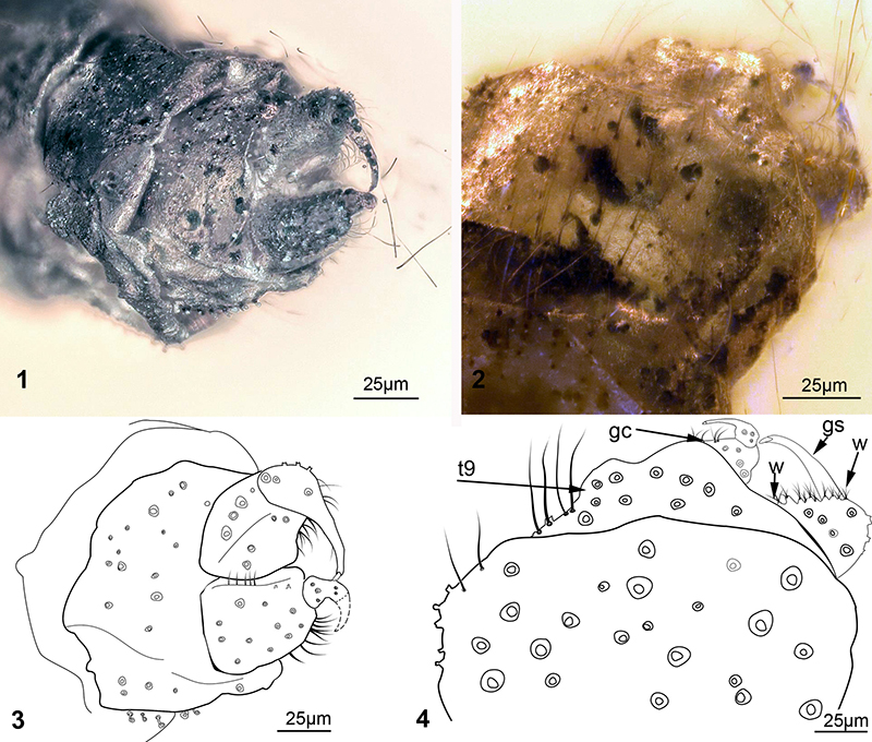
FIGURE 6. “Mushroom” sensilla of unknown function on tergite IX of Libanochlites eocenicus sp. nov. HT: Dip 00606. 1. Habitus, with arrow marking position of the sensilla. 2. Tergite IX with “mushroom” sensilla, white square marks position of the detail in shown figure 6.3, scalebar=20 µm. 3. “Mushroom” sensilla close-up.

FIGURE 7. Laser-confocal images of Libanochlites neocomicus Brundin, 1976. 1. Habitus of male representative. 2. Hypopigium of male representative. 3. Close-up of hypopigium of male representative. Abbreviations: w =warts of gonocoxite; gs = gonostylus; gc = gonocoxite; t9 = tergite IX.

FIGURE 8. Scanning Electron Microscopy image of female representative of Corethrella marksae Colles, 1986. 1. Head of the female, frontal view. 2. Maxilar palp of the same specimen. 3. Close-up of Maxilary palp with clavate sensilla.

