

FIGURE 1. Sketch map showing the fossil locality in Turpan-Hami Basin, Xinjiang, NW China.
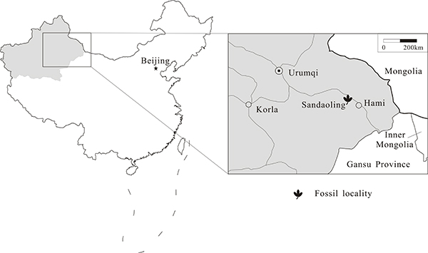
FIGURE 2. Stratigraphic section of the Middle Jurassic Xishanyao Formation in the Sandaoling open-cast coal mine, Hami, China. (Modified from figure 1 of Zhao et al., 2018)
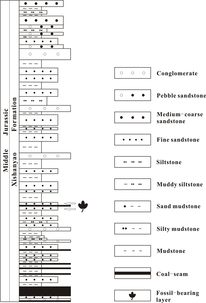
FIGURE 3. Thalli of Ricciopsis sandaolingensis sp. nov. (1-7) and extant Riccia sp. for comparison (8-9). 1, A colony of thalli, specimen LDGSW-2013-235. 2, Close-up of a in Figure 3.1 to show branch angles. 3, Counterpart of Figure 3.2 to show smooth surface of segments. 4, Close-up of b in Figure 3.1 to show branch angles. 5, Close-up of c in Figure 3.1 to show branching times. 6, Counterpart of Figure 3.4 to show apex shape. 7, Close-up of d in Figure 3.1 to show a single thallus and the base shape of the first segment before the first dichotomy. 8-9, Extant Riccia sp. for comparison (Photographed respectively by Jiming Liu and Binghua Chen and downloaded from the Plant Photo Bank of China). Sizes are unknown. Scale bar length: 1 = 2 cm, 2-4 = 1 cm, 5-7 = 0.5 cm.

FIGURE 4. Thalli of Ricciopsis sandaolingensis sp. nov. co-occur with ginkgoalean leaves. 1, Specimen showing thalli of R. sandaolingensis and ginkgoalean leaves preserved on the same bedding surface, specimen LDGSW-2013-208. 2, Specimen showing thalli of R. sandaolingensis and ginkgoalean leaves preserved on the same bedding surface, specimen LDGSW-2013-227. 3, Close-up of a in Figure 4.1 to show segment shape. 4, Counterpart of Figure 4.3 to show smooth surface of segments. 5, Close-up of b in Figure 3.1 to show segment shape. Scale bar length: 1-2 = 1 cm, 3-5 = 0.5 cm.
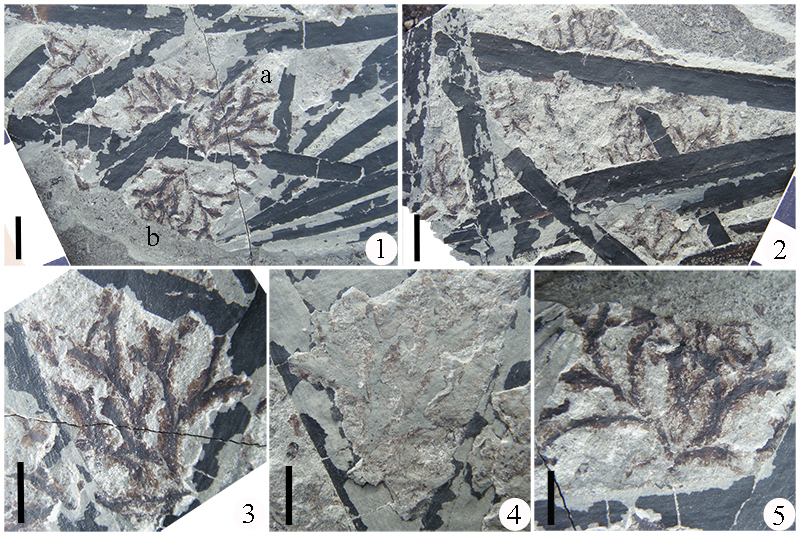
FIGURE 5. Microstructures of Ricciopsis sandaolingensis sp. nov. 1, Close of up of Figure 3.4 to show median ridge on the segment surface (indicated by white arrow) and entire margin (indicated by the black arrow). 2, Cuticle fragment showing the thick median ridge (indicated by the white arrow) and the thin wings (indicated by the black arrow). 3 and 5, Cuticle fragment showing rounded or polygonal, anticlinal epidermal cell walls. 4, Close-up of Figure 5.3 to show epidermal cell shape. 6, Rhizoids attached to cuticle fragment. 7-9, Rhizoids and ventral scales. Scale bar length: 1 = 1 mm; 2-3 = 0.2 mm; 4 and 7 = 0.1 mm; 5, 6, 8 = 0.05 mm; 9 = 0.025 mm.

