FIGURE 1. Optical images showing visual appearance of microfossils and micro-vertebrate remains before gold coating and after gold removal. 1, 2, Conodont element, Wurmiella excavata, Tramway Netherton, UK, Silurian: 1 before gold coating, 2 after removal. 3, 4, Sarcopterygian scale (probably Onychodus), Gogo Formation, Devonian, Australia: 3 before gold coating, 4 after removal. 5, 6, extant foraminiferan Globorotalia menardii (Caribbean Sea, 2900m water depth): 5 before gold coating, 6 after removal. 7, 8, Fragment of dermal armour, probably heterostracan, Devonian, Welsh Borders, UK, 7 before gold coating, 8 after removal. Scale bar equals 500µm.
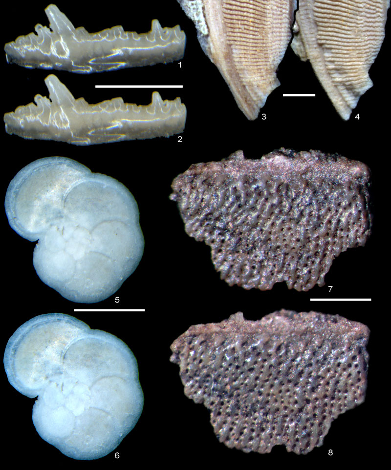
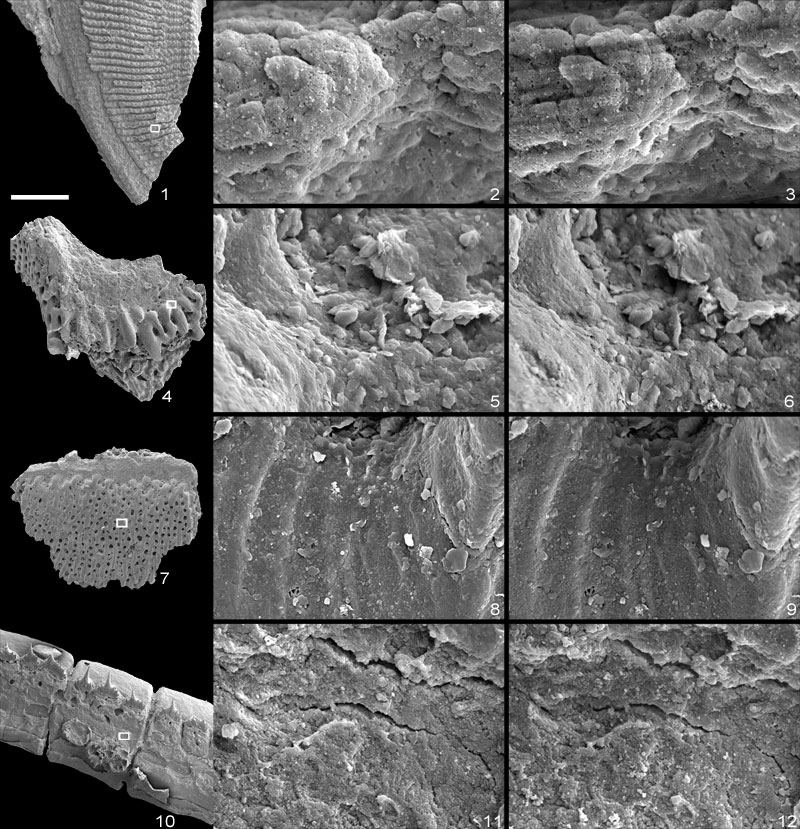
FIGURE 2. Scanning electron micrographs showing fine-scale surface textures of fossil microvertebrate remains before gold coating and after removal. 1-3, Sarcopterygian scale (probably Onychodus), Gogo Formation, Devonian, Australia: 2 before gold coating, 3 after removal (same specimen as Fig. 1.3,4). 4-6, Fragment of dermal armour, probably heterostracan (bony surface), Devonian, Welsh Borders, UK: 5 before gold coating, 6 after removal. 7-9, Fragment of dermal armour, probably heterostracan (dentine surface), Devonian, Welsh Borders, UK: 8 before gold coating, 9 after removal. 10-12, Sarcopterygian lepidotrichia (probably Onychodus), Gogo Formation, Devonian, Australia: 11 before gold coating, 12 after removal. White boxes in low-magnification images show location of high-magnification images. Low magnification scale bar equals 500 µm. Field of view of high-magnification images is 50µm.