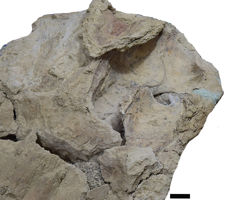
FIGURE S1. Photography of the ventral view with emphasis on the posterior region of the left mandible of Cochimicetus convexus. Scale bar 5 cm.

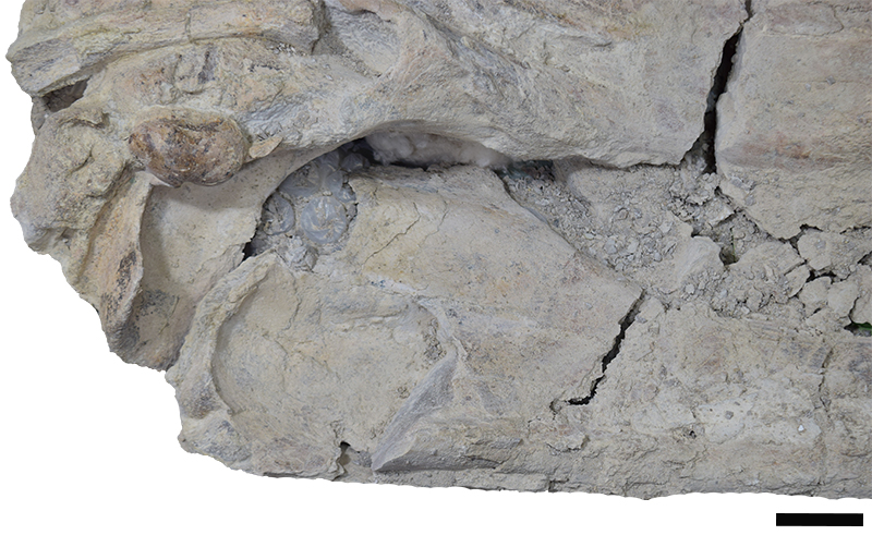
FIGURE S2. Photography of the dorsal-lateral view with emphasis on the coronoid process of the left mandible. Scale bar 5 cm.

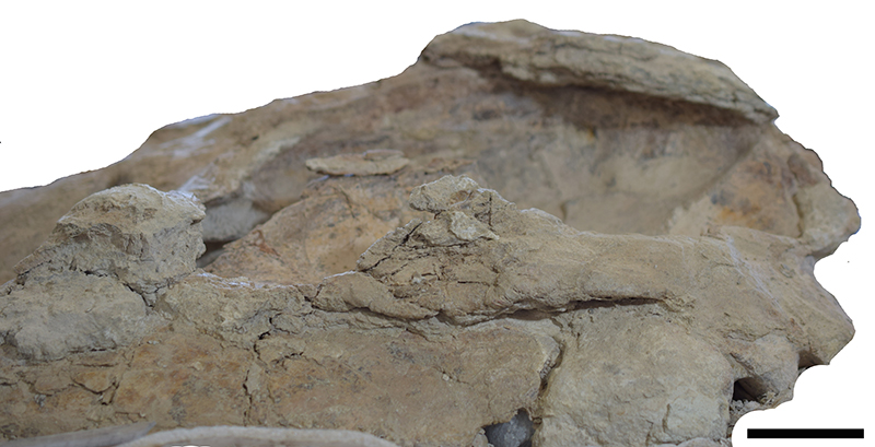
FIGURE S3. Photography of the posterior lateral view emphasizing the zygomatic process in the image. Scale bar 5 cm.

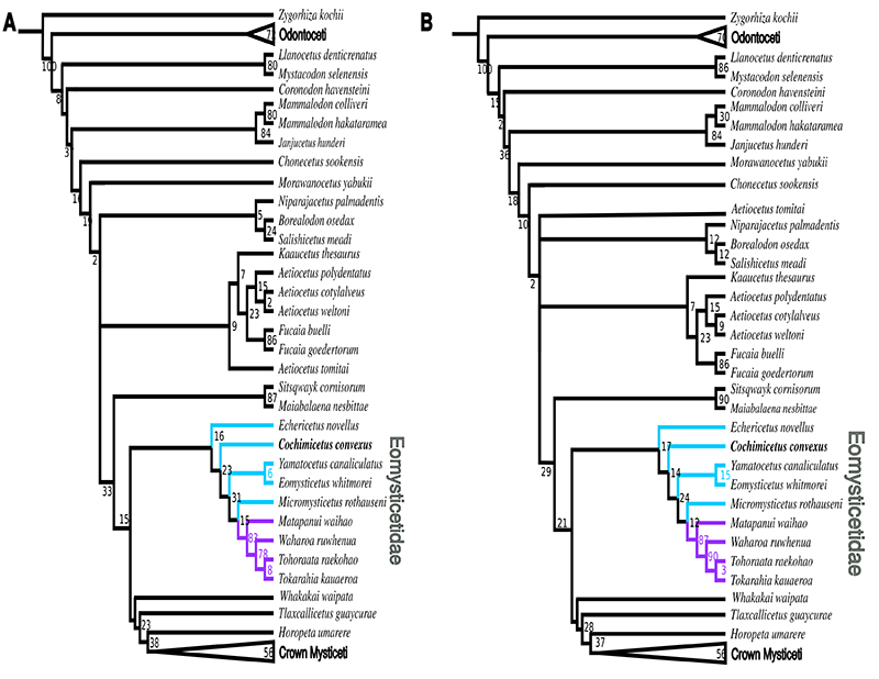
FIGURE S4. A, Implied weights with K 6; CI 0.259; RI 0.745; 89.35148 total length. B, Implied weights with K 12; CI 0.261; RI: 0.747; 57.74474 total length.


