|
|
|
METHODS
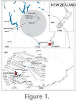
For the St Bathans Paleovalley samples a simple stratigraphic column is inappropriate as the sampled units are lensoid and widely-spread. However, a grid-reference and notes will allow relocation of the sample site. These details can be found in Appendix 1 and Appendix 2. The samples were disaggregated by covering with hot water and adding 40% hydrogen peroxide, fines were then removed by sieving through a 1 mm mesh. What remained was typically a "hash" of cuticle fragments, typically around 1–10 mm across. Cuticle could then be purified with further hydrogen peroxide treatment (which removes opaque cellular material) and hydrofluoric acid to remove silicates. It was then stained using either safranin or crystal violet. This concentrate was then scanned in a Petri dish under binocular microscope and individual cuticle fragments were isolated and mounted on microscope slides using thymol glycerine jelly for Transmitted Light Microscopy (TLM). When sufficient additional material was present, also mounted on stubs for scanning electron microscopy (SEM) using double-sided tape, and coated with platinum. Catalogue numbers for material mounted on microscope slides are prefixed with "SB" or "SL" and SEM stubs are prefixed with "S-". The task of distinguishing taxa was an iterative process that has taken over 15 years. This work mainly used TLM and enlarged TLM photographs were employed which could be laid side by side to facilitate comparison. SEM observation was useful to interpret the three-dimensional structure and view very fine details. However, for pragmatic purposes this study focuses on distinguishing characters which can be viewed under TLM. As a general observation, it is relatively simple to study one sample and separate out distinct taxa. However, as more and more samples are added, perhaps with slightly differing degrees of preservation, staining, environmental differences, etc, the issue of taxon distinction becomes critical. As more samples are studied, typically many 'provisional' taxa are merged with one another. The issue then becomes one of finding similarities rather than differences. The benefits of a large sample-base being that a more realistic concept of taxa emerges. However, it is certain that a few taxa distinguished here are likely to cover more than one "real" species, and thus the final number will be an underestimate. Where there may be some issues of distinction; these are addressed under the subheading "Distinguishing features". In this work, cuticle morphologies are described as parataxa. Each parataxon is prefixed by "CUT-", then a letter. This is a purely pragmatic subdivision of the cuticle taxa into large groups. For instance, "L" includes Lauraceae, "M" the Myrtaceae, and "P" for Proteaceae. Most parataxa described in this report were given a "Z" as they are the large group "left over" once the more immediately identifiable groups had been dealt with. Finally, each parataxon gets a unique string of three letters. This is meant as a flexible system to deal with the disparate and poorly hierarchical morphologies of cuticle fragments. The intention is that parataxa are equivalent to species. For each parataxon a reference specimen and sample is nominated, which are equivalent to the holotype and type locality of a Linnean species. A single specimen is also nominated from each other sample that the taxon is recorded from. For examples of this methodology see Pole (2007a). Standard epidermal terminology is used to describe the cuticle taxa and is based on authors such as Baranova (1987, 1992); Stace (1965); Dilcher (1974); Hewson (1988); Payne (1978); and Wilkinson (1979). Carpenter (2005) is followed in the use of 'stoma' (stomata pl.) to refer to the stomatal pore and the pair of guard cells, and 'stomatal complex' for the stoma plus subsidiary cells. The inadvisability of constructing a terminology with a mixture of purely morphological terms as well as terms based on developmental processes have been discussed by these authors. However, there are occasions where some evidence of developmental process is obvious in cuticular fragments (for instance where one cell has been divided in two by a new wall) and need to be indicated. For these instances I have used terms (heliocyctic and tangenticytic) from Timonin (1995). More recently Carpenter (2005) has introduced several terms which incorporate developmental considerations. He did not discuss these terms in the context of several previous authors (for instance Timonin 1995) but some of them are also clearly applicable to the fossils described here and so they are listed alongside Timonin's. The reader should be aware that what appear to be fundamentally different terms describing stomatal complex structure may result from the subjectivity involved in deciding what is and isn't a subsidiary cell. To clarify some further terms; OSL = outer stomatal ledge. By "outer stomatal ledge thickness" here is meant the combined thickness of ledges and any underlying cuticle as seen under TLM view. By "giant stomatal complexes" is meant any distinctly different population of complexes, either in terms of size, or by obviously increased development of subsidiary cells around them. "Networking" is used to describe the situation where contact or subsidiary cells are shared between stomatal complexes (Pole 1998b). When stomata are known to be on one leaf surface only the distribution is assumed to be hypostomatic. In all other cases the distribution with respect to leaf surfaces is unknown. Stomatal size classes follow Wilkinson (1979). "Texture" refers to a pattern on the outer or inner surfaces of cuticle, such as "granular" which is of much smaller dimensions than normal epidermal cells. Ornamentation refers to a pattern on the outer cuticular surface, such as ridging, which is comparable to the size of normal epidermal cells. It does not include papillae or scales. Taxonomic identification into the Linnean system is based on morphological characters which have been detailed in the published literature as well as a large cuticle reference collection developed by the author. To date this collection includes around 4000 species in about 1500 genera and 285 families of mostly rainforest taxa from around the world. In this paper cuticle preparations of extant herbarium material cite the original herbarium sheet number ("AQ" refers to catalogue numbers of specimens in the Queensland Herbarium, Brisbane; "CANB" of specimens in the Australian National Herbarium, Canberra; "OTA" of specimens in the Botany Department Herbarium, University of Otago, Dunedin) and material in the author's own reference herbarium is prefixed with "OPH". All material is stored in the Queensland Herbarium, Toowong. |
|