|
|
APPENDIX 3.In this publication identified fossil cuticle taxa are presented in order following the Angiosperm Phylogeny Group (APG 2003): Gnetales
Canellales
Laurales
Proteales
Ranunculales
Santalales
Fagales
Oxalidales
Sapindales
Ericales
Apiales
Asterales
The following key is a general guide for how the cuticle in this work has been allocated to families. Key to families1. Plant remains found as three-dimensional, four-sided articles. Stomatal complexes oriented at right angles to long axis (The cuticle is very delicate and difficult to prepare.) Gymnostoma 1. Remains found as dispersed cuticle and typically as sheets.2. 2. Raised, thick, peg-like attachment scars of deciduous trichomes common and ridges of cuticle commonly partially projecting over OSL. Argophyllaceae 2. Peg-like attachment scars absent, or OSL not obscured by ridges of cuticle. 3. 3. Stomatal complexes brachyparacytic 4. 3. Stomatal complexes not brachyparacytic 8. 4. Stomatal complexes with clearly visible guard cells, which are distinctly elongate, almost rectangular guard cells, outer stomatal ledges and separating walls between guard cells absent. Gnetales 4. Stomatal complexes with guard cells often partially obscured by an outer stomatal ledge, separating walls between guard cells present, guard cells irregular or rounded. 5. 5. Epidermis distinctly granular. Winteraceae 5. Epidermis not distinctly granular. 6. 6. Cuticle glabrous. Monimiaceae 6. Trichomes or trichome attachment sites present 7. 7. Multicelluar trichome attachment sites present (trichomes deciduous). Proteaceae 7. Persistent branching trichomes present. Santalaceae 8. Stomatal complexes anisocytic, ornamented by prominent ridging. Myrsinaceae 8. Stomatal complexes not anisocytic. 9. 9. Stomatal complexes with clear subsidiary cells. 10. 9. Stomatal complexes with no clear subsidiary cells (anomocytic, although there may be occasional divisions of contact cells). Stomatal complexes with prominent outer stomatal ledges. Strasburgeriacae 10. Stomatal complexes with a distinctive outer stomatal rim, which is typically ovate, of equal breadth around the stoma, and under TLM appears thicker than any other area of cuticle. Atherospermataceae 10. No distinct outer stomatal rim as above. 11. 11. Distinctive form of massively-thickened trichome attachment scars (probably poral) present. Menispermaceae 11. No massively-thickened trichome attachment scars as above. 12. 12. Stomatal complexes typically aligned. Ericaceae 12. Stomatal complexes randomly oriented 13. 13. Stomata size large (and with prominent outer stomatal ledges). Grisselinaeaceae 13. Stomata size medium 14. 14. Subsidiary cell flanges end abruptly at or overlap the outline of the guard cells. Sapindaceae. 14. Subsidiary cell flanges do not end abruptly or overlap the outline of the stomata 14. 15. Outer stomatal ledges so thin and smooth that they leave an unobstructed view of classic 'paired kidney' stomata. giant stomata (hydathodes), simple trichome attachment scars, and cork-warts may be present. Elaeocarpaceae – Cunoniaceae. 15. Margin of the guard cell pair is indistinct, while the outer stomatal ledge is well-defined but narrow, covering just the inner part of the guard cells. The contact zone between guard cells and subsidiary cells is often covered by very thin cuticle which leads to the appearance under TLM of the outer stomatal ledge seeming to 'float'. The pattern of subsidiary cells is often complex, the result of tangential cell division. Meliaceae This leaves a large group of taxa which could not be identified at all, or identified with reasonable confidence. To deal with this remainder they were grouped using morphological characters, but without any implication that they were taxonomically related. The morphological groups are segregated in this order (i.e. it is to be read sequentially as a dichotomising key): Group 1. Papillae present Group 2. Stomatal complexes generally aligned Group 3. Epidermal cells highly sinuous Group 4. Stomatal complexes surrounded by a prominent, broad peristomatal ring Group 5. Persistent trichomes present Group 6. Stomatal complexes paracytic Group 7. Stomatal complexes anisocytic Group 8. Multi cellular trichome attachment scars present Group 9. Fine surface ridging or striae around stomatal complexes Group 10. Stomatal complexes in islands Group 11. Outer stomatal ledges prominent or unusually shaped Group 12. Epidermal cells sinuous and stomatal complexes highly networked Group 13. Stomatal complexes typically having two rings of narrow subsidiary cells Group 14. Stomatal complexes prominently cyclocytic (developmentally tangeticytic) with prominent outer stomatal ledges Group 15. Stomatal complexes anomocytic Group 16. Cuticle either non-stomatiferous or stomatal complexes very inconspicuous. DESCRIPTIONS
Gnetalaceae Lindley 1834
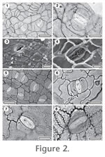
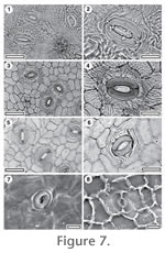
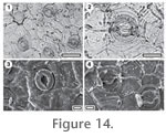
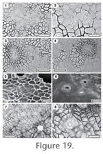
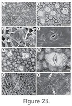
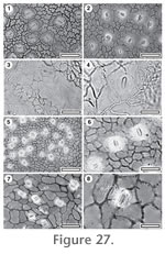
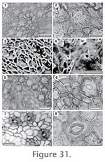
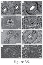
Referred specimens and occurrence: SL1309, BL-32; SB0841, GL-02; SL1740, Sthd-017; SL2434, Sthd-033; SL2076, Sthd-113. Stomatal complexes. Stomatal complexes evenly spread, isolated, randomly oriented, tetracytic (sometimes brachyparacytic), developmentally tangenticytic or stephanocytic bicylic, with distinct lateral subsidiary cells parallel to the stomatal axis and polar cells at right angles to the axis and which may 'enclose' the lateral cells. Sometimes subsidiary cells are modified by a tangential division, giving two lateral and two polar cells, size range unimodal. Subsidiary cells (4–6) typically elongate, or wedge-like, periclinal walls thicker than over normal epidermal cells, smooth, unornamented. Guard cell pair outline distinctly elongate, often with flattened poles, like a rounded rectangle, outlined by a well-defined anticlinal wall, length 27–37 µ (medium), at same level as subsidiary cells (exposed on surface), little polar development between guard cells (guard cells appear as continuous ring). Outer stomatal ledge possibly absent, with the visible cuticle lying directly over the guard cells, thinner than normal epidermal cells (often broken away), and with a narrowly elliptic pore. Epidermal Cells. Epidermal cell flanges clearly visible using TLM, normal cells highly variable from isodiametric to elongate (cells over major veins more isodiametric shape than normal epidermal cells), distinctly larger than the stomata, anticlinal walls straight to curved, unbuttressed, smoothly textured, unornamented. Indumentum. Glabrous. Distinguishing features. Distinguished by the elongate guard cell pair with no division at the polar ends, lack of outer stomatal ledges and subsidiary cells which include both typically paracytic forms as well as polar forms which have formed by tangential cut-off. Identification. On the basis of its highly distinctive stomatal complexes, this taxon is regarded as an extinct taxon in the Gnetaceae. Extant Gnetum species (for comparison G. microcarpum (Earl) Blume, G. gnemon L., G. tenuifolium Ridley, and G. latifolium Blume are illustrated; Figs 2.5-2.8), have uniquely-shaped stomata which are almost rounded rectangles. There is no clear division between the two guard cells and an outer stomatal ledge appears to be absent (the cuticle that is present lies directly over the guard cells). CUT-Z-ADE is distinct from Gnetum because it has distinct polar subsidiary cells, which are essentially absent in Gnetum. For further discussion see Pole (2007d) where CUT-Z-GDB from the Early Eocene of Tasmania is also regarded as Gnetalean. The clearly related cuticle of the Tasmanian specimens differs in the walls of the epidermal cells being distinctly wavy. Comments. Gnetum has no fossil record, but Won and Renner (2006) have used genetics to infer that the major divergences amongst the extant clades of the genus date from the Late Oligocene.
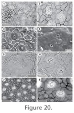
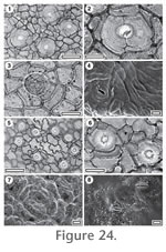
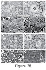
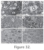
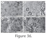
Magnoliopsida Cronquist 1981 Family identification: All extant Winteraceae genera are characterized by large paracytic stomata and a highly granular or ornamented cuticle (Bongers 1973). Key to Winteraceae1. Cuticle ornamented with closely-spaced, thick (5-10 m) discontinuous ridges. CUT-Z-FJF 1. Cuticle unornamented. 2. 2. Cuticle moderately granular (outline of outer stomatal ledge clearly visible under TLM) CUT-Z-FFD 2. Cuticle strongly granular (plugging and obscuring stomatal outline under TLM). CUT-Z-FJH
CUT-Z-FFD
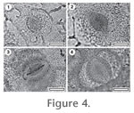
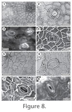
Stomatal complexes. Stomatal complexes evenly spread, isolated, randomly oriented, brachyparacytic, size range unimodal. Subsidiary cells elongate, periclinal walls of same thickness as normal epidermal cells, unornamented. Guard cell pair outline difficult or impossible to see in TLM view (obscured by outer stomatal ledge), but obviously elliptical, length 45–53 µ (large), at same level as subsidiary cells. Outer stomatal ledge elliptical, thicker than normal epidermal cells, extending over centre of stoma, with a broad, elliptical pore, plugged with cuticular material. Epidermal Cells. Epidermal cell flanges clearly visible using TLM, normal cells highly variable from isodiametric to elongate (cells over veins not distinguished by shape), approximately the same size as the stomata, anticlinal walls curved to wavy, unbuttressed, with moderately granular texture, unornamented. Indumentum. Glabrous. Identification. Winteraceae based on large paracytic stomata and moderately coarse cuticle. Compare with illustrations of extant Winteraceae (Belliolum haplopus (B.L. Burtt) A.C. Sm., B. burttianum A.C. Smith (1985), Exospermum stipitatum (Baillon) van Tieghem (1900) ex Morot, Zygogynum balansae Tiegh.; Figs 4.1-4.4).
CUT-Z-FJF Reference Specimen and locality: SL2538, GL-02. Stomatal complexes. Stomatal complexes evenly spread, isolated, randomly oriented, brachyparacytic, size range unimodal. Subsidiary cells with periclinal walls of same thickness as normal epidermal cells, smooth, ornamented with thick ridges generally parallel to the stomatal axis on either side of the stoma, and at right angles to it at either end. Guard cell pair outline difficult or impossible to see in TLM view (obscured by surface ornamentation), at same level as subsidiary cells, length 38–40 µ (large). Outer stomatal ledge elliptical, thinner than normal epidermal cells, extending over whole stoma, with a narrowly elliptic pore, plugged with cuticular matter.
Epidermal Cells. Epidermal cell flanges not clearly visible under TLM because of surface thickenings, normal cells elongate (cells over veins not distinguished by shape), approximately the same size as the stomata, anticlinal walls curved to wavy, unbuttressed, smoothly textured ornamented with closely-spaced, thick (5-10 m)
Indumentum. Glabrous. Distinguishing features. Distinguished from CUT-Z-FJH by the ornamentation of massive cuticular ridges. Identification. Winteraceae based on large paracytic stomata and highly ornamented cuticle. Compare with illustrations of extant Winteraceae (Figs 4.1-4.4).
CUT-Z-FJH Reference Specimen and locality: SL2185, GL-10. Referred specimens and occurrence: SL3163, BL-33. Stomatal complexes. Stomatal stomatal complexes evenly spread, isolated, randomly oriented, brachyparacytic, size range unimodal. Subsidiary cells with periclinal walls of same thickness as normal epidermal cells, texture granular, unornamented. Guard cell pair outline ovate, outer margin obscured under TLM by surface ornamentation, at same level as subsidiary cells, length 35–45 µ (large). Outer stomatal ledge elliptical, thicker than normal epidermal cells, extending over whole stoma with a narrowly elliptic pore, plugged with cuticular material. Epidermal Cells. Epidermal cell flanges somewhat diffuse, normal cells highly variable from isodiametric to elongate (cells over veins not distinguished by shape), approximately the same size as the stomata, anticlinal walls curved to wavy, unbuttressed, texture strongly granular, unornamented. Indumentum. Glabrous. Distinguishing features. Distinguished from CUT-Z-FJF and CUT-Z-FFD the strongly granular periclinal epidermal cell walls. Identification. Winteraceae based on large brachyparacytic stomata and highly granular cuticle. Compare with illustrations of extant Winteraceae (Figs 4.1-4.4). Atherospermataceae Brown 1814
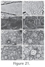
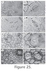
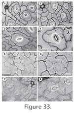
Key to Atherospermataceae1. Cuticle glabrous. CUT-Z-CEF 1. Trichome attachment scars present. 2. 2. Ornamented by cuticular ridges. CUT-Z-CFA 2. Unornamented. 3. 3. Massively thickened trichome attachment sites present. CUT-Z-CFC 3. Massively thickened trichome attachment sites absent. 4. 4. Epidermal cells distinctly smaller than the stomata. CUT-Z-ECG 4. Epidermal cells of roughly similar size to the stomata 5. 5. Epidermal cell anticlinal walls markedly sinuous, slightly buttressed, CUT-Z- CGB 5. Epidermal cell anticlinal walls not sinuous or buttressed, CUT-Z-JIF
CUT-Z-CEF
Referred specimens and occurrence: SL2409, BL-32; SB1392, BL-33; SL3136, GL-02. Stomatal complexes. Stomatal complexes evenly spread, isolated, randomly oriented, actinocytic, or staurocytic, size range unimodal, (some stomatal complexes have more subsidiary cell development, but they are no larger than normal stomatal complexes). Subsidiary cells (4–5) irregularly shaped, but often elongate radially to the stoma, periclinal walls thicker than over normal epidermal cells, smooth, unornamented. Guard cell pair outline distinctly circular, length c. 34 µ (medium), at same level as subsidiary cells (exposed on surface), with prominent T-piece thickenings at polar ends. Outer stomatal ledge subcircular, thicker than normal epidermal cells, extending over centre to inner edge of stoma, with a broad, sub-circular pore. Epidermal Cells. Epidermal cell flanges clearly visible using TLM, normal cells elongate (cells over veins not distinguished by shape), approximately the same size as the stomata, anticlinal walls curved to wavy, unbuttressed, smoothly textured, unornamented. Indumentum. Glabrous. Distinguishing features. Distinguished from CUT-Z-CFC by being glabrous and from CUT-Z-EHI by the subsidiary cells being level with the other epidermal cells.
CUT-Z-CFA
Referred specimens and occurrence: SL0289, BL-01; SL3064, BL-26; SL3151, GL-01; SL2543, GL-02; SL2682, GL-04; SL2130, GL-08; SL2157, GL-09; SL2919, GL-22; SL4799, GL-23; SL2960, GL-25. Stomatal complexes. Stomatal complexes evenly spread, isolated, randomly oriented, cyclocytic, with all some, or none of the subsidiary cells having been modified by a tangential division, size range unimodal. Subsidiary cells (4–6) typically elongate, or wedge-like, often including distinct polar cells (at right angles to the stomatal axis) which may 'enclose' the lateral cells, periclinal walls of same thickness as normal epidermal cells, smooth, ornamented with one or two peristomatal ridges and there may be varying degrees of ridges which tend to flow around the stoma. Guard cell pair outline ovate, outlined by a well-defined anticlinal wall, length 27–45 µ (large to medium), at same level as subsidiary cells (exposed on surface), with prominent T-piece thickenings at polar ends. Outer stomatal ledge sub circular, thicker than normal epidermal cells, extending over whole stoma, with a broad, sub-circular pore. Epidermal Cells. Epidermal cell flanges clearly visible using TLM, normal cells isodiametric (cells over major veins more elongate), distinctly smaller than the stomata, anticlinal walls curved to wavy, unbuttressed, smoothly textured, ornamented throughout with short ridges or just in the area of the stomatal complexes. Indumentum. Scars of deciduous trichomes sparse, scattered over venal and non-venal regions, inserted between (8–13, hard to count) epidermal cells modified by tangential divisions to form a sub-circular zone of foot cells with massive wall thickening, scar diameter much smaller than normal epidermal cell.
CUT-Z-CFC
Referred specimens and occurrence: SL0370, Mata-03. Stomatal complexes. Stomatal complexes evenly spread, isolated, randomly oriented, cyclocytic, with all some, or none of the subsidiary cells having been modified by a tangential division (developmentally tangenticytic or incomplete stephanocytic bicyclic), development size range unimodal. Subsidiary cells (5–6) irregularly-shaped, periclinal walls of same thickness as normal epidermal cells, smooth, unornamented. Guard cell pair outline circular, outer margin obscured under TLM by surface ornamentation, at same level as subsidiary cells, length 23–25 µ (medium). Outer stomatal ledge sub circular, circumscribed by a ring of very thin cuticle, the ledge itself thicker than normal epidermal cells, extending over centre of stoma, with a broad, sub-circular pore. Epidermal Cells. Epidermal cell flanges clearly visible using TLM, normal cells isodiametric (cells over veins not distinguished by shape), distinctly smaller than the stomata, anticlinal walls straight, unbuttressed, smoothly textured, unornamented. Indumentum. Attachment scars of deciduous trichomes common, diameter similar or larger in size than a normal epidermal cell. Epidermal cells around trichome attachment scar (4–11) modified with massive thickening to form a rim and radiating walls. Distinguishing features. Distinguished from CUT-Z-CEF by having massively thickened trichome attachment sites and from CUT-Z-EHI by the subsidiary cells being level with the other epidermal cells.
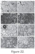
CUT-Z-CGB Reference Specimen and locality: SL0088, BL-08. Referred specimens and occurrence: SL0239, BL-04; SL3281, BL-05; SL3193, BL-32; SL1258, BL-33; SL1173, GL-01; SL2882, GL-20; SL3214, Sthd-158. Stomatal complexes. Stomatal complexes evenly spread, isolated, randomly oriented, cyclocytic, size range unimodal. Subsidiary cells (6–7) irregularly-shaped, periclinal walls of same thickness as normal epidermal cells, smooth, unornamented. Guard cell pair outline ovate, outlined by a well-defined anticlinal wall, at same level as subsidiary cells, length 30–45 µ (medium). Outer stomatal ledge sub circular, thicker than normal epidermal cells, extending over whole stoma, with a broad, sub-circular pore. Epidermal Cells. Epidermal cell flanges clearly visible using TLM, normal cells highly variable from isodiametric to elongate (cells over major veins more elongate), approximately the same size as the stomata, anticlinal walls markedly sinuous, slightly buttressed, smoothly textured, unornamented. Indumentum. Attachment scars of deciduous trichomes common, inserted between (5–7) epidermal cells modified into radially elongate foot cells, scar poral diameter much smaller than a normal epidermal cell. Distinguishing features. Distinguished from CUT-Z-CFC by the sinuous and slightly buttressed walls.
CUT-Z-JIF
Referred specimens and occurrence: SL1354, BL-05; SL3166, BL-33; SL2210, GL-12; SL1060, Mata-01. Stomatal complexes. Stomatal complexes in areoles, isolated, randomly oriented, anomocytic or with occasional thin subsidiary cells produced by tangential divisions (developmentally incomplete stephanocytic bicyclic), size range unimodal. Contact cells (4–6) irregularly-shaped, periclinal walls thicker than over normal epidermal cells, smooth, ornamentation sometimes a single discontinuous ridge around the outer stomatal ledge (periclinal wall thickness near this rim is much thinner than over epidermal cells). Guard cell pair outline circular, or ovate, outlined by a well-defined anticlinal wall, at same level as subsidiary cells (exposed on surface), length 37–45 µ (large), with prominent polar rods. Outer stomatal ledge sub circular, much thicker than normal epidermal cells, extending over whole stoma, with a broad, sub-circular pore. Epidermal Cells. Epidermal cell flanges clearly visible using TLM, normal cells isodiametric (cells over both major and fine venation more elongate), approximately the same size as the stomata, anticlinal walls curved to wavy, unbuttressed, smoothly textured, unornamented. Indumentum. Attachment scars of deciduous trichomes common, scattered over venal and non-venal regions, inserted between (5–7) epidermal cells modified by massive wall thickening to form a rim with radiating walls, scar diameter much smaller than a normal epidermal cell. Distinctive features. Comparable to CUT-Z-CGB but without the buttressed and sinuous epidermal cell walls.
CUT-Z-ECG Reference Specimen and locality: SL1954, Sthd-051. Referred specimens and occurrence: SL3042, BL-22; SL1563, Sthd-004; SL1730, Sthd-017; SL1716, Sthd-018; SL1706, Sthd-019; SL1776, Sthd-022; SL1779, Sthd-030; SL1937, Sthd-059; SL1894, Sthd-073; SL1906, Sthd-074; SL1833, Sthd-095. Stomatal complexes. Stomatal complexes evenly spread, isolated, randomly oriented, laterocyclic, all some, or none of the subsidiary cells may have been modified by a tangential division (developmentally incomplete stephanocytic bicyclic), size range unimodal (but with wide range of stomatal size). Subsidiary cells (7–8) irregularly-shaped, periclinal walls thicker than over normal epidermal cells, smooth, unornamented. Guard cell pair outline ovate, outlined by a well-defined anticlinal wall, at same level as subsidiary cells (exposed on surface), length 38–43 µ (large), with prominent T-piece thickenings at polar ends. Outer stomatal ledge sub circular, thicker than normal epidermal cells, extending over most of the stoma, with a broad, sub-circular pore. Epidermal Cells. Epidermal cell flanges clearly visible using TLM, normal cells isodiametric (cells over major veins vaguely or inconsistently more elongate), approximately the same size as the stomata, anticlinal walls straight, unbuttressed, smoothly textured, unornamented. Indumentum. Attachment scars of deciduous trichomes common, probably restricted to regions over veins (unclear), inserted between epidermal cells modified only slightly, forming a thickened rim, scar diameter similar in size to a normal epidermal cell. Distinguishing features. Differing from the other taxa in the group in having very thick outer stomatal ledges, and stomatal complexes with clear evidence of tangential divisions, and epidermal cells distinctly smaller than the stomata. Monimiaceae Jussieu 1809 Key to Monimiaceae1. Unornamented. CUT-Z-DDG 1. Ornamented with ridges. 2. 2. Ornamented with widely-spaced short ridges, outer stomatal ledge narrow. CUT-Z-DDH 2. Ornamented with prominent, closely-spaced long ridges, outer stomatal ledge prominent. CUT-Z-JIJ
CUT-Z-DDG (Hedycarya)
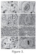
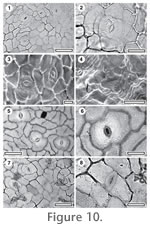
Stomatal complexes. Stomatal complexes evenly spread, isolated, randomly oriented, brachyparacytic or amphibrachyparacytic, size range unimodal. Subsidiary cells typically elongate, tangential to the guard cells, periclinal walls of same thickness as normal epidermal cells, smooth, unornamented. Guard cell pair outline elongate, outlined by a well-defined anticlinal wall, at same level as subsidiary cells (exposed on surface), length 20–25 µ (medium). Outer stomatal ledge elliptical, same thickness as normal epidermal cells, extending over inner edge of stoma, with a narrowly elliptic pore. Epidermal Cells. Epidermal cell flanges clearly visible using TLM, normal cells highly variable from isodiametric to elongate (cells over veins not distinguished by shape), distinctly larger than the stomata, anticlinal walls curved to wavy, unbuttressed, inner surfaces slightly granular, outer surfaces smooth and undulose, unornamented (though may have some faint ridging). Indumentum. Glabrous. Identification. The similarity to extant Hedycarya arborea Forster, J.R., and Forster, G. (1776) and H. dorstenoides A. Gray (1866) (Figs 10.5-10.8) is taken to indicate the genus.
CUT-Z-DDH (Hedycarya sp.)
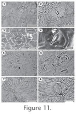
Referred specimens and occurrence: SL2105, GL-07; SL2145, GL-08. CUT-L-DDH, SL1193, GL-02; SL2927, GL-22; SL3293, Mata-03. Stomatal complexes. Stomatal stomatal complexes evenly spread, isolated, randomly oriented, brachyparacytic, size range unimodal. Subsidiary cells with periclinal walls thinner than that over normal epidermal cells, granular, unornamented normally, or with short ridges perpendicular to the stomatal axis. Guard cell pair outline elongate, not outlined by a clear anticlinal wall, at same level as subsidiary cells (exposed on surface), length 25–30 µ (medium). Outer stomatal ledge narrow-elliptical, thinner than normal epidermal cells, extending over inner edge of stoma. Epidermal Cells. Epidermal cell flanges somewhat diffuse, normal cells highly variable from isodiametric to elongate (cells over veins not distinguished by shape), distinctly larger than the stomata, anticlinal walls straight, unbuttressed, inner surface texture granular, outer surface mostly very smooth but with some ornamentation of short ridges perpendicular or parallel to some stomata. Indumentum. Glabrous. Distinguishing features. Distinguished by the rectangular outline of the stomatal complexes, the granular texture of the epidermal periclinal walls, the ridges of cuticle running at right angles to the stomatal pore on some stomatal complexes, and the obscure contact between guard cells and subsidiary cells. CUT-Z-JIJ differs by having distinct and broad outer stomatal ledges. Identification. The similarity to extant Hedycarya angustifolia A. Cunningham (1838) (AQ063593) is taken to indicate the genus (Figs 11.5-11.6).
CUT-Z-JIJ Reference Specimen and locality: SB1329, BL-32. Stomatal complexes. Stomatal complexes evenly spread, isolated, randomly oriented, brachyparacytic, size range unimodal. Subsidiary cells with periclinal walls of same thickness as normal epidermal cells, granular, ornamented with ridges projecting at right angles to the stomatal axis. Guard cell pair outline circular, or ovate, not outlined by a clear anticlinal wall, at same level as subsidiary cells (exposed on surface), length 26–30 µ (medium). Outer stomatal ledge elliptical, thicker than normal epidermal cells, extending over whole stoma, with a narrowly elliptic pore. Epidermal Cells. Epidermal cell flanges not clearly visible under TLM because of surface thickenings, normal cells elongate, cells over veins not distinguished by shape, anticlinal walls probably wavy, unbuttressed, inner surfaces coarsely granular, outer surfaces ornamented with bands of prominent, irregular ridges parallel to the stomata and radiating from trichomes. Indumentum. Glabrous. Distinguishing features. Distinguished by the brachyparacytic stomatal complexes with broad and distinct outer stomatal ledges in combination with highly granular cuticle. Specimen SL2647, which lacks the strong surficial ornamental of ridges found in the Reference Specimen, is included in this taxon. It comes from the same sample and agrees in the other major characters. CUT-Z-DDH differs in not having the broad outer stomatal ledges and narrow, slit-like stomatal apertures. Proteaceae Jussieu 1789 Family identification: The majority of Proteaceae have brachyparacytic stomatal complexes. They often have trichome scars associated with one to several epidermal cells (Lange 1978; Carpenter 1994). Key to Proteaceae1. Stomatal complexes aligned. 2. 1. Stomatal complexes randomly oriented. 3. 2. Outer stomatal ledge narrowly elliptic, epidermal cell anticlinal walls markedly sinuous, buttressed. CUT-P-EDA 2. Outer stomatal ledge subcircular, epidermal cell anticlinal walls straight, unbuttressed. CUT-P-FEE 3. Cuticle glabrous. CUT-P-EFB 3. Attachment scars of trichomes present. 4. 4. Trichome attachment scar with a thickened rim, subsidiary cells ornamented with many fine ridges. CUT-P-EHA 4. Trichome attachment scar thickened into a raised platform, subsidiary cells ornamented with 2-3 ridges. CUT-P-EHJ
CUT-P-EDA (Placospermum sp.)
Stomatal complexes. Stomatal distribution over leaf surfaces hypostomatic, stomatal complexes evenly spread, isolated, aligned more or less parallel to long axis of leaf, brachyparacytic, size range unimodal. Subsidiary cells elongate parallel to stomatal axis, with periclinal walls thinner than that over normal epidermal cells, smooth, unornamented. Guard cell pair outline narrow and elongate, outlined by a well-defined anticlinal wall, at same level as subsidiary cells (exposed on surface). Outer stomatal ledge narrowly elliptic, ridge-like, much thinner than normal epidermal cells (often broken away), extending over inner edge of stoma, with a narrowly elliptic pore. Epidermal Cells. Epidermal cell flanges clearly visible using TLM, normal cells isodiametric (cells over veins not distinguished by shape), approximately the same size as the stomata; anticlinal walls markedly sinuous; buttressed; smoothly textured, unornamented. Indumentum. Glabrous. Non-stomatal surface. Epidermal cells highly variable from isodiametric to elongate; sinuous; cells over veins not distinguished; trichomes absent. Identification. There are no multicelled trichome attachment scars which would make a stronger case for identification as Proteaceae. However, the morphology is comparable with Placospermum coriaceum (White and Francis 1924) (the sole member of the genus, and which also lacks or has very rare trichome attachment scars) in its generally aligned stomatal complexes, a strikingly distinctive elongate stomatal pore (cf. Carpenter 1994, figs 5-7) and sinuous epidermal cell walls. There are obvious differences in that the epidermal cells of the fossil are also buttressed, and the stomatal complexes of the extant species are often linked by large epidermal cells. The fossil is suggested to be an extinct Placospermum.
CUT-P-EFB (Lomatia sp.) Reference Specimen and locality: SL1828, Sthd-095. Referred specimens and occurrence: SL1556, Sthd-004; SL1766, Sthd-011; SL1843, Sthd-086. Stomatal complexes. Stomatal complexes evenly spread, isolated, randomly oriented, brachyparacytic, size range unimodal. Subsidiary cells isodiametric, periclinal walls of same thickness as normal epidermal cells, smooth, ornamented with 3–4 ridges parallel with the stomatal axis, and slightly longer than the stoma. Guard cell pair outline circular, outlined by a well-defined anticlinal wall, at same level as subsidiary cells (exposed on surface), length 18–23 µ (medium), with prominent T-piece thickenings at polar ends. Outer stomatal ledge sub circular, same thickness as normal epidermal cells, extending over whole stoma, with a broad elliptical or sub-circular pore. Epidermal Cells. Epidermal cell flanges clearly visible using TLM, normal cells isodiametric (cells over major veins more elongate), approximately the same size as the stomata, anticlinal walls sinuous, slightly buttressed, inner surfaces slightly granular, outer surfaces smooth and undulose, unornamented. Indumentum. Glabrous. Identification. There are no multicelled trichome attachment scars which would make a stronger case for identification as Proteaceae. In addition, the subsidiary cells are rather large, while in most extant Proteaceae they are relatively narrow. However, there is a general similarity with some extant species, such as Lomatia fraxinifolia F. Muell. ex Bentham (1870) (cf. Carpenter 1994, figs 65-66) in which trichome attachment scars may also be rare or absent.
CUT-P-EHA
Stomatal complexes. Stomatal complexes evenly spread, isolated, randomly oriented, brachyparacytic, size range unimodal. Subsidiary cells isodiametric, periclinal walls of same thickness as normal epidermal cells, ornamented with many fine ridges parallel with the stoma. Guard cell pair outline ovate, not outlined by a clear anticlinal wall, at same level as subsidiary cells (exposed on surface although sometimes partially obscured by adjacent epidermal cells), length 30–37 µ (medium), little polar development between guard cells (guard cells appear as continuous ring). Outer stomatal ledge sub circular, thinner than normal epidermal cells, extending over centre of stoma, with a broad, sub-circular pore. Epidermal Cells. Epidermal cell flanges clearly visible using TLM, normal cells isodiametric (cells over veins not distinguished by shape), approximately the same size as the stomata, anticlinal walls straight, unbuttressed, smoothly textured, unornamented. Indumentum. Attachment scars of deciduous trichomes common, scattered over venal and non-venal regions, inserted over several (2–5) modified epidermal cells selectively thickened to form a thickened rim, with a prominent central pore. Scar diameter similar in size to a normal epidermal cell. Identification. General morphology suggests the taxon has affinities with Tribe Macadamieae of Weston and Barker (2006) (see illustrations in Carpenter 1994). The surface ridging restricted to the subsidiary cells is similar to Carnarvonia although the morphology of the trichome scar is different (in Carnarvonia they are associated with mostly 1-2 epidermal cells and lack the central pore).
CUT-P-EHJ Reference Specimen and locality: SL1682, Sthd-033. Referred specimens and occurrence: SL0291, BL-01; SL2433, Sthd-051; SL2420, Sthd-078; SL2022, Sthd-087; SL1651, Sthd-110; SL3298. Stomatal complexes. Stomatal complexes evenly spread, isolated, randomly oriented, brachyparacytic, size range unimodal. Subsidiary cells isodiametric, periclinal walls of same thickness as normal epidermal cells, smooth, ornamented with 3–4 ridges parallel with, and on either side of the stoma. Guard cell pair outline ovate, outlined by a well-defined anticlinal wall, at same level as subsidiary cells (exposed on surface), length 20–22 µ (medium). Outer stomatal ledge sub circular, thinner than normal epidermal cells, extending over whole stoma, with an elliptical pore. Epidermal Cells. Epidermal cell flanges clearly visible using TLM, normal cells isodiametric (cells over veins not distinguished by shape), approximately the same size as the stomata, anticlinal walls markedly sinuous, unbuttressed, inner surfaces smooth, outer surfaces smooth and irregularly ridged with bands of irregular ridges parallel to stomatal complexes and radiating from trichomes. Indumentum. Attachment scars of deciduous trichomes common, inserted over several (3–4) epidermal cells modified to form a thick, raised circular platform, on top of which sits a smooth, thick hollow collar, scattered over venal and non-venal regions but slightly more common over veins. Pairs of scars may be joined by a narrow extension of the platform. Scar diameter similar in size to a normal epidermal cell. Identification. General morphology suggests the taxon has affinities with Tribe Macadamieae of Weston and Barker (2006) (Carpenter 1994). The surface ridging, more prominent on the subsidiary cells and the morphology of the trichome scars is similar to Sleumerodendron, Hicksbeachia and Turrillia vitensis (Turrill) A.C. Smith (1985) (or Kermadecia vitensis Turrill 1915).
CUT-P-FEE
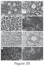
Stomatal complexes. Stomatal complexes in areoles, isolated, generally aligned, brachyparacytic, size range unimodal. Subsidiary cells narrow, periclinal walls of same thickness as normal epidermal cells, unornamented. Guard cell pair outline difficult or impossible to see in TLM view, outer margin obscured under TLM by surface ornamentation, at same level as subsidiary cells, length 28–35 µ (medium), with strong polar rod thickenings. Outer stomatal ledge prominent, circular to sub circular, thicker than normal epidermal cells, extending over whole stoma, with a broad, sub-circular pore. Epidermal Cells. Epidermal cell flanges clearly visible using TLM, normal cells isodiametric (cells over major veins more elongate), approximately the same size as the stomata, anticlinal walls straight, unbuttressed, smoothly textured, unornamented. Indumentum. Attachment scars of deciduous trichomes common, but not noted over veins. positioned over 1–2 epidermal cells modified to form an irregular platform (by thickening of the periclinal walls) which contains the trichome scar. Scar diameter similar in size to a normal epidermal cell. Identification. Identification with the Proteaceae seems clear, although further identification is uncertain. The generally aligned stomata and trichome attachment scars over only 1-2 epidermal cells are the most important characters and suggest affinities lie with Tribe Embothrieae of Weston and Barker (2006). Menispermaceae Jussieu 1789 Family identification: Based on the reference collection the Menispermanceae typically have distinct robust and very densely staining trichome attachment scars, although some species are glabrous. They also tend to have both guard and subsidiary cells with cuticle thinner than that over normal epidermal cells. Hill (1989) described a fossil genus of the family, Menispermaphyllum, although it is unclear how this genus differs from some extant genera, for instance the Legnephora with which it was compared. Hill compared the cuticle of his Eocene M, tomentosum with extant L. moorei (F.Muell.) Miers 1867, but the papillate cuticle of L. moorei in his fig. 2E looks nothing like any L. moorei specimen prepared for this database (which are not papillate), nor any other Menispermaceae in the database. Key to Menispermaceae1. Epidermal cell anticlinal walls curved to wavy, cuticle over stomatal complexes much thinner than normal epidermal cells (often broken away). CUT-Z-FCA 1. Epidermal cell anticlinal walls sinuous, cuticle over stomatal complexes thinner than normal epidermal cells. CUT-Z-FEC
CUT-Z-FCA
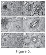
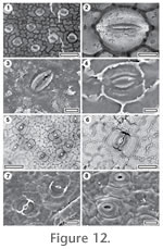
Stomatal complexes. Stomatal complexes evenly spread, isolated, randomly oriented, cyclocytic, size range unimodal. Subsidiary cells (5–6) irregularly shaped, but often with apparently distinct elongate lateral or polar cells, periclinal walls thinner than normal epidermal cells, smooth, unornamented. Guard cell pair outline ovate, outlined by a well-defined anticlinal wall, at same level as subsidiary cells (exposed on surface), length 20–23 µ (medium), some wall development between guard cells, and sometimes development of T-piece thickenings at the poles. Outer stomatal ledge sub circular, much thinner than normal epidermal cells (often broken away), extending over whole stoma, with an elliptical pore. Epidermal Cells. Epidermal cell flanges clearly visible using TLM, normal cells highly variable from isodiametric to elongate (cells over major veins more elongate), approximately the same size as the stomata, anticlinal walls curved to wavy, unbuttressed, smoothly textured, unornamented. Distinguishing features. The most distinct character is the massively-thickened trichome attachment scar.Indumentum. Bases of thick (densely staining), probably poral (with a very narrow pore) trichomes common, scattered over venal and non-venal regions, the bases persist, but in all cases the trichome has broken off near base, perhaps along a predefined zone of weakness, scar diameter similar in size to a normal epidermal cell. Epidermal cells around trichome attachment scar modified into (6–9) radially elongate foot cells, with no distinct thickening of walls. Identification. Broadly comparable with extant Pleiogyne australis Endlicher (1843) with respect to darkly staining trichome attachment scars, ovate shape of stoma, thin cuticle over guard and subsidiary cells, and the scattered presence of elongate lateral subsidiary cells (Figs 15.5-15.6).
CUT-Z-FEC Reference Specimen and locality: SB1321, BL-32. Stomatal complexes. Stomatal complexes in areoles, isolated, randomly oriented, anisocytic, size range unimodal. Subsidiary cells (3–4) isodiametric, with periclinal walls thinner than normal epidermal cells, granular, unornamented. Guard cell pair outline ovate, outlined by a well-defined anticlinal wall, at same level as subsidiary cells (exposed on surface), length 22–23 µ (medium). Outer stomatal ledge sub circular, thinner than normal epidermal cells, extending over whole stoma, with an elliptical pore. Epidermal Cells. Epidermal cell flanges clearly visible using TLM, normal cells elongate (cells over veins not distinguished by shape), distinctly larger than the stomata, anticlinal walls sinuous and sometimes rather sharply so, unbuttressed, texture granular, unornamented. Indumentum. Bases of thick (densely staining), probably poral (with a very narrow pore) trichomes common, scattered over venal and non-venal regions (but slightly more common over veins), the bases persist, but in all cases the trichome has broken off near base, perhaps along a predefined zone of weakness, base diameter similar in size to a normal epidermal cell. Epidermal cells around trichome attachment scar (4–6) unmodified. Identification. Compare with extant Pleiogyne australis (Figs 15.7-15.8) in terms of very thick trichome attachment scars, shape of stoma, thin guard and subsidiary cell cuticle. However, it does not have the occasional elongate subsidiary cells.
Santalaceae Brown 1814
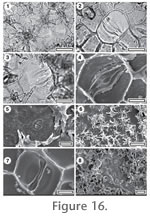
Stomatal complexes. Stomatal complexes evenly spread, isolated, randomly oriented, brachyparacytic, size range unimodal (but with wide range of stomatal size). Subsidiary cells length 32–40 µ (large), or (medium), periclinal walls thinner than normal epidermal cells. Guard cell pair very narrow, poles sharply reflexed outwards, overarched by the subsidiary cells (not exposed on the surface). Epidermal Cells. Epidermal cell flanges clearly visible using TLM, normal cells highly variable from isodiametric to elongate (cells over veins not distinguished by shape), approximately the same size as the stomata, anticlinal walls straight, unbuttressed, smoothly textured, unornamented. Indumentum. Hirsute, densely covered with stellate trichomes, inserted over, or the outgrowth of, a single modified epidermal cell, similar in size to a normal epidermal cell. Distinguishing features. Distinguished by the unique combination of brachyparacytic stomatal complexes (a pair of narrow guard cells flanked by a broad subsidiary cell on either side) and stellate/multibranched trichomes. Identification. The basic stomatal morphology is comparable with the Lauraceae. However, the stellate trichomes indicate it clearly belongs to Notothixos (Viscaceae), a mistletoe (see Barlow 1983 for more details on the trichomes). It may be compared closely with extant N. subaureus Oliver (1863) (Fig. 16.7-16.8).
Casuarinaceae Brown 1814
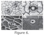
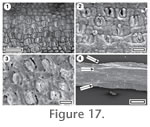
Referred specimens and occurrence: SL3232, GL-30; SL2705, Sthd-046; SL2702, Sthd-047, SL2703, Sthd-094; SL2706, Sthd-097; SL2704, Sthd-099. Description. Branchlets with four-sided articles, approximately square in cross-section, with four highly reduced leaves in a whorl at each junction. Leaves with acute apices, sharp sinuses, margin entire, length c. 400 m, width c. 200 m. Stomatal complexes in two compact zones within a shallow furrow on each side of the article, 4-7 files per zone, complexes brachyparacytic, oriented perpendicular to the long axis of the branchlet, length c 20 m. Mostly with 1-2 epidermal cells between each stomatal complex in a file. Glabrous. Epidermal cells isodiametric, walls distinctly thickened compared with the thinner cells of the stomatal zones. Identification. Clearly belongs to the Casuarinaceae on the basis of gross morphology of article branchlets with highly reduced leaves in whorls, and transversely oreiented stomata essentially restricted to the articles (terminology follows Scriven and Hill 1995). The identification as Gymnostoma is based on the four-sided articles and relatively surficial stomatal complexes (Scriven and Hill 1995). The stomatal length of c. 20 µ also supports Gymnostoma, as Scriven and Hill noted that stomatal length in Casuarina and Allocasuarina was usually more than 35 m. Based on Scriven and Hill's summary, the shape and thickness of the epidermal cells is unusual for Gymnostoma.
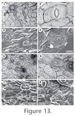
Strasburgeriaceae Solereder 1908
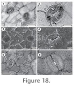
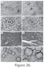
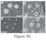
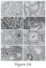
Stomatal complexes. Stomatal distribution over leaf surfaces hypostomatic; stomatal complexes evenly spread, mostly isolated although sometimes networked, possibly slightly aligned with one another, anomocytic (although there are 4–6 contact cells, and sometimes there have been tangential divisions around a few stomata which produce what could be called subsidiary cells), size range unimodal. Guard cell pair outline ovate, outlined by a well-defined anticlinal wall, at same level as subsidiary cells (exposed on surface), length 50–53 µ (large), with prominent T-piece thickenings at polar ends. Outer stomatal ledge sub circular, much thicker than normal epidermal cells, extending over whole stoma, with an elliptical pore. Epidermal Cells. Epidermal cell flanges clearly visible using TLM, normal cells highly variable from isodiametric to elongate (cells over veins not distinguished by shape), approximately the same size as the stomata, anticlinal walls straight, or curved to wavy, unbuttressed, smoothly textured, unornamented. Indumentum. Attachment scars of deciduous trichomes sparse, scattered over venal and non-venal regions, inserted between (6–7) epidermal cells modified only slightly, with a thickened rim. Fine ridges of cuticle radiate out from the trichome attachment scars, scar diameter much smaller than a normal epidermal cell. Distinguishing features. Distinguished by the strong thickening over the guard cells and the anomocytic stomatal complexes. Identification: The large size and general form of the stoma, the epidermal cells, and the anomocytic stomatal construction find a good match with extant New Caledonian Strasburgeria robusta (Vieill. ex Panch. & Sebert ) Guillaumin (1942) (Figs 18.5-18.6) The differences include scattered trichome attachment scars on the fossil, which the extant species appears to lack, and while stomata are common in this species and frequently networked, their density is much lower in the fossil although networking is still apparent. The modern specimen also shows much cuticularisation of the stomatal chamber which is absent on the fossil. The differences would be consistent with a different species of Strasburgeria. The genus has been recorded as pollen from the Gore Lignite Measures (Jarzen and Pocknall 1993). Cunoniaceae (Brown 1814) - Elaeocarpaceae (de Candolle 1816) Family identification: The guard cells of almost all Elaeocarpaceae – Cunoniaceae appear essentially 'naked' under TLM. The outer stomatal ledges are so thin and smooth that they leave an unobstructed view of classic 'paired kidney' stomata. The typical narrow and darkly-staining ring around the stomatal pore may be an inner ledge, or a thickened rim of the outer stomatal ledges. There is often a distinct notch at either pole of the guard cell pair. These characters are a good first step in suspecting Elaeocarpaceae – Cunoniaceae. Pole (1996) wrote "Presently, no cuticular characters are recognised which can separate these two, unrelated families" (DNA work has since shown them, to be sister taxa). Carpenter et al. (2004) identified an Australian fossil ("Weinmannia sp. 1") based on "areolar cyclocytic stomata with 5 or 6 subsidiary cells, hydathodes, numerous small, trichome base pores along veins that are surrounded by usually 7 or 8 radially modified cells, and occasional large diameter cavities presumably left by detached trichomes". They noted "This combination of characters is only known in Weinmannia (Barnes et al. 2001)". The Barnes et al. (2001) study was restricted to Cunoniaceae, so it was not possible to make this distinction, and unfortunately, this combination is also found in the Elaeocarpaceae. The author's reference collection contains 140 species of Elaeocarpus, of which 23 have hydathodes or lenticels (following Roth 1989) among which include E. altisectus Schlechter (1916), E grandis von Mueller (1861), E. kaniensis Schlechter (1916), and E. sericopetalus von Mueller (1868). The "large diameter cavities" are a feature of many species in both families. In some instances they may have had a trichome in their centre, in others it appears to have been a giant stomata, but in others the cavity appears primary. The number of subsidiary cells is more variable. The other characters are very widespread and variable throughout the two families. (Even the "Weinmannia sp. 1" described by Carpenter et al. (2004) shows several stomata with 4, whereas the extant W. paitensis Schlechter (1916) illustrates has at least one stomata with 3 subsidiary cells). It is still my opinion that differentiation of the two families on cuticular characters alone is not possible (although may be for a few genera). The following 20 fossil taxa display a broad syndrome of characters that suggest Elaeocarpaceae – Cunoniaceae. In my opinion, the stomatal form described above, along with giant stomata (hydathodes), simple trichome attachment scars, and cork-warts, are highly likely to indicate Elaeocarpaceae – Cunoniaceae. A variety of subsidiary cell arrangements are possible. Key to Cunoniaceae-Elaeocarpaceae1. Cuticle glabrous. 2. 1. Attachment scars of trichomes present. 5. 2. Epidermal cell anticlinal walls sinuous. CUT-Z-ADF 2. Epidermal cell anticlinal walls straight, curved or wavy. 3. 3. Lenticels or glands absent. 4. 3. Lenticels or glands present. 9. 4. Stomatal complexes anomocytic, size range bimodal, 'giant stomatal complexes' present. CUT-Z-FBG 4. Stomatal complexes cyclocytic (developmentally tangenticytic) 'giant stomatal complexes' absent. CUT-Z-EFA 5. Epidermal cell anticlinal walls sinuous. 6. 5. Epidermal cell anticlinal walls straight, curved or wavy. 12. 6. Bases of the trichomes are persistent, and balloon-out from the insertion hole. CUT-Z-ACC 6. Trichomes purely deciduous. 7. 7. Epidermal cell flanges clearly visible around stomatal complexes but become unclear further away. CUT-Z-CBI 7. Epidermal cell flanges clearly visible everywhere. 8. 8. Subsidiary cell periclinal walls thicker than normal epidermal cells, outer stomatal ledge thin. CUT-Z-FBE 8. Subsidiary cell periclinal walls same thickness as normal epidermal cells, outer stomatal ledge distinct. CUT-Z-CFJ 9. Stomatal complexes evenly spread. CUT-Z-CEB 9. Stomatal complexes clustered in areoles. 10. 10. Subsidiary cell periclinal walls same thickness as normal epidermal cells. CUT-Z-AJC 10. Subsidiary cell periclinal walls thicker than normal epidermal cells. 11. 11. Subsidiary cells elongate and tangential to stoma. CUT-Z-CAI 11. Subsidiary cells narrow to isodiametric. CUT-Z-FAJ 12. Epidermal cell anticlinal walls sinuous. CUT-Z-AJJ 12 Epidermal cell anticlinal walls straight, curved or wavy. 13. 13. Epidermal cell periclinal walls thicker than normal epidermal cells. CUT-Z-FCJ 13. Epidermal cell periclinal walls same thickness as normal epidermal cells. 14. 14. Trichome attachment sites massively thickened CUT-Z-FAA 14. Trichome attachment sites not massively thickened. 15. 15. Trichomes inserted over several cells. CUT-Z-FBC 15. Trichomes inserted between several cells. 16. 16. Foot cells around trichome attachment site with a distinctly circular outer margin, and thickened periclinal walls. CUT-Z-EDG 16. Foot cells around trichome attachment site without a distinctly circular outer margin, and without thickened periclinal walls. 17. 17. Cuticle ornamented with flowing fine ridges. CUT-Z-EDJ 17. Cuticle unornamented. 18. 18. Trichome attachment sites distinctly frilled. CUT-Z-CCE 18. Trichome attachment sites not frilled. 19. 19. Stoma typically distinctly subcircular. CUT-Z-EGA 19. Stoma typically distinctly broader than long. CUT-Z-CDJ
CUT-Z-AJC
Stomatal complexes. Stomatal complexes clustered in areoles, typically isolated though networking may occur, randomly oriented, cyclocytic, size range bimodal, with distinct 'giant stomatal complexes' present. Subsidiary cells (6–8) irregularly-shaped, periclinal walls of same thickness as normal epidermal cells, smooth, unornamented. Guard cell pair outline circular, outlined by a well-defined anticlinal wall, at same level as subsidiary cells (exposed on surface), length 19–20 µ (medium), with prominent T-piece thickenings at polar ends. Outer stomatal ledge sub circular, thinner than normal epidermal cells, extending over whole stoma. Epidermal Cells. Epidermal cell flanges clearly visible using TLM, normal cells isodiametric (cells over both major and fine venation more elongate), approximately the same size as the stomata, anticlinal walls straight, unbuttressed, inner and outer surfaces smooth, unornamented. Indumentum. Glabrous. Other structures. Lenticels present, with a distinct outer zone where the periclinal walls are thicker than those of normal epidermal cells, and a central zone where they are thinner.
CUT-Z-CEB Reference Specimen and locality: SL0235, BL-04. Referred specimens and occurrence: SL3191, BL-32. Stomatal complexes. Stomatal complexes evenly spread, isolated, randomly oriented, cyclocytic, developmentally stephanocytic bicyclic size range unimodal. Subsidiary cells (6–8) irregularly-shaped, periclinal walls of same thickness as normal epidermal cells, smooth, unornamented. Guard cell pair outline circular, outlined by a well-defined anticlinal wall, clearly separated by polar walls, at same level as subsidiary cells (exposed on surface). Outer stomatal ledge sub circular, same thickness as normal epidermal cells, extending over centre of stoma, with a broad, sub-circular pore. Epidermal Cells. Epidermal cell flanges clearly visible using TLM, normal cells isodiametric (cells over veins not distinguished by shape), distinctly smaller than the stomata, anticlinal walls straight, unbuttressed, smoothly textured, unornamented. Indumentum. Glabrous. Other structures. Lenticels present, as large circular areas of very thinly cutinised cells (often broken away), surrounded by radiating files of normal-thickness epidermal cells.
CUT-Z-CCE
Referred specimens and occurrence: CUT-Z-CCE, SL0353, BL-05; SL3044, BL-22; SL1220, BL-30; SL3190, BL-32; SL2683, GL-04; SL2982, GL-26; SL0384, Mata-01; SL2575, Mata-06; SL1754, Sthd-012; SL3216, Sthd-158. Stomatal complexes. Stomatal distribution over leaf surfaces hypostomatic, stomatal complexes in areoles, isolated, randomly oriented, cyclocytic, developmentally incomplete stephanocytic bicyclic, all some, or none of the subsidiary cells have been modified by a tangential division, size range bimodal, with distinct 'giant stomatal complexes' present. Subsidiary cells (4–8) irregularly-shaped, periclinal walls of same thickness as normal epidermal cells, smooth, unornamented. Guard cell pair outline circular, or ovate, outlined by a well-defined anticlinal wall, at same level as subsidiary cells (exposed on surface), length 15–20 µ (medium), at same level as subsidiary cells (exposed on surface), little polar development between guard cells (guard cells appear as continuous ring). Outer stomatal ledge sub circular, thinner than normal epidermal cells, extending over whole stoma and just thickened along inner edge, with a broad, sub-circular pore. Epidermal Cells. Epidermal cell flanges clearly visible using TLM, normal cells isodiametric (cells over both major and fine venation more elongate), approximately the same size as the stomata, anticlinal walls straight, or curved to wavy, unbuttressed, inner surfaces slightly granular, outer surfaces smooth, unornamented. Indumentum. Attachment scars of deciduous trichomes sparse, restricted to regions over veins, inserted between (7–9) epidermal cells modified to form a ring of more or less isodiametric foot-cells (sometimes with a distinctly circular outer margin) and with a 'frilly' thickened rim and radial walls, scar diameter similar in size to a normal epidermal cell. Distinguishing features. Clear and naked guard cells and distinctly frilled trichome attachment scars.
CUT-Z-FBG Reference Specimen and locality: SL3058, BL-25. Stomatal complexes. Stomatal complexes in areoles, isolated, randomly oriented, anomocytic, size range bimodal, with distinct 'giant stomatal complexes' present (restricted to venal regions). Subsidiary cells (5–7). Guard cell pair outline circular, or ovate, outlined by a well-defined anticlinal wall, at same level as subsidiary cells (exposed on surface), length 19–27 µ (medium), little polar development between guard cells (guard cells appear as continuous ring). Outer stomatal ledge sub circular, thinner than normal epidermal cells, with an elliptical pore. Epidermal Cells. Epidermal cell flanges clearly visible using TLM, normal cells highly variable from isodiametric to elongate (cells over both major and fine venation more elongate), approximately the same size as the stomata, anticlinal walls straight, or curved to wavy, unbuttressed, smoothly textured, unornamented. Indumentum. Glabrous.
CUT-Z-ADF
Stomatal complexes. Stomatal complexes in areoles, isolated, randomly oriented. Subsidiary cells (5–7) typically elongate, or wedge-like, often with distinct polar cells (at right angles to the stomatal axis) which may 'enclose' the lateral cells, developmentally incomplete stephanocytic bicyclic periclinal walls of same thickness as normal epidermal cells, smooth, unornamented. Guard cell pair outline circular, outlined by a well-defined anticlinal wall, at same level as subsidiary cells (exposed on surface), length 20–22 µ (medium), at same level as subsidiary cells (exposed on surface). Outer stomatal ledge sub circular, thinner than normal epidermal cells, extending over whole stoma, with a broad, sub-circular pore. Epidermal Cells. Epidermal cell flanges clearly visible using TLM, normal cells highly variable from isodiametric to elongate (unclear if cells over veins are distinguished), approximately the same size as the stomata, anticlinal walls markedly sinuous, unbuttressed, inner surface moderately granular, outer surfaces smooth, unornamented. Indumentum. Glabrous. Distinguishing features. OSL thin and approximates the guard cell outlines very closely, so appears almost invisible under TLM. Sinuous epidermal cells.
CUT-Z-EDG Reference Specimen and locality: SL2026, Sthd-087. Epidermal Cells. Epidermal cell flanges clearly visible using TLM, normal cells isodiametric (cells over major veins more elongate), approximately the same size as the stomata, anticlinal walls straight, unbuttressed, smoothly textured, unornamented.Stomatal complexes. Stomatal complexes evenly spread, isolated, randomly oriented, cyclocytic, all some, or none of the subsidiary cells may have been modified by a tangential division (developmentally stephanocytic bicyclic), size range unimodal (but with wide range of stomatal size). Subsidiary cells (6–7) irregularly-shaped, periclinal walls of same thickness as normal epidermal cells, smooth, unornamented. Guard cell pair outline circular, outlined by a well-defined anticlinal wall, length 14–20 µ (small – medium), underthrust by subsidiary cells (subsidiary cell anticlinal walls bisect outline of the guard cells), with prominent T-piece thickenings at polar ends. Outer stomatal ledge sub circular, same thickness or slightly thinner than normal epidermal cells, extending over whole stoma, with a broad, sub-circular pore. Indumentum. Attachment scars of deciduous trichomes common, scattered over venal and non-venal regions, inserted between epidermal cells modified into a distinct ring of foot cells with a distinctly circular outer margin, with thickened periclinal walls, scar diameter much larger than a normal epidermal cell.
CUT-Z-FCJ Reference Specimen and locality: SL3085, BL-28. Stomatal complexes. Stomatal complexes in areoles, isolated, randomly oriented, cyclocytic, size range bimodal, with distinct 'giant stomatal complexes' present (restricted to venal regions). Subsidiary cells (6–8) of varying size, curved and generally tangential to the guard cells, developmentally incomplete stephanocytic bicyclic, periclinal walls thicker than over normal epidermal cells, smooth, unornamented. Guard cell pair outline ovate, outlined by a well-defined anticlinal wall, at same level as subsidiary cells (exposed on surface), length 23–38 µ (medium), some wall development between guard cells, and sometimes development of T-piece thickenings at the poles. Outer stomatal ledge sub circular, thinner than normal epidermal cells, extending over centre of stoma, with a broad, sub-circular pore. Epidermal Cells. Epidermal cell flanges clearly visible using TLM, normal cells isodiametric (cells over both major and fine venation more elongate), approximately the same size as the stomata, anticlinal walls straight, unbuttressed, smoothly textured, unornamented. Indumentum. Attachment scars of deciduous trichomes sparse, restricted to regions over major veins, inserted between (6–8) epidermal cells modified into radially elongate foot cells, scar diameter much smaller than a normal epidermal cell.
CUT-Z-ACC
Referred specimens and occurrence: SL1201, GL-01; SB1302, Mata-03. Stomatal complexes. Stomatal stomatal complexes in areoles, isolated, randomly oriented; anomocytic; size range bimodal, with distinct 'giant stomatal complexes' present. Subsidiary cells (4–6, difficult to tell under TLM) with periclinal walls of same thickness as normal epidermal cells; smooth; unornamented. Guard cell pair outline circular; outlined by a well-defined anticlinal wall; at same level as subsidiary cells (exposed on surface), length 20–31 µ (medium); with prominent T-piece thickenings at polar ends. Outer stomatal ledge sub circular, same thickness as normal epidermal cells, extending over whole stoma, with a broad, sub-circular pore. Epidermal Cells. Epidermal cell flanges clearly visible using TLM; normal cells highly variable from isodiametric to elongate (cells over veins not distinguished by shape); approximately the same size as the stomata; anticlinal walls markedly sinuous; unbuttressed; inner surfaces very finely granular, outer surfaces smooth, although ornamented with a few bands of irregular ridges parallel to stomatal complexes and radiating from trichomes. Indumentum. Attachment scars of deciduous trichomes common; scattered over venal and non-venal regions; inserted between (5–9) epidermal cells modified into radially elongate foot cells, with no distinct thickening of walls; scar diameter much smaller than a normal epidermal cell, but the trichome expanded abruptly above the insertion point. Trichome-bases over the midvein are distinct in having a massively-thickened rim. Distinguishing features. This is reasonably thin cuticle, where the guard cells are raised above the general surface, and have distinct polar T-pieces. The very bases of the trichomes are persistent, and balloon-out from the insertion hole. In TLM this means the insertion hole appears surrounded by a ring, or perhaps having a circular cap to it. The reality only becomes clear when a trichome base is found which has been flattened obliquely.
CUT-Z-FAA Reference Specimen and locality: SL2574, Mata-06. Stomatal complexes. Stomatal distribution over leaf surfaces hypostomatic, stomatal complexes in areoles, sometimes networked, randomly oriented, laterocytic, developmentally incomplete stephanocytic bicyclic, size range unimodal (but with wide range of stomatal size). Subsidiary cells (4–7) with periclinal walls of same thickness as normal epidermal cells, smooth, unornamented. Guard cell pair outline ovate, outlined by a well-defined anticlinal wall, at same level as subsidiary cells (exposed on surface), length 20–25 µ (medium), clearly separated by polar walls. Outer stomatal ledge sub circular, thinner than normal epidermal cells, extending over whole stoma, with a broad, sub-circular pore. Epidermal Cells. Epidermal cell flanges clearly visible using TLM, normal cells highly variable from isodiametric to elongate (cells over both major and fine venation more elongate), approximately the same size as the stomata, anticlinal walls straight, unbuttressed, smoothly textured, unornamented. Indumentum. Attachment scars of deciduous trichomes common, scattered over venal and non-venal regions, inserted between (6–10) epidermal cells modified by many tangential divisions and massively thickened to form a frilly rim, scar diameter smaller than a normal epidermal cell. Distinguishing features. Distinguished from CUT-Z-EDC in having straight epidermal walls (even distal to stomatal complexes), subsidiary cells that do not staining darker than normal epidermal cells, and common trichome scars which have massively thickened poral rims.
CUT-Z-EGA Reference Specimen and locality: SL1887, Sthd-073. Referred specimens and occurrence: SL2245, BL-14; SL2109, GL-07; SL2165, GL-09; SL1963, Sthd-054; SL1538, Sthd-067. Stomatal complexes. Stomatal complexes in areoles, isolated, randomly oriented, anomocytic (and sometimes incompletely cyclocytic), developmentally tangenticytic, size range unimodal, (but with wide range of stomatal size). Subsidiary cells (4–5) irregularly-shaped, (hard to count as radial flanges of subsidiary cells are not well developed), periclinal walls of same thickness as normal epidermal cells, smooth, unornamented. Guard cell pair outline circular, not outlined by a clear anticlinal wall, at same level as subsidiary cells (exposed on surface), length 16–22 µ (medium), at same level as subsidiary cells (exposed on surface), little polar development between guard cells (guard cells appear as continuous ring). Outer stomatal ledge sub circular, thinner than normal epidermal cells, extending over whole stoma, with a broad, sub-circular pore. Epidermal Cells. Epidermal cell flanges clearly visible using TLM, normal cells isodiametric (cells over major veins more elongate), approximately the same size as the stomata, anticlinal walls curved to wavy, unbuttressed, smoothly textured, unornamented. Indumentum. Attachment scars of deciduous trichomes common, restricted to regions over veins, inserted between epidermal cells modified by tangential divisions to form a sub-circular zone of foot cells, scar diameter similar in size to a normal epidermal cell.
CUT-Z-CDJ
Referred specimens and occurrence: SL0354, BL-05. Stomatal complexes. Stomatal distribution over leaf surfaces hypostomatic, stomatal complexes in areoles, isolated, randomly oriented, cyclocytic, developmentally incomplete stephanocytic bicyclic, size range bimodal, with distinct 'giant stomatal complexes' present. Subsidiary cells (5–8) irregularly-shaped, periclinal walls of same thickness as normal epidermal cells, smooth, with a narrow, discontinuous peristomatal ridge, otherwise unornamented. Guard cell pair outline varies from broadly elliptic (wider than long), to circular, to narrowly elliptic, outlined by a well-defined anticlinal wall, length 12–18 µ (small), at same level as subsidiary cells (exposed on surface), clearly separated by polar walls. Outer stomatal ledge sub circular, thinner than normal epidermal cells, extending over centre of stoma, with a broad, sub-circular pore. Epidermal Cells. Epidermal cell flanges clearly visible using TLM, normal cells isodiametric (cells over major veins more elongate), distinctly smaller than the stomata, anticlinal walls straight, unbuttressed, inner and outer surfaces smooth, unornamented except for a few subdued ridges. Indumentum. Scars of deciduous trichomes sparse, scattered over venal and non-venal regions but slightly more common over veins, simple, inserted between (5–8) epidermal cells modified to form a broadly thickened rim, scar diameter similar in size to a normal epidermal cell. Distinguishing features. The presence of broadly elliptical (wider than long) stomata and the relatively broad thickening of the trichome scar are both distinctive characters.
CUT-Z-EDJ Reference Specimen and locality: SL1902, Sthd-073. Referred specimens and occurrence: SL1647, Sthd-109. Stomatal complexes. Stomatal complexes evenly spread, isolated, randomly oriented, cyclocytic, size range unimodal. Subsidiary cells (4–5) irregularly-shaped, with periclinal walls thicker than over normal epidermal cells, smooth, unornamented. Guard cell pair outline ovate, at same level as subsidiary cells, outer margin obscured under TLM by surface ornamentation, length 35–40 µ (medium -large). Outer stomatal ledge elliptical, thinner than normal epidermal cells, extending over whole stoma, with a narrowly elliptic pore. Epidermal Cells. Epidermal cell flanges clearly visible using TLM, irregularly extended, normal cells isodiametric (unclear if cells over veins are distinguished), approximately the same size as the stomata, anticlinal walls straight, unbuttressed, inner surfaces smooth, outer surfaces smooth and ornamented with a flowing pattern of many fine ridges. Indumentum. Deciduous trichome scars sparse, not clear.
CUT-Z-FBC
Reference Specimen and locality: SL3022, GL-29.
Stomatal complexes. Stomatal complexes in areoles, isolated, randomly oriented, cyclocytic, There are usually two rings of subsidiary cells, an inner with periclinal walls thinner than epidermal cells, and an outer with periclinal walls thicker than epidermal cells, developmentally stephanocytic bicyclic, size range unimodal (but with wide range of stomatal size). Subsidiary cells (4–7) typically elongate, or wedge-like, often with distinct polar cells (at right angles to the stomatal axis) which may 'enclose' the lateral cells, smooth, unornamented. Guard cell pair outline circular, or ovate, outlined by a well-defined anticlinal wall, at same level as subsidiary cells (exposed on surface), length 30–37 µ (medium), with prominent T-piece thickenings at polar ends. Outer stomatal ledge sub circular, thinner than normal epidermal cells, extending over whole stoma, with a broad, sub-circular pore. Epidermal Cells. Epidermal cell flanges clearly visible using TLM, normal cells isodiametric (cells over major veins more elongate), approximately the same size as the stomata, anticlinal walls curved to wavy, unbuttressed, inner and outer surfaces smooth, unornamented, but with sharp grooves over the anticlinal walls. Indumentum. Attachment scars of deciduous trichomes common, scattered over venal and non-venal regions, inserted between epidermal cells, with the distinctly circular and thickened base expanding over (6–7) surrounding cells and similar in size to a normal epidermal cell. Distinguishing features. The multicellular trichome attachment scar with its central insertion point is distinctive.
CUT-Z-FBE Reference Specimen and locality: SL3007, GL-28. Stomatal complexes. Stomatal complexes in areoles, isolated, randomly oriented, cyclocytic, size range bimodal, with distinct 'giant stomatal complexes' present (restricted to venal regions). Subsidiary cells (5–7) irregularly-shaped, periclinal walls thicker than over normal epidermal cells, developmentally incomplete stephanocytic bicyclic, smooth, unornamented. Guard cell pair outline circular, or ovate, outlined by a well-defined anticlinal wall, at same level as subsidiary cells (exposed on surface), length 22–25 µ (medium), with T-piece thickenings at polar ends (often just the cross-bar of the T). Outer stomatal ledge sub circular to elliptical, thinner than normal epidermal cells, extending over whole stoma, with a broad, sub-circular pore. Epidermal Cells. Epidermal cell flanges clearly visible using TLM, normal cells highly variable from isodiametric to elongate (cells over both major and fine venation more elongate), distinctly larger than the stomata, anticlinal walls markedly sinuous, unbuttressed, inner surfaces slightly granular, outer surfaces smooth, unornamented. Indumentum. Attachment cars of deciduous trichomes sparse, restricted to regions over major veins, inserted between (5–7) epidermal cells modified into a distinct ring of foot cells, with thickened periclinal walls, scar diameter similar in size to a normal epidermal cell.
CUT-Z-AJJ
Reference Specimen and locality: SB1369, BL-32.
Stomatal complexes. Stomatal complexes isolated, randomly oriented, cyclocytic, all some, or none of the subsidiary cells have been modified by a tangential division, size range unimodal. Subsidiary cells (4–5) sometimes forming 2 distinct polar cells (right angles to stomatal orientation) and 2 lateral cells (parallel to stomatal orientation), developmentally incomplete stephanocytic bicyclic, periclinal walls of same thickness as normal epidermal cells, smooth, unornamented. Guard cell pair outline circular, outlined by a well-defined anticlinal wall, length 35–40 µ (medium), at same level as subsidiary cells (exposed on surface), some wall development between guard cells, and sometimes development of T-piece thickenings at the poles, slightly granular. Outer stomatal ledge sub circular, same thickness as normal epidermal cells, extending over whole stoma, with a broad, sub-circular pore. Epidermal Cells. Epidermal cell flanges clearly visible using TLM, normal cells elongate (cells over major veins more elongate), approximately the same size as the stomata, anticlinal walls curved to wavy, unbuttressed, smoothly textured, unornamented. Indumentum. Attachment scars of deciduous trichomes sparse, restricted to regions over major veins, simple, inserted between (c. 8) epidermal cells modified into radially elongate foot cells, scar diameter much smaller than a normal epidermal cell. Distinguishing features. Subsidiary cells having distinctly straight tangential walls and with clear polar and lateral forms. Epidermal cells highly sinuous.
CUT-Z-CFJ Reference Specimen and locality: SL0347, BL-05. Stomatal complexes. Stomatal complexes in areoles, isolated, randomly oriented, anomocytic, size range unimodal. Subsidiary cells (4–7) when distinct are typically elongate, or wedge-like, often with distinct polar cells (at right angles to the stomatal axis) which may 'enclose' the lateral cells, developmentally incomplete stephanocytic bicyclic, periclinal walls of same thickness as normal epidermal cells, smooth, unornamented. Guard cell pair outline ovate, outlined by a well-defined anticlinal wall, length 25–30 µ (medium); at same level as subsidiary cells (exposed on surface); with prominent T-piece thickenings at polar ends. Outer stomatal ledge sub circular, same thickness as normal epidermal cells, extending over centre of stoma, with a broad, sub-circular pore. Epidermal Cells. Epidermal cell flanges clearly visible using TLM; normal cells elongate (cells over major veins more elongate); approximately the same size as the stomata; anticlinal walls markedly sinuous; unbuttressed; smoothly textured, unornamented. Indumentum. Attachment scars of deciduous, poral trichomes sparse; restricted to regions over veins; inserted between (c. 7) epidermal cells modified into radially elongate foot cells; scar diameter much smaller than a normal epidermal cell.
CUT-Z-EFA Reference Specimen and locality: SL1899, Sthd-073. Stomatal complexes. Stomatal complexes evenly spread, isolated, randomly oriented, cyclocytic, developmentally tangenticytic or stephanocytic bicyclic, size range unimodal. Subsidiary cells (4–5) elongate, periclinal walls thicker than over normal epidermal cells, smooth, unornamented. Guard cell pair outline ovate, not outlined by a clear anticlinal wall, length c. 32 µ (medium), at same level as subsidiary cells (exposed on surface), little polar development between guard cells (guard cells appear as continuous ring). Outer stomatal ledge elliptical, much thinner than normal epidermal cells (often broken away), extending over whole stoma, with a narrowly elliptic pore. Epidermal Cells. Epidermal cell flanges clearly visible using TLM, normal cells isodiametric (cells over veins not distinguished by shape), approximately the same size as the stomata, anticlinal walls straight, or curved to wavy, unbuttressed, smoothly textured, unornamented. Indumentum. Glabrous.
CUT-Z-CAI Reference Specimen and locality: SL3294, BL-04. Referred specimens and occurrence: SL3280, BL-05; SL2848, GL-18. Stomatal complexes. Stomatal complexes in areoles, isolated, randomly oriented, cyclocytic, developmentally tangenticytic or stephanocytic bicyclic, size range unimodal. Subsidiary cells (5–8) isodiametric or elongate and tangential to the guard cells, periclinal walls thicker than over normal epidermal cells, smooth, unornamented. Guard cell pair outline circular, outlined by a well-defined anticlinal wall, at same level as subsidiary cells (exposed on surface), length 23–28 µ (medium), clearly separated by polar walls. Outer stomatal ledge sub circular, thicker than normal epidermal cells, extending over centre of stoma, with a broad, sub-circular pore. Epidermal Cells. Epidermal cell flanges clearly visible using TLM, normal cells elongate (cells over major veins more elongate), approximately the same size as the stomata, anticlinal walls curved to wavy, unbuttressed, smoothly textured, unornamented. Indumentum. Glabrous. Other structures. Lenticels present, formed of large circular areas of very thinly cutinised cells (often broken away), surrounded by radiating files of epidermal cells with relatively thick periclinal walls.
CUT-Z-CBI
Stomatal complexes. Stomatal complexes small clusters, isolated, randomly oriented, anomocytic, or brachyparacytic, developmentally incomplete stephanocytic bicyclic or amphibrachyparacytic, size range unimodal. Subsidiary cells (5–7 contact cells) irregularly-shaped, or typically elongate, tangential to the guard cells, periclinal walls of same thickness as normal epidermal cells, smooth, unornamented. Guard cell pair outline circular, outlined by a well-defined anticlinal wall, at same level as subsidiary cells (exposed on surface), length 15–25 µ (medium). Outer stomatal ledge sub circular, thicker than normal epidermal cells, extending over inner edge of stoma, with a broad, sub-circular pore. Epidermal Cells. Epidermal cell flanges clearly visible around stomatal complexes but become unclear further away (cells over veins not distinguished by shape), approximately the same size as the stomata, anticlinal walls sinuous, unbuttressed, inner surfaces slightly granular, outer surfaces smooth, unornamented. Indumentum. Attachment scars of deciduous trichomes common, epidermal cells around trichome attachment scar (6–7) modified into radially elongate foot cells.
CUT-Z-FAJ Reference Specimen and locality: SL2108, GL-07. Referred specimens and occurrence: SL2252, BL-14; SL1242, BL-32; SL2166, GL-09; SL2175, GL-10. Epidermal Cells. Epidermal cell flanges clearly visible using TLM, normal cells elongate (cells over both major and fine venation more elongate), distinctly larger than the stomata, anticlinal walls straight, unbuttressed, smoothly textured, unornamented.Stomatal complexes. Stomatal complexes in areoles, isolated (very rarely networked), randomly oriented, cyclocytic, developmentally tangenticytic or stephanocytic bicyclic, size range bimodal, with distinct 'giant stomatal complexes' present. Subsidiary cells (4–7) with periclinal walls thicker than over normal epidermal cells, smooth, unornamented, shape varying from narrow to isodiametric. Guard cell pair outline circular, outlined by a well-defined anticlinal wall, at same level as subsidiary cells (exposed on surface), length 20–32 µ (medium), with prominent T-piece thickenings at polar ends. Outer stomatal ledge sub circular, thinner than normal epidermal cells, extending over whole stoma, with a broad, sub-circular pore. Indumentum. Glabrous. Glands. Glands present, distinguished by patches of thin cuticle (often broken away) located over veins, surrounded by relatively small epidermal cells with thickly cutinised walls. Meliaceae Jussieu 1789 Family identification: Based on the reference collection, one Meliaceae "syndrome" that stands out is a tendency to have stomata in which the margin of the guard cell pair is indistinct, while the outer stomatal ledge is well-defined but narrow, covering just the inner part of the guard cells. The contact zone between guard cells and subsidiary cells is often covered by very thin cuticle which leads to the appearance under TLM of the outer stomatal ledge seeming to 'float'. The pattern of subsidiary cells is often complex, the result of tangential cell division (see Figs 27.1-27.2 for two examples of extant Meliaceae cuticle, Dysoxylum spectabile (G.Forst.) Hooker (1864) and Reinwardtiodendron celebicum Koorders (1898). The following five taxa are offered as likely examples of fossil Meliaceae. Key to Meliaceae1. Cuticle glabrous. CUT-Z-CCH 1. Trichome attachment sites present. 2. 2. Ornamented with 'flowing' pattern of many fine ridges. CUT-Z-CDD 2. Cuticle unornamented. 3. 3. Periclinal walls of epidermal cells distinctly thinner than normal epidermal cells. CUT-Z-FFC 3. Periclinal walls of epidermal cells same thickness (or sometimes slightly thicker) than normal epidermal cells. 4. 4. Subsidiary cells (4-8) typically isodiametric, anticlinal walls typically slight indistinct. CUT-Z-CDE 4. Subsidiary cells (3-5) very irregularly shaped, anticlinal walls typically distinct. CUT-Z-AAB
CUT-Z-AAB Reference Specimen and locality: SB1344, BL-32. Stomatal complexes. Stomatal complexes in areoles, isolated, randomly oriented, anisocytic, size range unimodal. Subsidiary cells (3–5) with periclinal walls of same thickness as normal epidermal cells, smooth, unornamented. Guard cell pair outline ovate, not outlined by a clear anticlinal wall, length 17–20 µ (medium), at same level as subsidiary cells (exposed on surface), little polar development between guard cells (guard cells appear as continuous ring). Outer stomatal ledge sub circular, same thickness as normal epidermal cells, with an elliptical pore. Epidermal Cells. Epidermal cell flanges clearly visible using TLM, normal cells elongate (cells over major veins more elongate), approximately the same size as the stomata, anticlinal walls curved to wavy, unbuttressed, smoothly textured, unornamented. Indumentum. Glabrous. Distinguishing features. Distinguished by having highly variable subsidiary cells (although generally on an anisocytic pattern).
CUT-Z-CCH
Stomatal complexes. Stomatal surfaces, stomatal complexes in areoles, isolated, randomly oriented, cyclocyctic, size range unimodal, (but with a wide range of stomatal size). Subsidiary cells (5–9), isodiametric, periclinal walls thicker than over normal epidermal cells, smooth, unornamented. Guard cell pair outline ovate, outlined by a well-defined anticlinal wall, length 15–20 µ (medium), at same level as subsidiary cells (exposed on surface). Outer stomatal ledge narrowly elliptical, thinner than normal epidermal cells, extending over inner edge of stomate. Pore narrowly elliptic. Epidermal Cells. Epidermal cell flanges clearly visible using TLM, normal cells isodiametric, distinctly smaller than the stomata, cells over major veins more elongate, anticlinal walls curved - wavy, unbuttressed, texture smooth, unornamented. Indumentum. Glabrous. Distinguishing features. Distinguished from CUT-Z-CDE and CUT-Z-EID by the absence of trichome attachment scars, by subsidiary cells which are not clearly defined, and by a simple and thin OSL.
CUT-Z-FFC Reference Specimen and locality: SL3009, GL-28. Stomatal complexes. Stomatal complexes evenly spread, isolated, randomly oriented, actinocytic, or staurocytic, size range unimodal. Subsidiary cells (3–5) irregularly-shaped, periclinal walls thicker than over normal epidermal cells, unornamented. Guard cell pair outline difficult or impossible to see in TLM view, not outlined by a clear anticlinal wall, at same level as subsidiary cells (exposed on surface), length 15–25 µ (medium). Outer stomatal ledge elliptical, thinner than normal epidermal cells, extending over whole stoma. Pore narrowly elliptic. Epidermal Cells. Epidermal cell flanges clearly visible using TLM, normal cells highly variable from isodiametric to elongate (cells over veins not distinguished by shape), approximately the same size as the stomata, anticlinal walls curved to wavy, slightly buttressed, smoothly textured, unornamented. Indumentum. Attachment scars of deciduous trichomes sparse, mostly over, or close to, major veins, inserted between (5–7) epidermal cells modified only slightly, with a thickened rim, scar diameter much smaller than a normal epidermal cell.
CUT-Z-CDE
Referred specimens and occurrence: SL2281, BL-18; SL3070, BL-26; SL2131, GL-08; SL2151, GL-09; SL2197, GL-11; SL2853, GL-18; SL2894, GL-20; SL2938, GL-23; SL3099, GL-24; SL2961, GL-25; SL2984, GL-27; SL2990, GL-27. Stomatal complexes. Stomatal complexes in areoles, isolated, randomly oriented, cyclocytic, all some, or none of the subsidiary cells may have been modified by a tangential division, size range unimodal. Subsidiary cells (4–8) irregularly-shaped, periclinal walls same thickness or sometimes thicker than over normal epidermal cells, with a slightly more granular inner surface than normal epidermal cells, unornamented. Guard cell pair outline ovate, not outlined by a clear anticlinal wall, length 23–25 µ (medium), at same level as subsidiary cells (stomatal complex slightly sunken so that outer stomatal ledges are at same level as surface of epidermal cells), little polar development between guard cells (guard cells appear as continuous ring). Outer stomatal ledge elliptical, much thicker than normal epidermal cells, extending over inner edge of stoma. Pore narrowly elliptic. Epidermal Cells. Epidermal cell flanges clearly visible using TLM, normal cells isodiametric (cells over both major and fine venation more elongate), distinctly smaller than the stomata, anticlinal walls straight, unbuttressed, inner surfaces smooth, outer surfaces irregular, slightly rough and some cells raised up in a slightly papillose manner, otherwise unornamented. Indumentum. Attachment scars of deciduous trichomes sparse, scattered over venal and non-venal regions but slightly more common over veins, simple, inserted between (5–8) epidermal cells modified only slightly, with a thickened rim, scar diameter much smaller than a normal epidermal cell. Distinguishing features. Distinguished from CUT-Z-CCH by having clearly defined subsidiary cells (based on texture), prominent OSL, and stomatal complexes which are about twice as large. CUT-Z-CDE differs in having many subsidiary cells and no peristomatal ridge.
CUT-Z-CDD Reference Specimen and locality: SL0342, Mata-23. Referred specimens and occurrence: SL3130, GL-02; SL2208, GL-12; SL2930, GL-22; SL2992, GL-27. Stomatal complexes. Stomatal complexes evenly spread, isolated, randomly oriented, cyclocytic, size range unimodal (but with wide range of stomatal size). Subsidiary cells (4–5), irregularly-shaped, difficult to count under TLM; periclinal walls thicker than over normal epidermal cells; smooth; ornamented with groups of ridges radiating from the stoma. Guard cell pair outline ovate; not outlined by a clear anticlinal wall; at same level as subsidiary cells (exposed on surface), length 20–22 µ (medium). Outer stomatal ledge elliptical, thinner than normal epidermal cells, extending over inner edge of stoma. Pore narrowly elliptic. Epidermal Cells. Epidermal cell flanges clearly visible using TLM; normal cells isodiametric (cells over major veins more elongate); distinctly larger than the stomata; anticlinal walls markedly sinuous; buttressed, inner surfaces smooth, outer surfaces smooth and ornamented with 'flowing' pattern of many fine ridges, areas over anticlinal walls marked by a groove flanked on either side by a ridge. Indumentum. Attachment scars of deciduous trichomes sparse, probably restricted to regions over veins (unclear), inserted between (5–6) epidermal cells modified into a distinct ring of foot cells, with thickened periclinal walls, scar diameter much smaller than a normal epidermal cell. Sapindaceae Jussieu 1789 Family identification: The extant Sapindaceae have a very wide range of epidermal morphology. The epidermis of the Australasian species is under review by the author and although some genera and groups of genera are distinct, there is no simple set of epidermal features which will identify the family. However, stomata which are at the small end of the medium size class, have thin-walls, and subsidiary cells with flanges which abruptly end or even overlap the outline of the stomata appear to be common and distinctive in the Sapindaceae. The following five parataxa are suggested as Sapindaceae based on broad resemblance to extant taxa. Key to Sapindaceae1. Epidermal cell flanges buttressed. CUT-Z-EDD 1. Epidermal cell flanges unbuttressed. 2. 2. Epidermal cell walls sinuous. CUT-Z-GDC 2. Epidermal cell walls straight or wavy. 3. 3. Cuticle unornamented, subsidiary cell number up to 5. CUT-Z-FJI 3. Cuticle ornamented with ridges, subsidiary cell number up to 6. 4. 4. Subsidiary cells elongate tangential to stoma, often wedge-like. CUT-Z-CEI 4. Subsidiary cells typically isodiametric, not wedge-like. CUT-Z-ABF
CUT-Z-EDD
Referred specimens and occurrence: SL2540, GL-02; SL1733, Sthd-017; SL1713, Sthd-018; SL1965, Sthd-054; SL1993, Sthd-072; SL1622, Sthd-106; SL1595, Sthd-108. Stomatal complexes. Stomatal complexes evenly spread, sometimes networked, randomly oriented, staurocytic, developmentally tangenticytic, size range bimodal, with distinct 'giant stomatal complexes' present. Subsidiary cells (4–5) typically elongate, or wedge-like, or skewed tangentially, often forming distinct polar cells (at right angles to the stomatal axis) which may 'enclose' the lateral cells, periclinal walls thicker than over normal epidermal cells, smooth, unornamented. Guard cell pair outline ovate, not outlined by a clear anticlinal wall, at same level as subsidiary cells (stomatal complexes sunken so that outer stomatal ledges are at same level as surface of epidermal cells), length 22–30 µ (medium), little polar development between guard cells (guard cells appear as continuous ring). Outer stomatal ledge sub circular, thinner than normal epidermal cells, extending over inner edge of stoma, with an elliptical pore. Epidermal Cells. Epidermal cell flanges clearly visible using TLM, normal cells isodiametric (cells over major veins more elongate), approximately the same size as the stomata, anticlinal walls markedly sinuous, buttressed, inner surfaces smooth, outer surfaces smooth except ornamented with narrow bands of fine ridges joining some stomatal complexes and radiating from trichome attachment scars. Indumentum. Scars of deciduous trichomes sparse, scattered over venal and non-venal regions but slightly more common over veins, inserted between epidermal cells modified into radially elongate foot cells, scar diameter much smaller than a normal epidermal cell. Distinguishing features. One of a group of taxa which have relatively small stomatal complexes without a distinct rim around the guard cells, and where the flanges of the subsidiary cells sometimes project over the stomatal outline. CUT-Z-EDD is distinct in having buttressed epidermal cells and straight subsidiary cell anticlinal walls. CUT-Z-FJI differs in having unbuttressed epidermal cell walls, and curved subsidiary cell anticlinal walls. CUT-Z-ABF differs in having large diameter, hollow trichome attachment scars, which are surrounded by a ring of many foot cells, each distinctly smaller than the trichome diameter.
I
CUT-Z-FJI Reference Specimen and locality: SL2539, GL-02. Referred specimens and occurrence: SL2260, BL-15; SL2270, BL-16. Stomatal complexes. Stomatal complexes in areoles, isolated, randomly oriented, actinocytic, or staurocytic, size range unimodal. Subsidiary cells (4–5) with periclinal walls of same thickness as normal epidermal cells, smooth, unornamented. Guard cell pair outline ovate, not outlined by a clear anticlinal wall, at same level as subsidiary cells (stomatal complexes sunken so that outer stomatal ledges do not protrude above normal epidermal cells), length 18–20 µ (medium), little polar development between guard cells (guard cells appear as continuous ring). Outer stomatal ledge sub circular, thinner than normal epidermal cells, extending over whole stoma, with an elliptical pore. Epidermal Cells. Epidermal cell flanges clearly visible using TLM, normal cells elongate (cells over both major and fine venation more elongate), approximately the same size as the stomata, anticlinal walls curved to wavy, unbuttressed, inner and outer surfaces smooth, unornamented. Indumentum. Scars of deciduous trichomes sparse, simple, inserted between (4–7) epidermal cells modified into a distinct ring of foot cells, with thickened periclinal walls, scar diameter much smaller than a normal epidermal cell. Distinguishing features. One of a group of taxa which have relatively small stomatal complexes without a distinct rim around the guard cells, and where the flanges of the subsidiary cells sometimes project over the stomatal outline. CUT-Z-FJI is distinct in having unbuttressed epidermal cell walls, and curved subsidiary cell anticlinal walls. CUT-Z-EDD differs in having buttressed epidermal cells and straight subsidiary cell anticlinal walls. CUT-Z-ABF differs in having large diameter, hollow trichome attachment scars, which are surrounded by a ring of many foot cells, each distinctly smaller than the trichome diameter. Identification. Suggested as Sapindaceae based on the thin-walled stomata surrounded by subsidiary cells which have prominent flanges which terminate abruptly at the stomatal outline, and in some cases bisect the outline. It may be compared in a very broad way with extant Cupaniopsis anacardioides (Fig. 30.1), although it differs in shape of the subsidiary cells (which are more elongate in the fossil) and thickness of the subsidiary cell flanges (thinner in the fossil).
CUT-Z-ABF
Referred specimens and occurrence: SB0879, GL-01; SL2233, GL-12; SL2875, GL-20; SL3106, GL-24; SL2951, GL-25; SL2995, GL-27; SL3019, GL-29; SL1218, BL-30; SL1855, Sthd-088; SL1861, Sthd-089. Stomatal complexes. Stomatal complexes evenly spread, isolated, randomly oriented, actinocytic, size range bimodal, with distinct 'giant stomatal complexes' present. Subsidiary cells (4–6) irregularly-shaped, typically isodiametric, periclinal walls thicker than over normal epidermal cells, smooth, ornamented with 5-6 bands of fine ridges radiating from the pore. Guard cell pair outline circular, outlined by a well-defined anticlinal wall, length c. 20 µ (medium), underthrust by subsidiary cells (subsidiary cell anticlinal walls bisect outline of the guard cells in TLM view), little polar development between guard cells (guard cells appear as continuous ring). Outer stomatal ledge sub circular, thinner than normal epidermal cells, extending over whole stoma, with a broad, sub-circular pore. Epidermal Cells. Epidermal cell flanges clearly visible using TLM, normal cells isodiametric (cells over veins not distinguished by shape), approximately the same size as the stomata; anticlinal walls curved to wavy, unbuttressed, inner and outer surfaces smooth, unornamented. Indumentum. Attachment scars of deciduous trichomes common, scattered over venal and non-venal regions, inserted between (8–11) epidermal cells modified by tangential divisions to form a sub-circular zone of foot cells, scar diameter similar in size to a normal epidermal cell. Distinguishing features. One of a group of taxa which have relatively small stomatal complexes without a distinct rim around the guard cells, and where the flanges of the subsidiary cells sometimes project over the stomatal outline. CUT-Z-ABF is distinct in having large diameter, hollow trichome attachment scars, which are surrounded by a ring of many foot cells, each distinctly smaller than the trichome diameter. Identification. Suggested as Sapindaceae based on the thin-walled stomata surrounded by subsidiary cells which have prominent flanges which terminate abruptly at the stomatal outline, and in some cases bisect the outline. It compares closely with extant Cupaniopsis anacardioides in terms of stoma shape, thin outer stomatal ledges, flanges of the subsidiary cells abruptly abutting against and sometimes project over the stomatal outline, and thickened subsidiary cell periclinal walls (Fig. 30.1).
CUT-Z-CEI Reference Specimen and locality: SL0364, BL-09. Stomatal complexes. Stomatal complexes evenly spread, isolated, randomly oriented, cyclocytic, all some, or none of the subsidiary cells have been modified by a tangential division, size range unimodal. Subsidiary cells (5–6) typically elongate tangential to stoma, or wedge-like, often with distinct polar cells (at right angles to the stomatal axis) which may 'enclose' the lateral cells, periclinal walls thicker than over normal epidermal cells, smooth, ornamented with groups of ridges radiating from the stoma. Guard cell pair outline circular, outlined by a well-defined anticlinal wall, length 20–25 µ (medium), underthrust by subsidiary cells (subsidiary cell anticlinal walls bisect outline of the guard cells). Outer stomatal ledge sub circular, same thickness as normal epidermal cells, extending over centre of stoma, with a broad, sub-circular pore. Epidermal Cells. Epidermal cell flanges clearly visible using TLM; normal cells elongate (cells over major veins more elongate); approximately the same size as the stomata; anticlinal walls straight, unbuttressed, smoothly textured except for an irregular ridge on either side of stoma. Indumentum. Attachment scars of deciduous trichomes common, scattered over venal and non-venal regions, inserted between (6–10) epidermal cells modified into radially elongate foot cells, with no distinct thickening of walls, scar diameter much smaller than a normal epidermal cell. Distinguishing features. CUT-Z-CEI can be compared with CUT-Z-ABF, CUT-Z-EDD, and CUT-Z-FJI, where the subsidiary cell flanges sometimes intersect the outline of the guard cells. However, in those cases, the guard cell outline is not rimmed, and the outer stomatal ledges are much thinner. CUT-Z-CEI has clearly rimmed guard cells, and well-developed outer stomatal ledges. The stomatal complexes also have distinctly more subsidiary cells. Identification. Suggested as Sapindaceae based on subsidiary cells which have prominent flanges which terminate abruptly at the stomatal outline, and in some cases bisect the outline. The stomata have a more developed outer stomatal ledge than CUT-Z-EDD, CUT-Z-FJI, or CUT-Z-ABF, but may still be compared with some extant Sapindaceae, for instance Diploglottis diphyllostegia Mueller, F.J.H. von ex Bailey (1885), (Fig. 30.2), Rhysotoechia bifoliolata Radlkofer (1879) (Fig. 31.3), and Mischocarpus stipitatus S.T. Reynolds (1985) (Fig. 30.4).
CUT-Z-GDC Reference Specimen and locality: SL1222, BL-30. Stomatal complexes. Stomatal complexes in areoles, isolated (but very rare networking noted), randomly oriented, actinocytic, or staurocytic, size range unimodal. Subsidiary cells (4–5) isodiametric, periclinal walls thicker than over normal epidermal cells, smooth, unornamented. Guard cell pair outline circular, or ovate, outlined by a well-defined anticlinal wall, at same level as subsidiary cells (exposed on surface), length 28–32 µ (medium), with prominent T-piece thickenings at polar ends. Outer stomatal ledge sub circular, much thicker than normal epidermal cells, extending over whole stoma, with a broad, sub-circular pore. Epidermal Cells. Epidermal cell flanges clearly visible using TLM, normal cells elongate (cells over major veins more elongate, but those over finer veins inconsistently so), approximately the same size as the stomata, anticlinal walls markedly sinuous, unbuttressed, smoothly textured, unornamented. Indumentum. Attachment scars of deciduous trichomes common, scattered over venal and non-venal regions, inserted between (5–7) epidermal cells modified by several tangential divisions, with a massively thickened rim, scar diameter much smaller than a normal epidermal cell. Identification. Suggested as Sapindaceae based on broad similarity with extant species such as Rhysotoechia bifoliolata inn terms of stomatal and subsidiary cell shape and degree of thickening (Fig. 30.3). Ericaceae Jussieu 1789 Family identification: The fossil record of the Epacridaceae, which is now regarded as included within the Ericaceae (Angiosperm Phylogeny Group 2003) was summarized by Jordan and Hill (1996) and leaf cuticle of some fossil and extant species was illustrated by Jordan and Hill (1995). The stomatal complexes range from cyclocytic, to paracytic to anomocytic and are generally aligned. The epidermal cells are often (not always) highly sinuous. The tree-heath Trochocarpa has generally aligned anomocytic stomata and highly sinuous epidermal cells. For comparison two species of extant Trochocarpa, T. laurina Brown (1814) and T. bellendenkerensis Domin (1930) are illustrated (Figs 32.5 -32.6).
CUT-Z-ECD
Stomatal complexes. Stomatal distribution over leaf surfaces hypostomatic, stomatal complexes evenly spread, isolated, showing a clear trend towards alignment, cyclocytic, size range unimodal. Subsidiary cells (5–7) irregularly-shaped, difficult to count under TLM, periclinal walls of same thickness as normal epidermal cells, smooth, unornamented. Guard cell pair outline ovate, outlined by a well-defined anticlinal wall, at same level as subsidiary cells (exposed on surface), length 20–25 µ (medium), with prominent T-piece thickenings at polar ends. Outer stomatal ledge sub circular, thinner than normal epidermal cells, extending over whole stoma, with a broad, sub-circular pore. Epidermal Cells. Epidermal cell flanges often indistinct using TLM, normal cells isodiametric (cells over veins not distinguished by shape), approximately the same size as the stomata, anticlinal walls markedly sinuous, buttressed, inner surfaces slightly granular, outer surfaces strongly granular, unornamented. Indumentum. Glabrous. Glands. Possible sparse multicellular glands present. Distinguishing features. Distinguished from CUT-M-EFJ in having stomatal complexes which are approximately twice as large. Lid cells, which are present in CUT-M-EFJ, have not been found in CUT-Z-ECD. Myrsinaceae Brown 1814
Key to Mysinaceae1. Cuticle unornamented. CUT-Z-AJG 1. Cuticle ornamented. 2. 2. Ornamentation consisting of a single ridge on either side of the stoma. CUT-Z-CGG 2. Ornamentation consisting of multiple ridges. 3. 3. Ornamentation of fine ridges extending all over cuticle (epidermal and subsidiary cells). 4. 3. Ornamentation of fine ridges restricted to subsidiary cells. CUT-Z-CDH 4. Stomatal complexes slightly depressed, portions of subsidiary cell cuticle extending over parts of the outer stomatal ledge. CUT-Z-EDF 4. Stomatal complexes not depressed, not obscured by subsidiary cells. CUT-Z-CGC
CUT-Z-AJG Reference Specimen and locality: SB1290, Mata-03. Referred specimens and occurrence: SL3303, Sthd-078. Stomatal complexes. Stomatal complexes evenly spread, isolated, randomly oriented, anisocytic, heliocyctic, size range unimodal. Subsidiary cells (3–4) irregularly-shaped, periclinal walls of same thickness as normal epidermal cells, smooth, ornamented with many ridges which tend to flow around the stoma. Guard cell pair outline ovate, outlined by a well-defined anticlinal wall, at same level as subsidiary cells (exposed on surface), length 22–23 µ (medium). Outer stomatal ledge sub circular, same thickness as normal epidermal cells, extending over inner edge of stoma, with an elliptical pore. Epidermal Cells. Epidermal cell flanges clearly visible using TLM, normal cells highly variable from isodiametric to elongate (cells over veins not distinguished by shape), distinctly larger than the stomata, anticlinal walls curved to wavy, unbuttressed, smoothly textured, unornamented. Indumentum. Attachment scars of deciduous trichome common, not clearly rimmed with thicker cuticle. Distinguishing features. Distinguished from CUT-Z-CDH by the absence of a thick rim around the trichome scars.
CUT-Z-CDH Reference Specimen and locality: SL0241, BL-04. Stomatal complexes. Stomatal complexes evenly spread, isolated, randomly oriented, anisocytic, heliocyctic, size range unimodal. Subsidiary cells (3–4) irregularly-shaped, periclinal walls of same thickness as normal epidermal cells, smooth, ornamented with ridges concentric about the stoma. Guard cell pair outline ovate, outlined by a well-defined anticlinal wall, at same level as subsidiary cells (exposed on surface), length 20–25 µ (medium). Outer stomatal ledge sub circular, thinner than normal epidermal cells, extending over inner edge of stoma, with an elliptical pore. Epidermal Cells. Epidermal cell flanges clearly visible using TLM, normal cells highly variable from isodiametric to elongate (cells over veins not distinguished by shape), distinctly larger than the stomata, anticlinal walls curved to wavy, unbuttressed, smoothly textured ornamented with fine ridges over subsidiary cells which flow around the stoma. Indumentum. Attachment scars of deciduous trichomes common, restricted to regions over major veins, inserted between (5–7) epidermal cells modified into radially elongate foot cells, and with a distinct rim, scar diameter similar in size to a normal epidermal cell. Distinguishing features. Distinguished from CUT-Z-EDF in that the stomatal complexes not depressed (so outer stomatal ledge is not obscured by portions of subsidiary cell cuticle), and both anticlinal and radial walls of foot cells are thickened. Distinguished from CUT-Z-AJG by the presence of a distinct poral rim around the trichome scar.
CUT-Z-CGG
Referred specimens and occurrence: SB0870, BL-30; SL3171, BL-33; SL1962, Sthd-054. Stomatal complexes. Stomatal complexes evenly spread, isolated, randomly oriented, anisocytic, heliocyctic, size range unimodal. Subsidiary cells (3–4) of varying size, curved and generally tangential to the guard cells, periclinal walls thicker than over normal epidermal cells, smooth, ornamented with 1–2 ridges parallel with, and on either side of the stoma. Guard cell pair outline ovate, outer margin obscured under TLM by surface ornamentation, at same level as subsidiary cells (exposed on surface), length 20–30 µ (medium). Outer stomatal ledge sub circular, thinner than normal epidermal cells, extending over whole stoma, with a broad, sub-circular pore. Epidermal Cells. Epidermal cell flanges clearly visible using TLM, normal cells elongate (cells over major veins more elongate), distinctly larger than the stomata, anticlinal walls markedly sinuous, unbuttressed, inner surfaces very finely granular, outer surfaces smooth, ornamented with an irregular ridge on either side of the stoma. Indumentum. Glabrous. Distinguishing features. Distinguished from other taxa with anisocytic stomatal complexes in having sinuous anticlinal walls, and ornamentation restricted to a ridge or two on either side of the stomata, and being glabrous.
CUT-Z-CGC Reference Specimen and locality: SL0324, Harliwich-3. Referred specimens and occurrence: SL3198, BL-32. Stomatal complexes. Stomatal complexes evenly spread, isolated, randomly oriented, anisocytic, heliocyctic, size range unimodal. Subsidiary cells (3) irregularly-shaped, periclinal walls of same thickness as normal epidermal cells, smooth, ornamented with many ridges which tend to flow around the stoma. Guard cell pair outline ovate, outlined by a well-defined anticlinal wall, length 22–23 µ (medium); at same level as subsidiary cells (exposed on surface). Outer stomatal ledge sub circular, thinner than normal epidermal cells, extending over inner edge of stoma, with an elliptical pore. Epidermal Cells. Epidermal cell flanges clearly visible using TLM; normal cells elongate (cells over veins not distinguished by shape); distinctly larger than the stomata; anticlinal walls markedly sinuous; unbuttressed; smooth, ornamented with 'flowing' pattern of many fine ridges. Indumentum. Attachment scars of deciduous trichomes common; scattered over venal and non-venal regions, inserted over a single epidermal cell with a strongly thickened periclinal wall and a central pore, epidermal cells around trichome attachment scar (6–7) unmodified, scar diameter similar in size to a normal epidermal cell. Distinguishing features. Distinguished from CUT-Z-EDF and CUT-Z-CDH in having an ornamentation of fine ridges extending all over cuticle (both epidermal and subsidiary cells).
CUT-Z-EDF Reference Specimen and locality: SL2070, Sthd-047. Referred specimens and occurrence: SL1875, Sthd-078. Stomatal complexes. Stomatal complexes evenly spread, isolated, possibly slightly aligned with one another, anisocytic, heliocyctic, size range unimodal. Subsidiary cells (3) of varying size, curved and generally tangential to the guard cells, periclinal walls of same thickness as normal epidermal cells, smooth, ornamented with fine ridges, often radiating laterally from guard cells, and partially obscuring them. Guard cell pair outline ovate, outer margin obscured under TLM by surface ornamentation, length 25–34 µ (medium), overarched irregularly by subsidiary cells, little polar development between guard cells (guard cells appear as continuous ring). Outer stomatal ledge sub circular, thinner than normal epidermal cells, extending over inner edge of stoma, with an elliptical pore. Epidermal Cells. Epidermal cell flanges clearly visible using TLM, normal cells highly variable from isodiametric to elongate (cells over veins not distinguished by shape), approximately the same size as the stomata, anticlinal walls curved to wavy, unbuttressed, smoothly textured ornamented with 'flowing' pattern of many fine ridges. Indumentum. Attachment scars of deciduous trichomes common, scattered over venal and non-venal regions, inserted between epidermal cells, epidermal cells around trichome attachment scar modified into radially-elongate foot-cells (some tangential divisions result in concentric rings), with a massively thickened rim, scar diameter much smaller than a normal epidermal cell. Distinguishing features. Distinguished from CUT-Z-CDH in that the stomatal complexes are slightly depressed, with portions of subsidiary cell cuticle extending over parts of the outer stomatal ledge. In addition, only the anticlinal walls around trichome insertion points are thickened to form a poral ring (no thickening of radial walls).
Grisseliniaceae Cunningham 1839
Stomatal complexes. Stomatal complexes evenly spread, isolated, randomly oriented, cyclocytic, developmentally tangenticytic, size range unimodal. Subsidiary cells (mostly appears to be 5, but difficult to count) of varying size, curved and generally tangential to the guard cells, periclinal walls slightly thicker than over normal epidermal cells, smooth, unornamented. Guard cell pair outline ovate, outlined by a well-defined anticlinal wall, at same level as subsidiary cells (exposed on surface), length 68–83 µ (large). Outer stomatal ledge elliptical, much thicker than normal epidermal cells, extending over whole stoma, surrounded by a deep, narrow groove, pore elliptical. Epidermal Cells. Epidermal cell flanges clearly visible using TLM, with a smooth junction with the periclinal walls, normal cells isodiametric (cells over veins not distinguished by shape), distinctly smaller than the stomata, anticlinal walls straight, unbuttressed, inner and outer surfaces smooth, unornamented. Indumentum. Glabrous. Identification: CUT-Z-GBF is identified as Griselinia because of the basic similarity, especially with respect to the large stomata and prominent outer stomatal ledges (Fig. 35.5-35.8). The extant species G. lucida Forster. (1786), has peristomatal rings which are not present on the fossil.
Argophyllaceae Takhtajan 1987
Stomatal complexes. Stomatal complexes evenly spread, isolated, randomly oriented, number of subsidiary cells unclear, size range unimodal. Irregularly-shaped, periclinal walls of same thickness as normal epidermal cells, smooth, ornamented with irregular rims of cuticle, which partially project over the OSL. Guard cell pair outline circular, at same level as subsidiary cells, outer margin obscured under TLM by surface ornamentation, length c. 35 µ (medium). Outer stomatal ledge sub circular, same thickness as normal epidermal cells, extending over whole stoma, with an elliptical pore. Epidermal Cells. Epidermal cell flanges not clearly visible under TLM because of surface thickenings, approximately the same size as the stomata, (cells over major veins more elongate), anticlinal walls curved to wavy, unbuttressed, ornamented with irregular, broad concentric ridges which cut irregularly across the subsidiary cells. Indumentum. Raised, thick, peg-like attachment scars of deciduous trichomes common, scattered over venal and non-venal regions, simple, inserted between (c. 7) unmodified epidermal cells, scar diameter similar in size to a normal epidermal cell. Distinguishing features. Distinguished by the ornamentation of irregular ridges and trichome attachment scars. Identification. CUT-Z-ACD can be compared directly with Argophyllum nullumense R.T.Baker (1899) especially with respect to the thick, irregular ridges of cuticle and prominent trichome attachment scars (Fig. 36.3-36.4). |