FIGURE 1. Geographic (1) and stratigraphic (2) location of the new species. 1, geological map of the Monte San Giorgio area showing the Middle Triassic carbonate sequence and the location of the excavation sites in which specimens of Eosemionotus have been collected (modified from Stockar and Kustatscher, 2010). Location of ‘Prà degli Spiriti’ according to the information provided by local people through Alberto Marchi (pers. comm., 12.06.2018). 2, Middle Triassic stratigraphic units of the Monte San Giorgio area and occurrences of Eosemionotus. Stratigraphy after Commissione scientifica transnazionale Monte San Giorgio 2014, modified. U-Pb ages after Mundil et al. (2010) and Stockar, Baumgartner and Condon (2012).
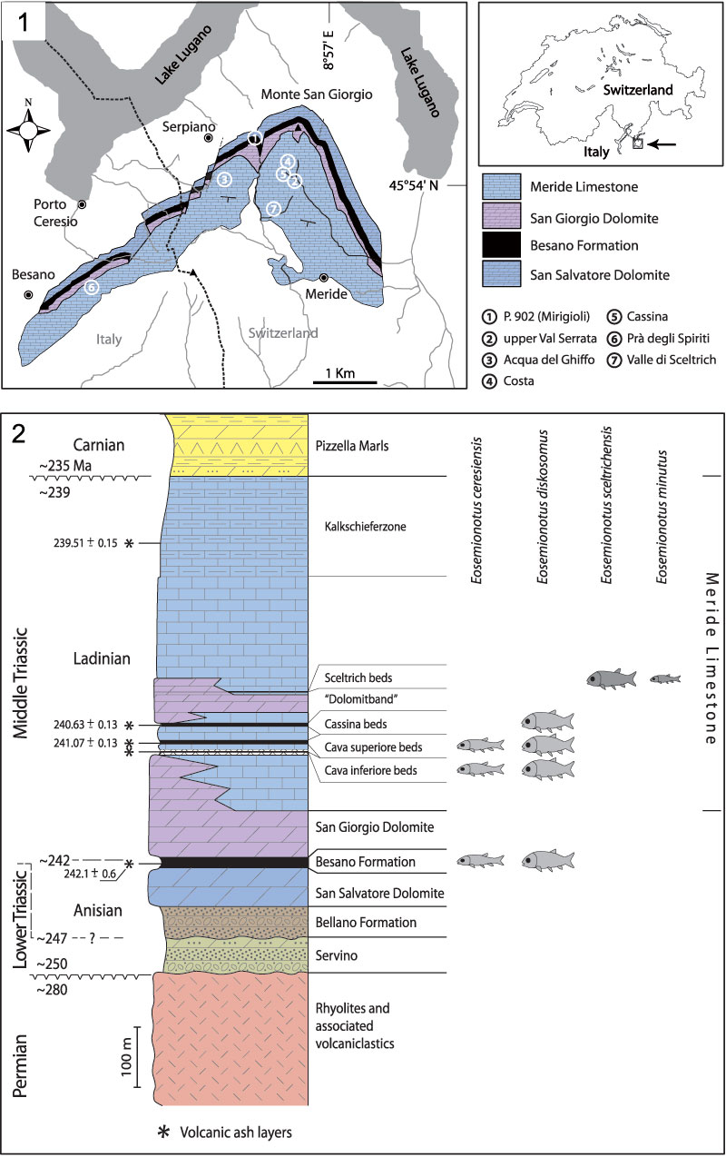
FIGURE 2. Eosemionotus vogeli. 1, holotype (IGWuG L2; 57 mm SL) from Förderstedt bei Staßfurt. 2, largest juvenile (GZG.G6.1202.1; 24.5 mm SL) amongst the specimens from Gebenberg. 3, Most complete and best-preserved specimen (MBf 14894; 30 mm SL) from Rüdersdorf bei Berlin (Photograph © Rudolf Gold, Wien). Scale bars equal 5 mm.
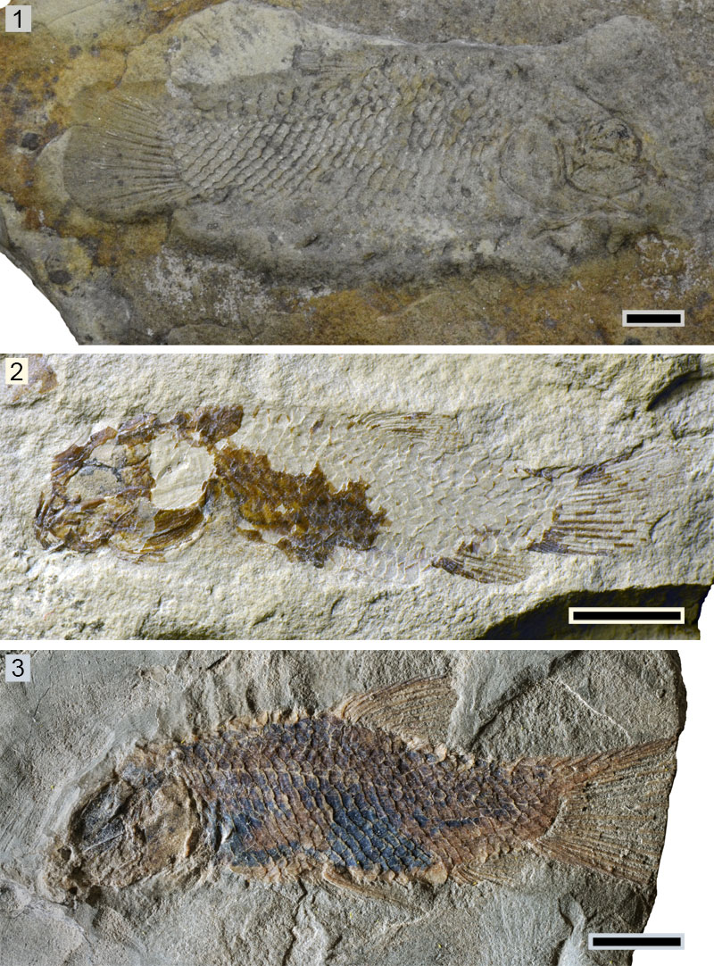
FIGURE 3. Rüdersdorf specimen MBf 14894 of Eosemionotus vogeli. 1, photograph of the posterior abdominal and caudal region showing the squamation and the pelvic, dorsal, anal and caudal fins (© Rudolf Gold, Wien). Scale bar equals 2 mm. 2, detailed photograph showing the most posterior portion of the lateral line. White arrow points towards the 27th scale traversed by the lateral line; black arrow points towards the 28th scale in this series. Scale bar equals 1 mm.
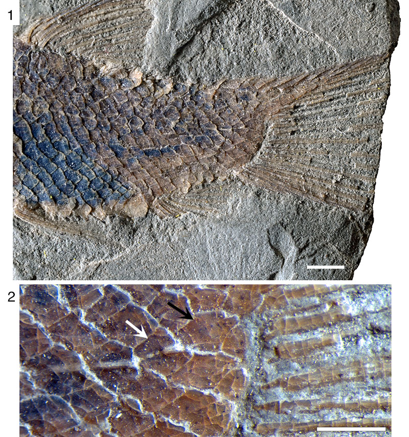
FIGURE 4. Caudal fin of Eosemionotus vogeli. 1, line drawing of the counterslab of the holotype (IGWuG L2) from Förderstedt bei Staßfurt, mirrored; all elements preserved as impressions. 2, line drawing of the specimen MBf 14894 from Rüdersdorf bei Berlin. Distinct dermal structures are indicated with colours: scales traversed by the lateral line (yellow), scales along the last vertical row at the hinge line (light blue), scales along the marginal row of the body lobe (orange), scutes (blue), dorsal caudal fulcra (red), ventral basal fulcra (purple), fringing fulcra (green). Note that the scutes in ‘1’ are preserved as imprint and, thus, the rounded shape might be an artefact of preservation. Scale bars equal 2 mm.
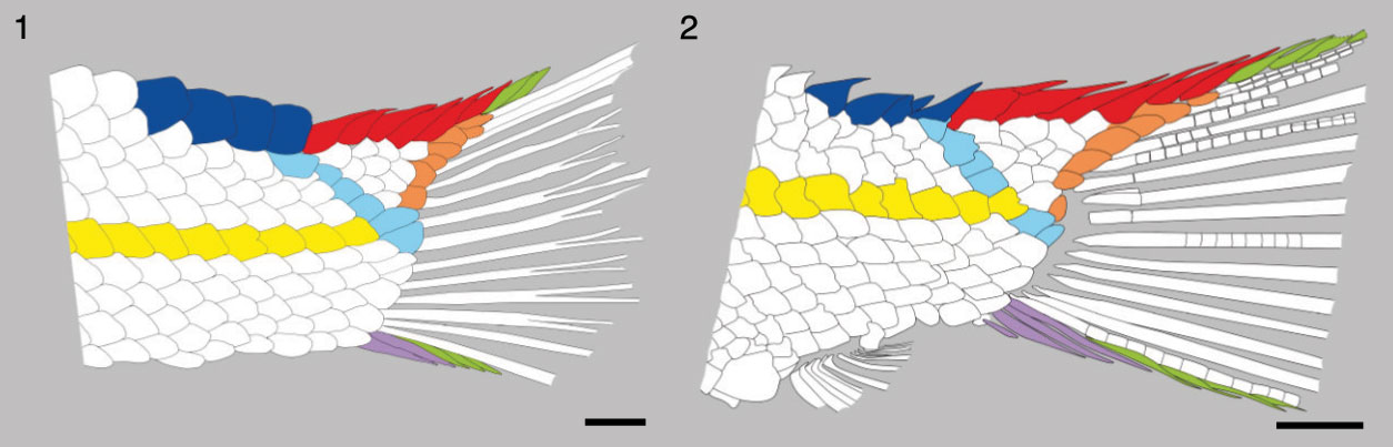
FIGURE 5. Eosemionotus ceresiensis. Holotype (PIMUZ T 357) preserved in left lateral view, in part and counterpart (39 mm SL). Scale bar equals 5 mm.
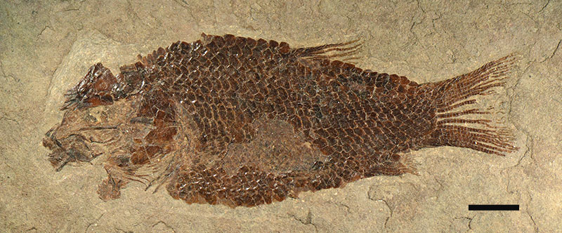
FIGURE 6. Eosemionotus diskosomus. Complete specimens. 1, holotype MCSN 8082 (45 mm SL); 2, MCSN 8006 (60.5 mm SL); 3, PIMUZ T 2924 (22.5 mm SL); 4, MCSN 5617 (46.7 mm SL). Scale bars equal 5 mm.
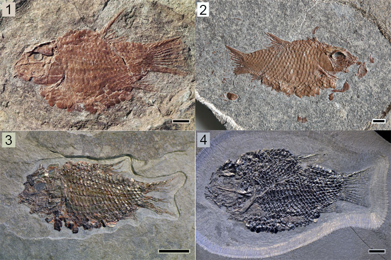
FIGURE 7. Eosemionotus diskosomus. Skull of the holotype MCSN 8082. 1, photograph. 2, line drawing. Dash-dotted lines indicate the trajectory of sensory canals. Scale bars equal 1 mm.
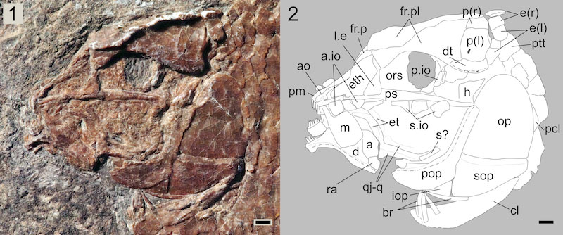
FIGURE 8. Eosemionotus diskosomus. Skull of MCSN 5617. 1, photograph. 2, line drawing. Dash-dotted lines indicate the trajectory of sensory canals. Scale bars equal 2 mm.
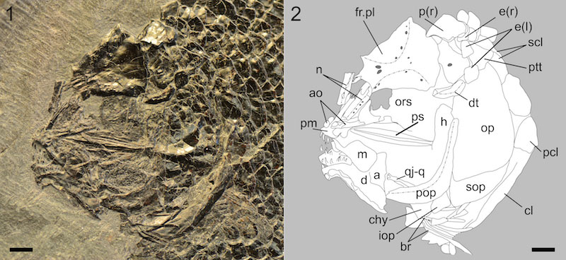
FIGURE 9. Eosemionotus diskosomus. Skull of MCSN 8006. 1, photograph. Black arrow points to the isolated hyomandibula. 2, line drawing. The line drawing of the isolated hyomandibula has been relocated and reoriented (anterior to the right). White arrow points to the small process at the base of the anterior border of the vertical arm of the preopercle. Dash-dotted lines indicate the trajectory of sensory canals. Scale bars equal 5 mm.
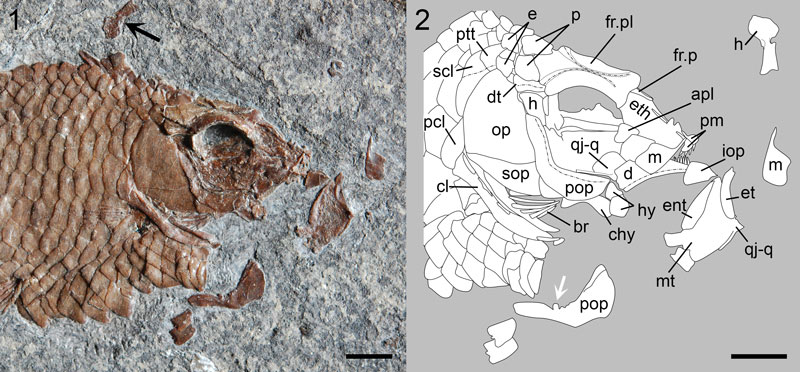
FIGURE 10. Eosemionotus diskosomus. Fins. 1, pelvic fin (MCSN 5617); 2, dorsal fin (MCSN 8006); 3, anal fin (MCSN 8082). Scale bars equal 2 mm.
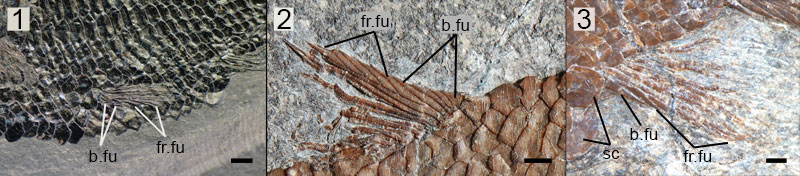
FIGURE 11. Eosemionotus diskosomus. Caudal fin of the holotype MCSN 8082. 1, photograph; 2, line drawing. Distinct dermal structures are indicated with colours: scales traversed by the lateral line (yellow), scales along the last vertical row at the hinge line (light blue), scales along the marginal row of the body lobe (orange), scutes (blue), dorsal caudal fulcra (red), ventral basal fulcra (purple), fringing fulcra (green). Scale bars equal 1 mm.

FIGURE 12. Eosemionotus diskosomus, specimen MCSN 5617. 1, photograph of the caudal fin; scale bar equals 5 mm. 2 and 3, detailed photograph (2) and line drawing (3) of the most posterior scales in the horizontal row along the lateral line; scale bar equals 2 mm.
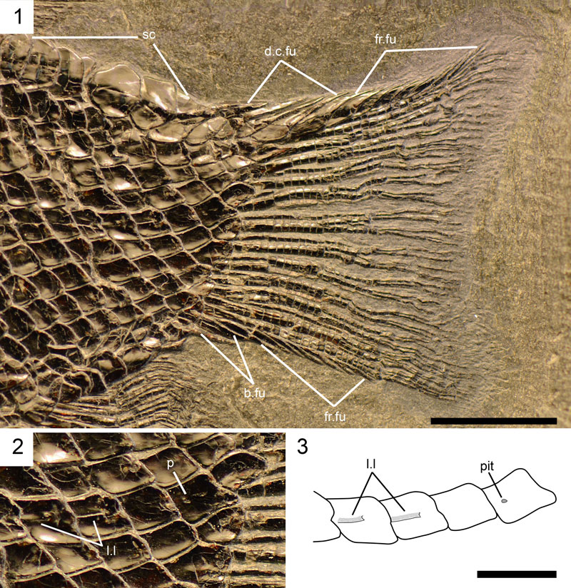
FIGURE 13. Eosemionotus diskosomus. Specimen MCSN 8006 showing the squamation pattern. Scale bar equals 5 mm.
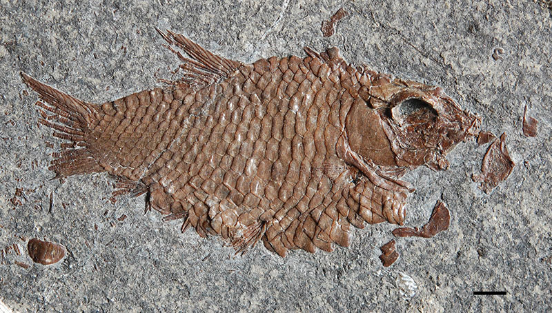
FIGURE 14. Eosemionotus sceltrichensis. 1, Holotype MCSN 8418 (52.8 mm SL) preserved in right lateral view. 2, MCSN 8497 (39.14 mm) preserved in right lateral view. Scale bars equal 5 mm.
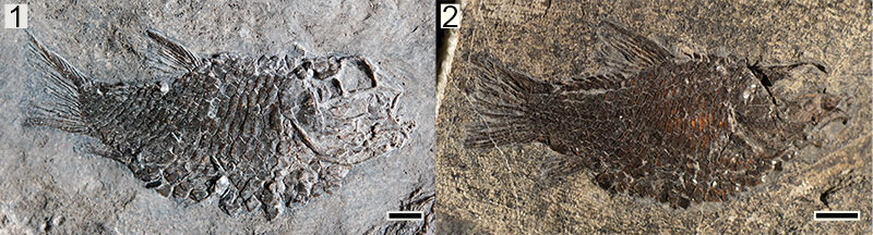
FIGURE 15. Ontogenetic changes in body proportions in the Monte San Giorgio species. Plot of the ratio between body depth (BD) and standard length (SL) in the y-axis vs. the standard length (SL) in the x-axis. Data in Table 2.
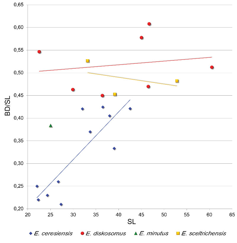
FIGURE 16. Additional specimens of Eosemionotus sceltrichensis: 1, photograph of the mostly disarticulated head of MCSN 8491 (33.16 mm SL); 2, photograph of the disarticulated and displaced skull roof in right latero-dorsal view of MCSN 8494; 3, line drawing of the latter specimen. Scale bars equal 2 mm.
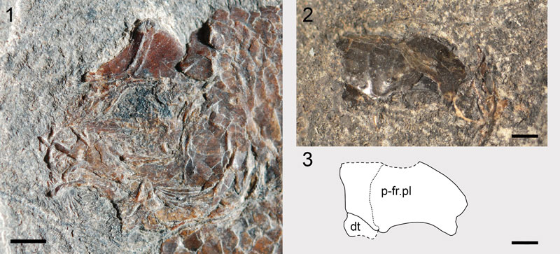
FIGURE 17. Eosemionotus sceltrichensis. Skull of the holotype MCSN 8418. 1, photograph. 2, line drawing. Scale bars equal 2 mm.
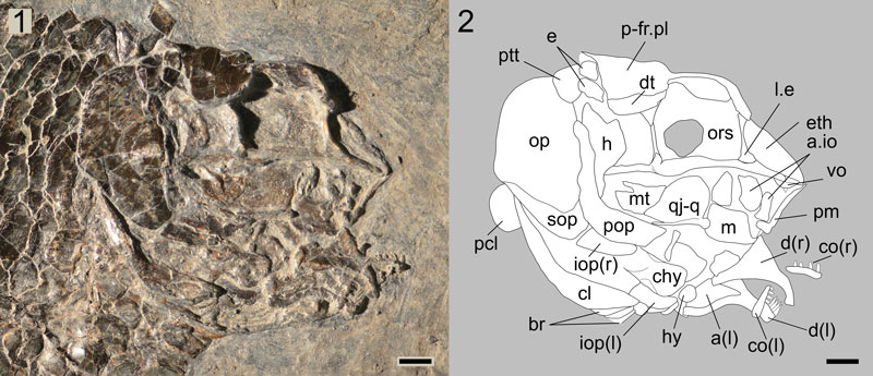
FIGURE 18. Eosemionotus sceltrichensis. Dorsal fin. 1, specimen MCSN 8418. 2, specimen MCSN 8497. Scale bars equal 2 mm.

FIGURE 19. Eosemionotus sceltrichensis. Anal fin. 1, specimen MCSN 8491; specimen MCSN 8418; 3, specimen MCSN 8497. Scale bars equal 2 mm.
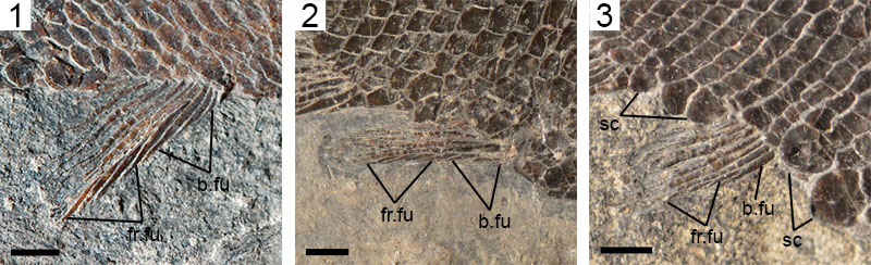
FIGURE 20. Eosemionotus sceltrichensis. Caudal fin of the holotype MCSN 8418 1, photograph. 2, line drawing. Distinct dermal structures are indicated with colours: scales traversed by the lateral line (yellow), scales along the last vertical row at the hinge line (light blue), scales along the marginal row of the body lobe (orange), scutes (blue), dorsal caudal fulcra (red), ventral caudal fulcra (purple), fringing fulcra (green). Scale bars equal 2 mm.

FIGURE 21. Eosemionotus minutus. Holotype MCSN 8482 (25 mm SL), preserved in left lateral view. Scale bar equal 5 mm.
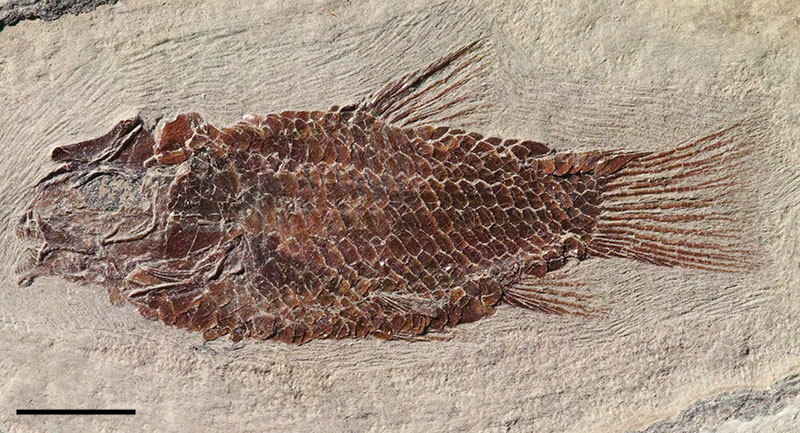
FIGURE 22. Eosemionotus minutus. Skull of the holotype MCSN 8482. 1, photograph. 2, line drawing. Scale bars equal 2 mm.
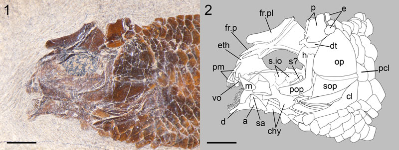
FIGURE 23. Eosemionotus minutus. Caudal fin of the holotype MCSN 8482. 1, photograph. 2, line drawing. Distinct dermal structures are indicated with colours: scales traversed by the lateral line (yellow), scales along the last vertical row at the hinge line (light blue), scales along the marginal row of the body lobe (orange), scutes (blue), dorsal caudal fulcra (red), ventral caudal fulcra (purple), fringing fulcra (green). Scale bars equal 2 mm.
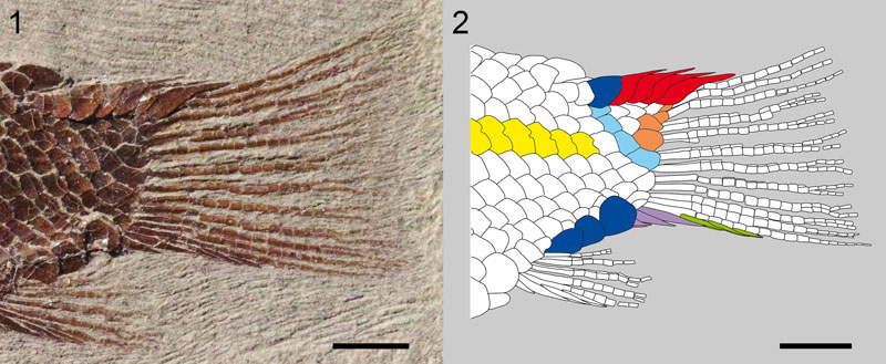
FIGURE 24. Strict consensus tree of 16 most parsimonious trees. Tree length = 968 steps, consistency index = 0.380 and retention index = 0.645. Bremer indexes and bootstrap values larger than 50 are indicated with red and black numbers at the corresponding nodes, respectively. Macrosemiid branches are indicated in blue, with the branches corresponding to the five species of Eosemionotus in light blue.
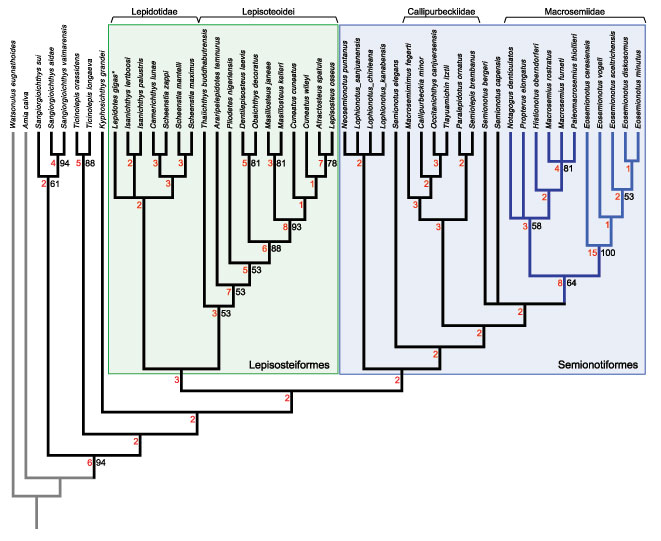
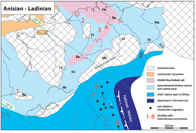
FIGURE 25. Simplified palaeogeography of the Alpine and the Germanic Triassic at the Anisian-Ladinian boundary with the Triassic position of the localities with Eosemionotus occurrences: 1 Alcover, Spain; 2 Winterswijk, The Netherlands; 3 Grebenberg, Germany; 4 Förderstedt, Germany; 5 Rüdersdorf, Germany; 6 Ducan and Landwasser areas, Switzerland; 7 Monte San Giorgio, Switzerland/Italy; 8 Velika Planina, Slovenia. BG Burgundy Gate; SMG Silesian-Moravian Gate; ECG eastern Carpathian Gate (modified from Brack et al. 1999). Small letters indicate location of major cities: Ba, Barcelona; Be, Berlin; Bs, Basel; Bu, Budapest; Fr, Frankfurt am Main; Ha, Hamburg; Kr, Cracow; Ly, Lyon; Mr, Marseilles; Mü, Munich; Pr, Prague; Wa, Warsaw.