FIGURE 1. The holotype of Rahonavis ostromi (UA 8656): 1, the 1995 quarry, Level 4, at locality MAD 93-18. The sub-triangular Rahonavis ostromi holotype block (outlined at the rear of the quarry) is shown after trenching but before jacketing and removal from the quarry. The bones in the foreground are those of a disarticulated juvenile specimen of the titanosaurian sauropod dinosaur Rapetosaurus krausei; 2, the right ulna of Rahonavis being uncovered with a brush in the field; the head of the right femur is exposed just below the ulna and demonstrates the position of these two elements relative to one another.
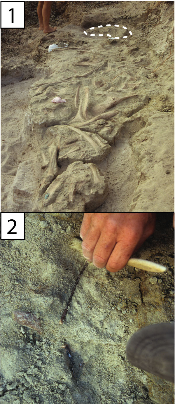
FIGURE 2. The holotype of Rahonavis ostromi (UA 8656): 1, Rahonavis block in the lab midway through its preparation; the right ulna has been removed from the jacket. The dotted rectangles depict the main body of the specimen (left upper rectangle) and the pes and scapula (right lower rectangle) shown in Figure 3; 2, placement of the skeletal elements as found, based on photos taken in the field and during preparation in the lab (modified from Forster et al., 1998, figure 1B).
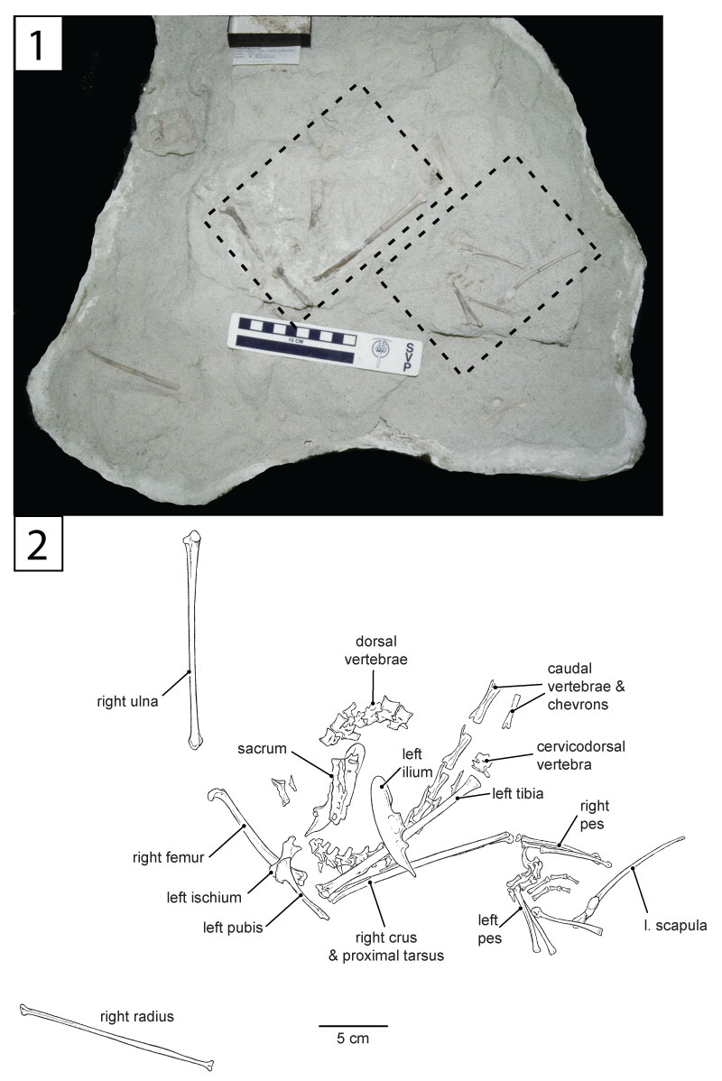
FIGURE 3. The holotype of Rahonavis ostromi (UA 8656): 1, main body in an early stage of preparation showing the head of the right femur, sacrum, and left ilium; 2, main body in an intermediate stage of preparation with the left pubis and left tibia exposed; 3, main body in a late stage of preparation with the axial column, chevrons, right tibia, and right ilium exposed. The left pubis, left ilium, and left tibia have been removed; 4, pedes and left scapula showing the reversal of the first digit on the left pes. The acromion process of the scapula lies just under metatarsal III of the left pes. The distal end of the right tibiotarsus can be seen in articulation with the proximal end of the right metatarsals.
Scale bars are shown in each photograph. Abbreviations: ch, chevrons; cdv, caudal vertebrae; dv, dorsal vertebrae; li, left ilium; lp, left pes; lpu, left pubis; ls, left scapula; lt, left tibia; rf, right femur; ri, right ilium; rp, right pes; rt, right tibia; sa, sacrum.
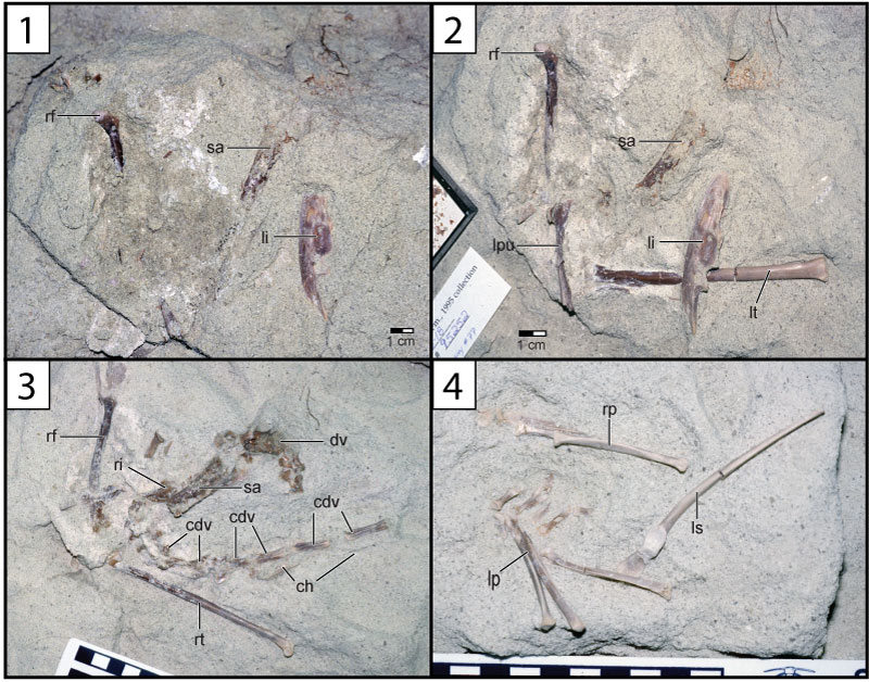
FIGURE 4. Stacked plots showing the relative proportions of the ulna, femur, and tibia of Rahonavis ostromi compared to (1) avialan and (2) non-avialan theropods. In each set of plots, the taxa are organized, left to right, from shortest to longest relative ulna. Aurornis and Anchiornis are considered basal avialans in Godefroit et al. (2013a) and Lefèvre et al. (2014), but non-avialan theropods in Brusatte et al. (2014) and Lefèvre et al. (2017). Cau et al. (2015) placed Anchiornis as a basal bird but Aurornis as a theropod. See Table 1 for measurements.
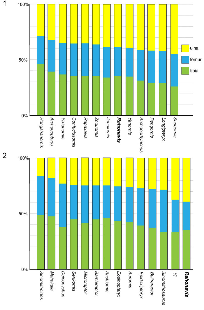
FIGURE 5. The alternative placements of Rahonavis ostromi within Paraves based on the phylogenetic analyses of (1) Lefèvre et al. (2017) and (2) Brusatte et al. (2014). Avialae are shown in red and unenlagiines are boxed in pale blue. Not all taxa included in these analyses are shown here. In Brusatte et al. (2014), Epidexipteryx is an oviraptorosaur and lies outside of Paraves; Lefèvre et al. (2017) recovered this taxon as a basal paravian.
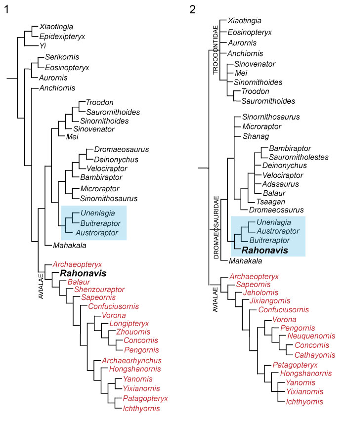
FIGURE 6. Partial left dentary (FMNH PA 740) assigned to Rahonavis ostromi. 1, medial view; 2, lateral view; 3, dorsal view (rostral to left); 4, ventral view (rostral to left); 5, transparent left lateral projection of dentary with included teeth in dark gray (caudal end not shown); enlarged µCT renderings of tooth 6 (above) and tooth 12 (below) the transparent dentary. The small numbers in (3) refer to alveolar positions. Scale bar equals 1 cm. Morphosource link to mesh file: https://doi.org/10.17602/M2/M81403. See Appendix for complete list of digital data associated with this project. Abbreviations: ag, alveolar groove; ap, apical; dia, diastema; ds, distal; lab, labial; lin, lingual; me, mesial; mf, sulcus for mental foramina; mg, Meckelian groove; sf, symphyseal surface; to, tooth; tr, tooth root.
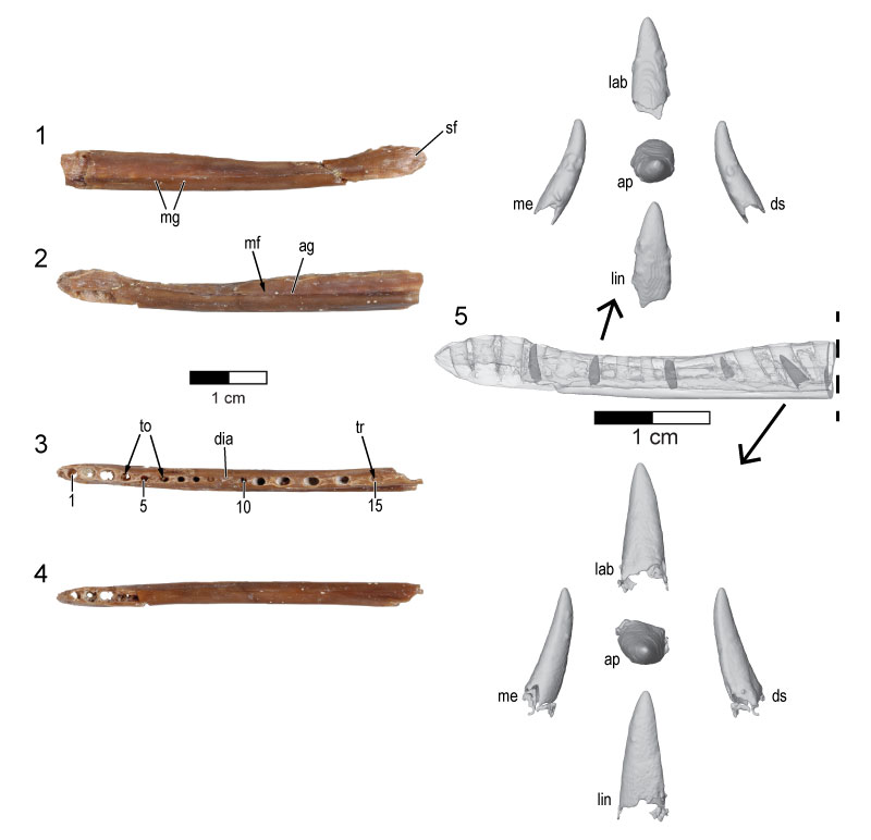
FIGURE 7. Cervicodorsal vertebra of the holotype (UA 8656) of Rahonavis ostromi. 1, left lateral view; 2, right lateral view; 3, dorsal view (cranial to left); 4, ventral view (cranial to left); 5, cranial view; 6, caudal view. Scale bar equals 1 cm. Morphosource link to mesh file: https://doi.org/10.17602/M2/M80693. Abbreviations: h, hypapophysis; ipref, infraprezygapophyseal fossa; ipostf, infrapostzygapophyseal fossa; nc, neural canal; ns, neural spine; pcdl, posterior centrodiapophyseal lamina; pf, pneumatic foramen; pp, parapophysis; ppdl, paradiapophyseal lamina; prz, prezygapophysis; tvp, transverse process.
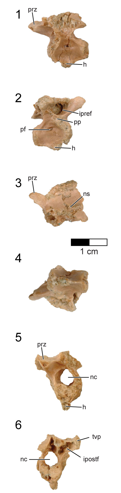
FIGURE 8. CT slices through the cervicodorsal vertebra of the holotype (UA 8656) of Rahonavis ostromi. CT slices (1) through (8) extend from the cranial to the caudal end of the vertebra; the position of each individual CT slice is shown on the left lateral view of the cervicodorsal vertebra in (9). Scale bar equals 1 cm. Image in (9) not to the same scale. Morphosource link to stack data: https://doi.org/10.17602/M2/M80694. Abbreviations: h, hypapophysis; nc, neural canal; ns, neural spine; pf, pneumatic foramen; prz, prezygapophysis; tvp, transverse process.
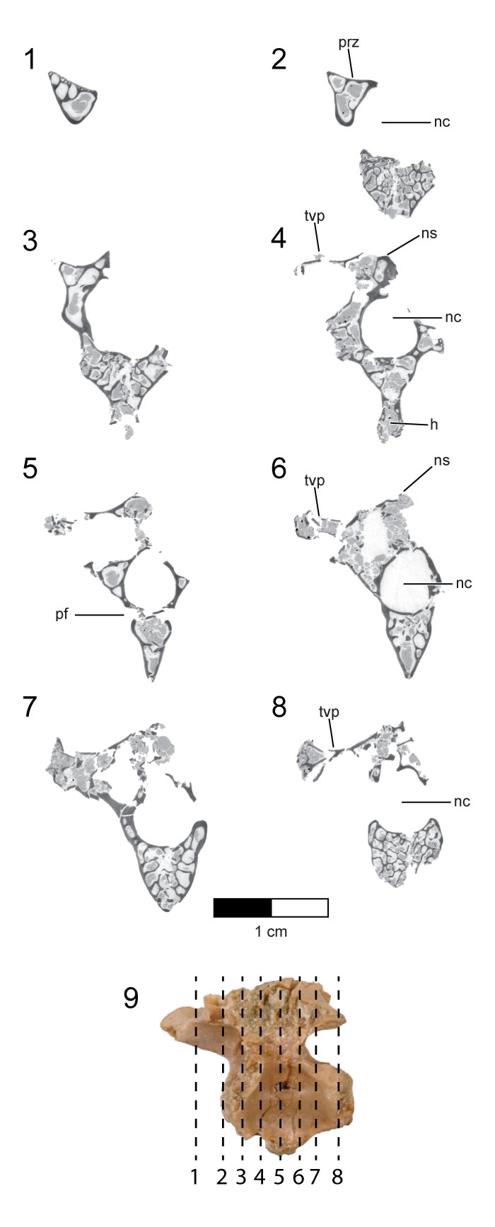
FIGURE 9. First preserved dorsal vertebra of the holotype (UA 8656) of Rahonavis ostromi. 1, left lateral view; 2, right lateral view; 3, dorsal view (cranial to left); 4, ventral view (cranial to left); 5, cranial view; 6, caudal view. Scale bar equals 1 cm. Morphosource link to mesh file: https://doi.org/10.17602/M2/M80695. Abbreviations: ipref, infraprezygapophyseal fossa; ipostf, infrapostzygapophyseal fossa; nc, neural canal; ns, neural spine; pcdl, posterior centrodiapophyseal lamina; pf, pneumatic foramen; pp, parapophysis; ppdl, paradiapophyseal lamina; prz, prezygapophysis; poz, postzygapophysis; tvp, transverse process.
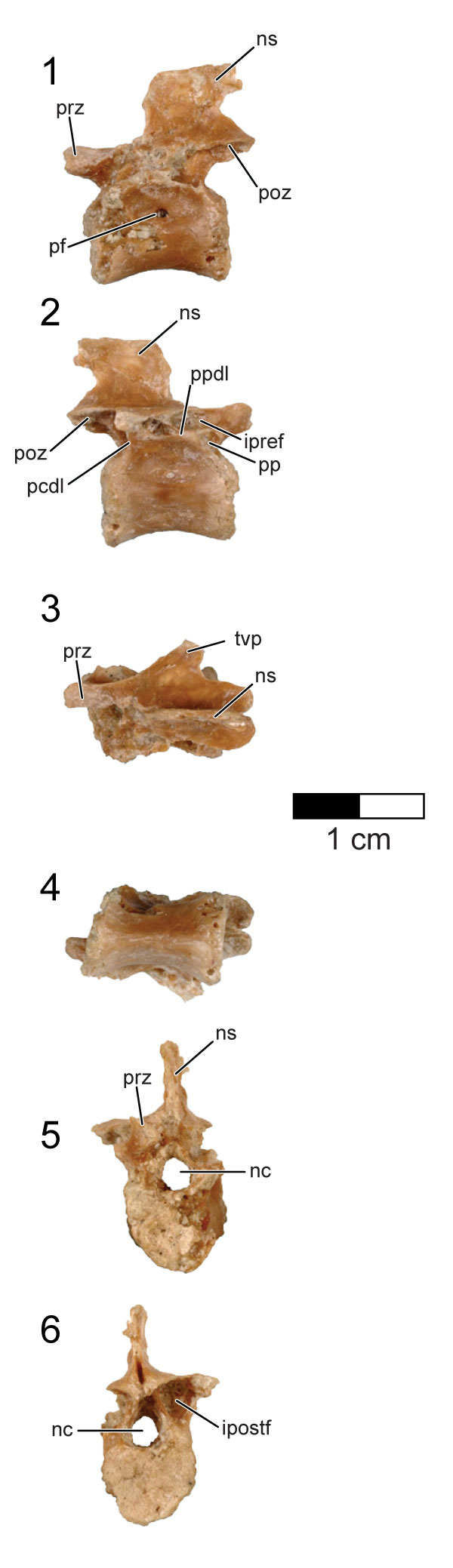
FIGURE 10. CT slices through the first preserved dorsal vertebra of the holotype (UA 8656) of Rahonavis ostromi. CT slices (1) through (9) extend from the cranial to the caudal end of the vertebra; the position of each CT slice is shown on the left lateral view of the vertebra shown in (10). Scale bar equals 1 cm. Image in (10) not to same scale. Morphosource link to stack data: https://doi.org/10.17602/M2/M80696. Abbreviations: ns, neural spine; pf, pneumatic foramen; prz, prezygapophysis; poz, postzygapophysis; tvp, transverse process.
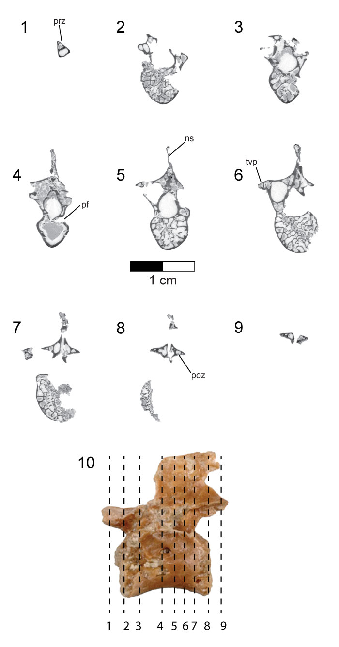
FIGURE 11. Four dorsal vertebrae of the holotype (UA 8656) of Rahonavis ostromi. 1, left lateral view; 2, right lateral view; 3, dorsal view (cranial to left); 4, ventral view (cranial to left); 5, cranial view; 6, caudal view. These vertebrae are positioned between the first preserved dorsal vertebra shown in Figure 6, and the last preserved dorsal vertebra shown in Figure 8. Scale bar equals 1 cm. Morphosource link to mesh file: https://doi.org/10.17602/M2/M80141. Abbreviations: e, epoxy; h-h, hyposphene-hypantra articulation; nc, neural canal; ns, neural spine; pf, pneumatic foramen; tvp, transverse process.
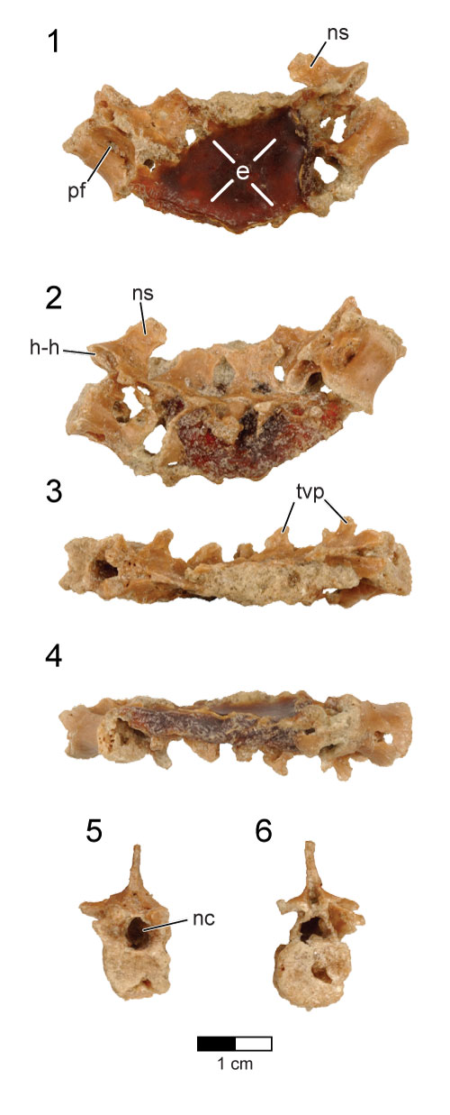
FIGURE 12. Last dorsal vertebra of the holotype (UA 8656) of Rahonavis ostromi. 1, left lateral view; 2, right lateral view; 3, dorsal view (cranial to left); 4, ventral view (cranial to left); 5, cranial view; 6, caudal view. Scale bar equals 1 cm. Morphosource link to mesh file: https://doi.org/10.17602/M2/M84077. Abbreviations: nc, neural canal; pf, pneumatic foramen.
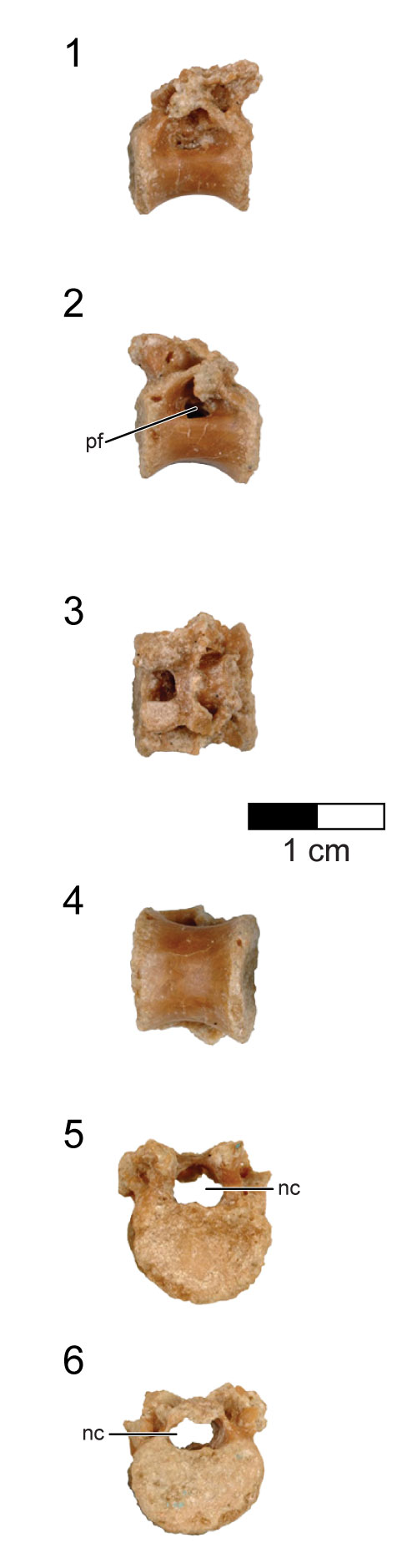
FIGURE 13. CT slices through the last dorsal vertebra of the holotype (UA 8656) of Rahonavis ostromi. CT slices (1) through (7) extend from the cranial to the caudal end of the vertebra; the position of each CT slice is shown on the left lateral view of the vertebra shown in (8). Scale bar equals 1 cm. Image in (8) not to same scale. Morphosource link to stack data: https://doi.org/10.17602/M2/M84078. Abbreviations: nc, neural canal; pf, pneumatic foramen; tvp, transverse process.
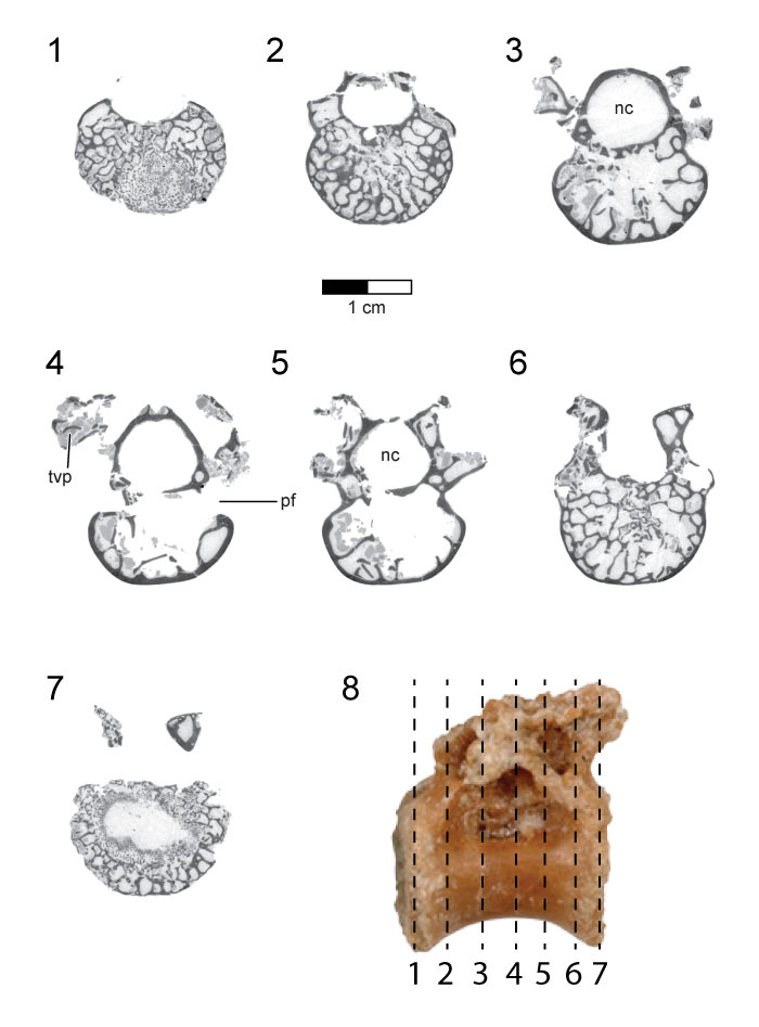
FIGURE 14. Sacrum of the holotype (UA 8656) of Rahonavis ostromi. 1, left lateral view; 2, right lateral view; 3, dorsal view (cranial to left); 4, ventral view (cranial to left); 5, cranial view; 6, caudal view. Scale bar equals 1 cm. Roman numerals in figures refer to the position of selected vertebral centra. Morphosource link to mesh file: https://doi.org/10.17602/M2/M80143. Abbreviations: dns, neural spine of last dorsal vertebra; nc, neural canal; pnf, pneumatic fossa; sr, sacral spinal ridge; tvp, transverse process; vs, ventral sulcus.
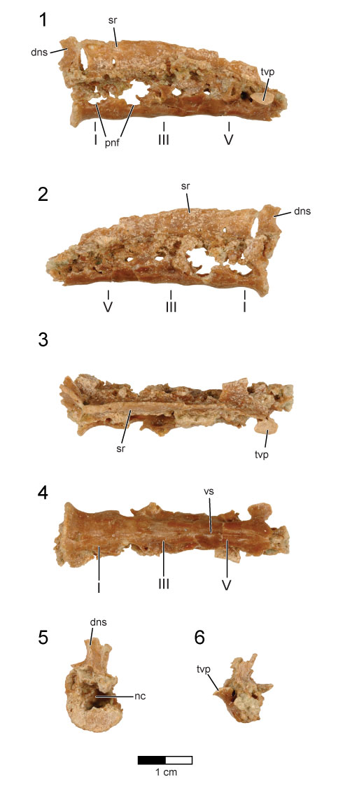
FIGURE 15. CT slices through the sacrum of the holotype (UA 8656) of Rahonavis ostromi. CT slices (1) through (11) extend from the cranial to the caudal end of the sacrum; the position of each individual CT slice is shown in the left lateral view of the sacrum in (12). Scale bar equals 1 cm. Image in (12) not to same scale. Morphosource link to stack data: https://doi.org/10.17602/M2/M80144. Abbreviations: nc, neural canal; pnf, pneumatic fossa; sr, sacral spinal ridge; tvp, transverse process.
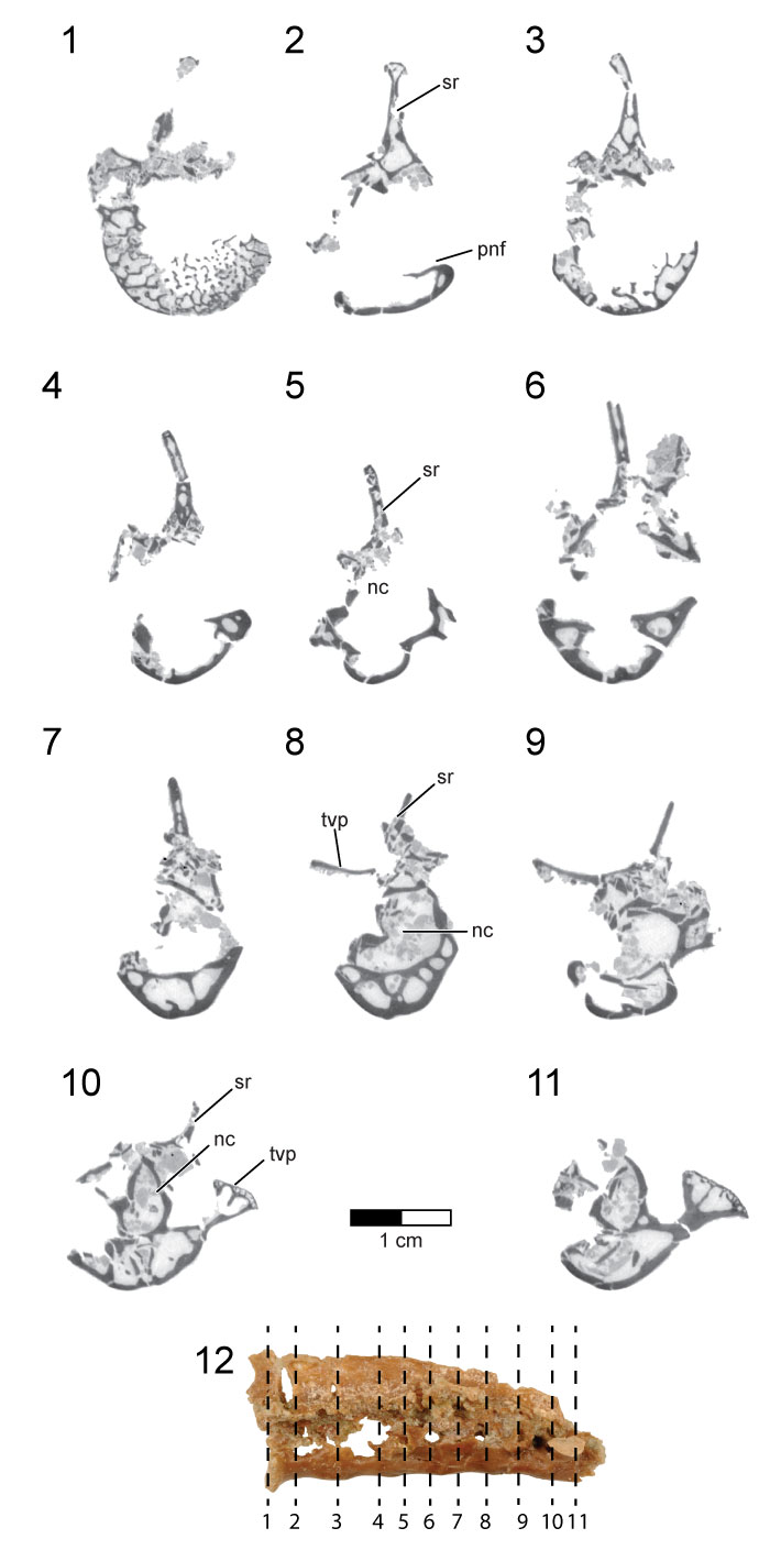
FIGURE 16. Thirteen caudal vertebrae of the holotype (UA 8656) of Rahonavis ostromi. 1, left lateral view; 2, right lateral view; 3, dorsal view (cranial to the left); 4, ventral view (cranial to the left). In all views, the vertebrae are aligned with caudal vertebrae one through nine in the upper row, and caudal vertebrae 10 through 13 in the lower row. Caudal vertebrae 1 through 3 are preserved in articulation. Scale bar equals 1 cm. See Appendix for complete list of Morphosource links for mesh and stack data for caudal vertebrae.
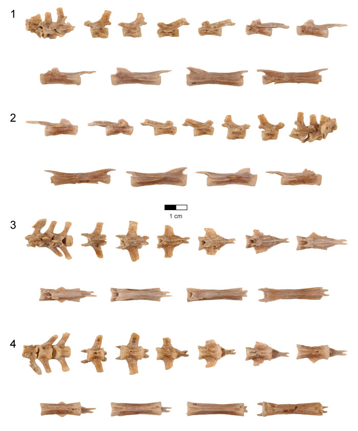
FIGURE 17. Digital surface reconstructions from CT scans of disarticulated caudal vertebrae 1 (Cd1, left column), caudal vertebra 2 (Cd2, center column), and caudal vertebra 3 (Cd3, right column) of the holotype (UA 8656) of Rahonavis ostromi. 1, left lateral views; 2, right lateral views; 3, dorsal views (cranial to the left); 4, ventral views (cranial to the left); 5, cranial views; 6, caudal views. Scale bar equals 1 cm. See Appendix for complete list of Morphosource links for mesh and stack data for caudal vertebrae. Abbreviations: ns, neural spine; poz, postzygapophysis; prz, prezygapophysis; tvp, transverse process.
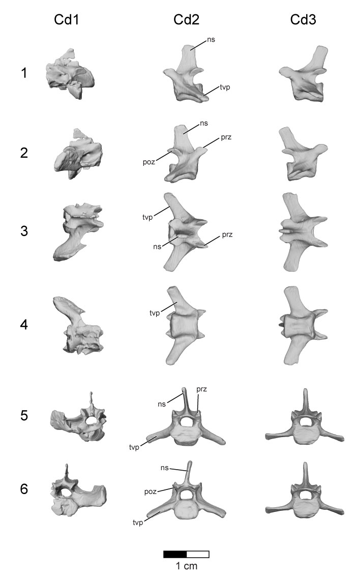
FIGURE 18. Digital surface reconstructions from CT scans of caudal vertebrae 5 (Cd5, left column), 7 (Cd7, center column), and 11 (Cd11, right column) of the holotype (UA 8656) of Rahonavis ostromi. 1, left lateral views; 2, right lateral views; 3, dorsal views (cranial to the left); 4, ventral views (cranial to the left); 5, cranial views; 6, caudal views. Scale bar equals 1 cm. See Appendix for complete list of Morphosource links for mesh and stack data for caudal vertebrae. Abbreviations: ns, neural spine; poz, postzygapophysis; prz, prezygapophysis; tvp, transverse process.
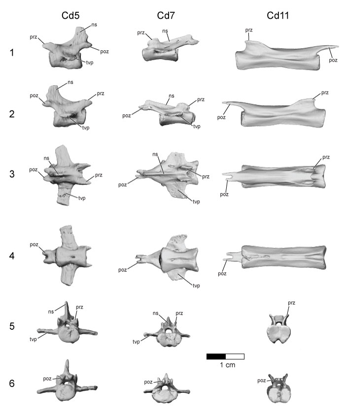
FIGURE 19. CT slices through the seventh caudal vertebra (Cd7) of the holotype (UA 8656) of Rahonavis ostromi. CT slices (1) through (10) extend from the cranial end to the caudal end of the vertebra. The position of each individual CT slice is shown in the left lateral view of Cd7 in (11). Scale bar equals 1 cm. Image in (11) not to same scale as slice images. Morphosource link to stack data: https://doi.org/10.17602/M2/M80679. Abbreviations: nc, neural canal; ns, neural spine; poz, postzygapophysis; tvp, transverse process.
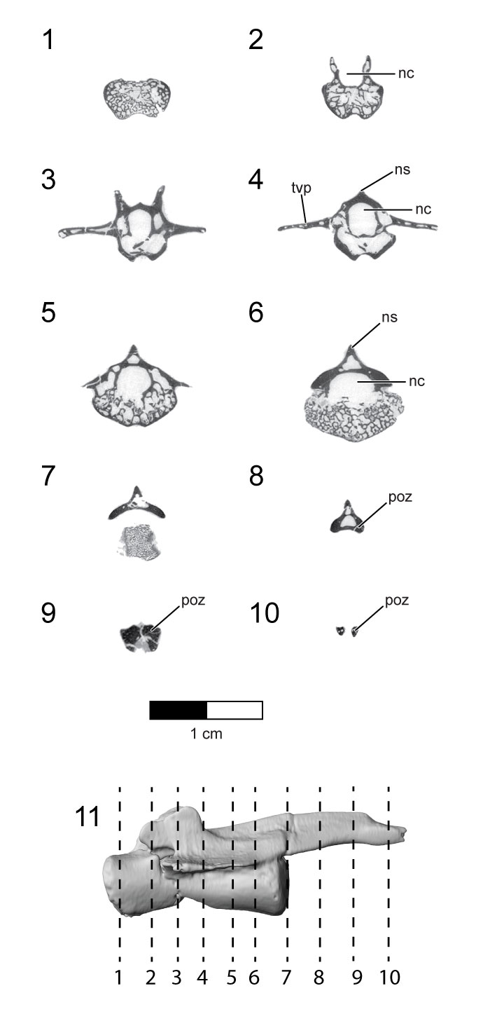
FIGURE 20. CT slices through the eleventh caudal vertebra (Cd11) of the holotype (UA 8656) of Rahonavis ostromi. CT slices (1) through (11) extend from the cranial end to the caudal end of the vertebra. The position of each individual CT slice is shown in the left lateral view of Cd11 in (12). Scale bar equals 1 cm. Image in (12) not to same scale as slice images. Morphosource link to stack data: https://doi.org/10.17602/M2/M80688. Abbreviations: nc, neural canal; poz, postzygapophysis.
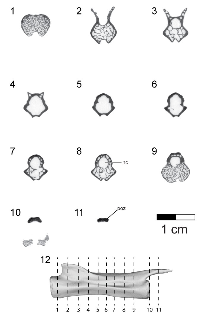
FIGURE 21. Chevron series of the holotype (UA 8656) of Rahonavis ostromi in left lateral views (left column), ventral views (second column, cranial to the right), right lateral views (third column), and dorsal views (right column, cranial to the right). 1, first chevron (articulated with caudoventral margin of Cd2); 2, second chevron; 3, third chevron; 4, fourth chevron; 5, fifth chevron; 6, sixth chevron; 7, seventh chevron; 8, eighth chevron; 9, ninth chevron; 10, tenth chevron; 11, eleventh(?) chevron; 12, twelfth(?) chevron. See Appendix for complete list of Morphosource links for mesh and stack data for chevrons.
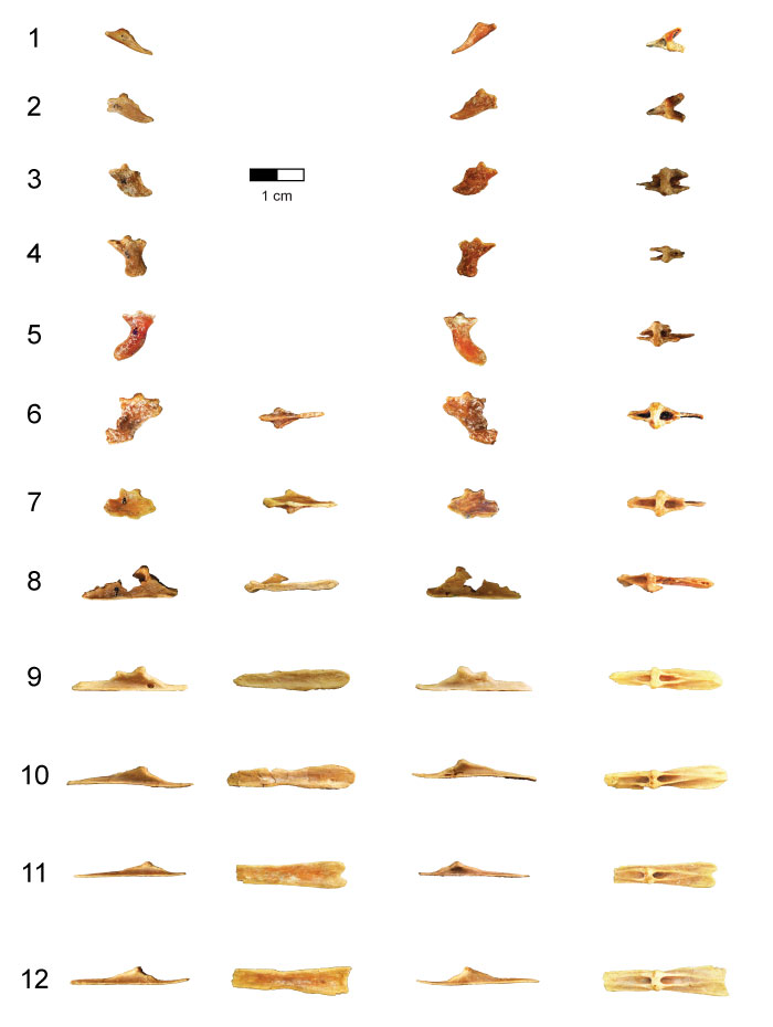
FIGURE 22. Left scapula of the holotype (UA 8656) of Rahonavis ostromi. 1, lateral view; 2, medial view (dorsal side down); 3, dorsal view; 4, ventral view; 5, proximal view (dorsal side up). The distal (posterior) end of the scapula is oriented to the right in (1) to (4). Scale bar equals 5 cm. Morphosource link to mesh file: https://doi.org/10.17602/M2/M81206. Abbreviations: ac, acromial crest; af, acromial flange; ap, acromion process; ca, coracoid articulation; gf, glenoid fossa; tt, triceps tubercle.
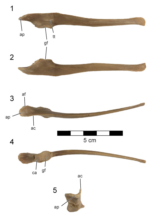
FIGURE 23. CT slices through the left scapula of the holotype (UA 8656) of Rahonavis ostromi. CT slices (1) through (15) extend from the cranial to the caudal (distal) end of the scapula; the position of each individual CT slice is shown in the left lateral view of the scapula in (16). Image in (16) not to same scale. Scale bar equals 5 cm. Morphosource link to stack data: https://doi.org/10.17602/M2/M81207. Abbreviations: af, acromial flange; ap, acromion process; ca, coracoid articulation; gf, glenoid.
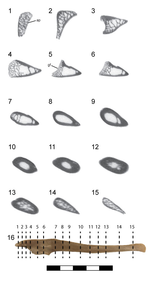
FIGURE 24. Left (FMNH PA 746) and right (UA 9604) distal humeri referred to Rahonavis ostromi. 1, caudal views; 2, cranial views; 3, ventral (medial) views; 4, dorsal (lateral) views; 5, distal views (caudal surfaces facing each other). Scale bar equals 1 cm. See Appendix for complete list of Morphosource links for mesh and stack data for humeri. Abbreviations: dco, dorsal condyle; dep, dorsal epicondyle; fp, flexor process; ici, intercondylar incisure; olf, olecranon fossa; vco, ventral condyle; vep, ventral epicondyle.
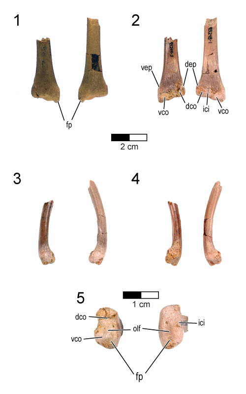
FIGURE 25. Right ulnae of the holotype (UA 8656) and referred (FMNH PR 2821) specimen of Rahonavis ostromi. 1, ventral (medial) views; 2, dorsal (lateral) views; 3, cranial (interosseal) views; 4, caudal views; 5, proximal views (medial to the left); 6, distal views (medial to the left). In all views, the ulna on the left is the referred specimen (FMNH PR 2821) and the ulna on the right is the holotype (UA 8656). Scale bar equals 1 cm. Morphosource link to mesh files: https://doi.org/10.17602/M2/M83162 (UA 8656) and https://doi.org/10.17602/M2/M84083 (FMNH PR 2821). Abbreviations: bf, brachialis fossa; dct, dorsal cotyle; is, intercondylar sulcus; nf, neurovascular foramen; vct, ventral cotyle.
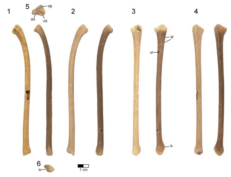
FIGURE 26. CT slices through the right ulna of the holotype (UA 8656) of Rahonavis ostromi. CT slices extend from the proximal to the distal end of the ulna in (1) through (14). The position of each individual slice is shown on the ulna in (15). Scale bar equals 1 cm. Image in (15) is not to same scale as slice images. Morphosource link to stack data: https://doi.org/10.17602/M2/M84084.
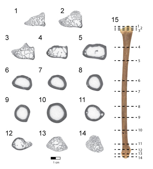
FIGURE 27. Right radius of the holotype (UA 8656) of Rahonavis ostromi. 1, cranial view; 2, caudal (interosseal) view; 3, ventral (medial) view; 4, dorsal (lateral) view; 5, proximal view (dorsal to the left); 6, distal view (dorsal to the right). Scale bar equals 1 cm. Morphosource link to mesh file: https://doi.org/10.17602/M2/M81201. Abbreviations: nf, neurovascular foramen; tub, tubercle.
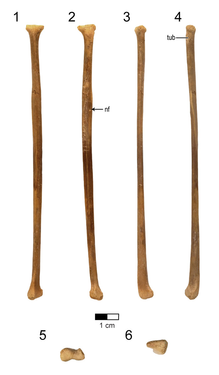
FIGURE 28. CT slices through the right radius of the holotype (UA 8656) of Rahonavis ostromi. CT slices extend from the proximal to the distal end of the radius in (1) through (17). The position of each individual slice is shown on the radius in (18). Scale bar equals 1 cm. Image in (17) not to same scale as slice images. Morphosource link to stack data: https://doi.org/10.17602/M2/M81202.
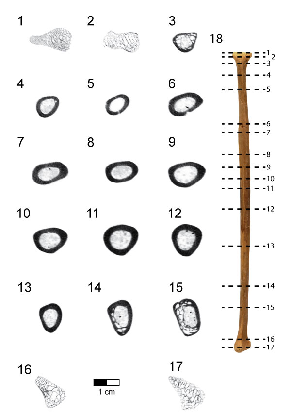
FIGURE 29. Left and right ilia of the holotype (UA 8656) of Rahonavis ostromi. Images in the left column (1, 3, 5, 7) depict the left ilium, and those in the right column (2, 4, 6, 8) depict the right ilium. 1 and 2, lateral views; 3 and 4, medial views; 5 and 6, dorsal views (caudal ends facing each other as in 1 and 2); 7 and 8, ventral views (caudal ends facing each other as in 1 and 2). Scale bar equals 1 cm. See Appendix for complete list of Morphosource links for mesh and stack data for ilia. Abbreviations: act, acetabulum; at, antitrochanter; bf, brevis fossa; ishp, ischial peduncle; mcf, m. cuppedicus fossa; pubp, pubic peduncle; stp supratrochanteric process; ve, ventral expansion.
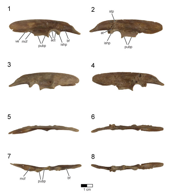
FIGURE 30. Left and right ischia of the holotype (UA 8656) of Rahonavis ostromi. Photographs in the top row (1-3) are of the right ischium, and photographs of the bottom row (4-6) are of the left ischium. 1 and 4, lateral views; 2 and 5, cranial views; 3 and 6, medial views. Scale bar equals 1 cm. See Appendix for complete list of Morphosource links for mesh and stack data for ischia. Abbreviations: act, acetabulum; at, antitrochanter; ia, iliac articulation; op, obturator process; pa, pubic articulation; pdp, proximodorsal process.
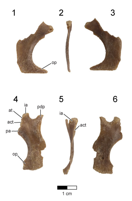
FIGURE 31. Left and right pubes of the holotype (UA 8656) of Rahonavis ostromi. Photographs in the top row (1-5) are of the right pubis, and the bottom row (6-10) the left pubis. 1 and 6, lateral views; 2 and 7, caudal views; 3 and 8, medial views; 4 and 9, cranial views; 9 and 10, proximal views (cranial end pointing down). Scale bar equals 1 cm. See Appendix for complete list of Morphosource links for mesh and stack data for pubes. Abbreviations: act, acetabulum; ila, iliac articulation; isa, ischial articulation; mpc, medial pubic crest; pf, pubic foot; ppf, postpubic fossa; ps, pubic symphysis.
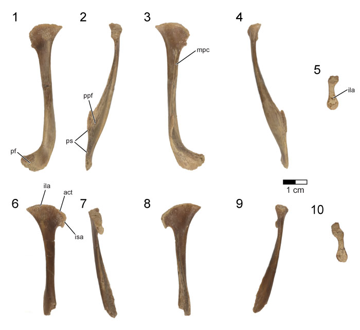
FIGURE 32. Femora of the holotype (UA 8656) of Rahonavis ostromi. Photos in the top row (1-4) are of the right femur, and in the bottom row (5-8) are of the left femur. 1 and 5, cranial views; 2 and 6, lateral views; 3 and 7, caudal views; 4 and 8, medial views. Proximal and distal views of each femur are shown above (proximal; medial side to right in 1 and medial side to left in 5) and below (distal; cranial end up) that element, respectively, in (1) and (5). Scale bar equals 1 cm. See Appendix for complete list of Morphosource links for mesh and stack data for femora. Abbreviations: et, ectocondylar tuber; fh, femoral head; lc, lateral condyle; mc, medial condyle; pt, posterior trochanter; set, supraectocondylar ridge; tc, trochanteric crest; ?4t, ?fourth trochanter.
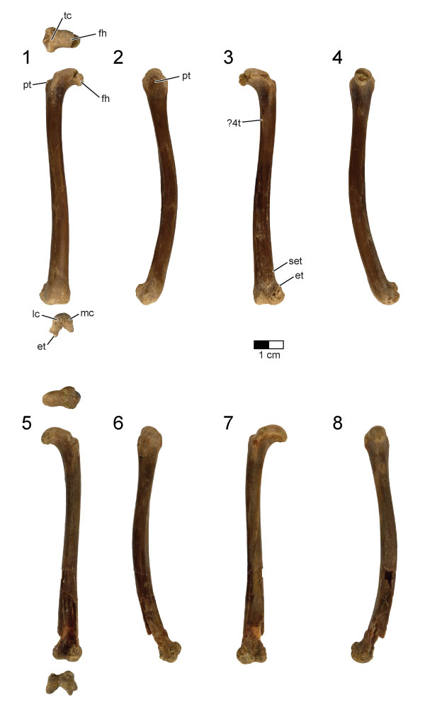
FIGURE 33. CT slices through the right femur of the holotype (UA 8656) of Rahonavis ostromi. CT slices (1) through (16) extend from the proximal to the distal end of the element; the cranial surface is up. The position of each individual CT slice is shown on the cranial view of the right femur in (17). Scale bar equals 1 cm. Image in (17) not to same scale as slice images. Morphosource link to stack data: https://doi.org/10.17602/M2/M81190.
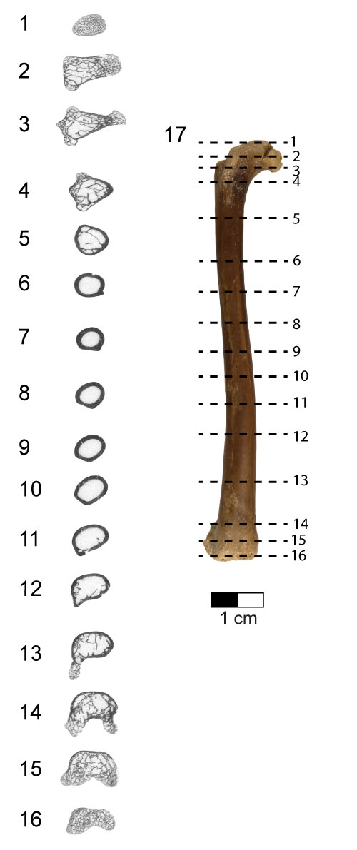
FIGURE 34. Tibiae of the holotype (UA 8656) of Rahonavis ostromi. In images (1-4), the right tibia is on the left and the left tibia is on the right. 1, cranial views; 2, lateral views; 3, caudal views; 4, medial views; 5, proximal view of left tibia; 6, distal view of left tibia. The cranial surface is facing the bottom of the image in both (5) and (6). Scale bar equals 1 cm. See Appendix for complete list of Morphosource links for mesh and stack data for tibiae. Abbreviations: cc, cnemial crest; es, extensor sulcus; fc, fibular crest; fcd, fibular condyle.
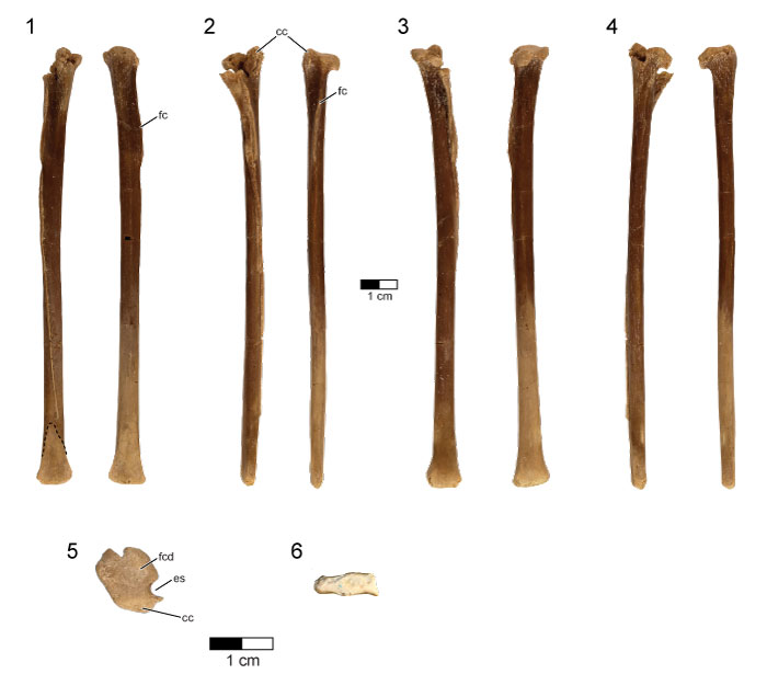
FIGURE 35. CT slices through the left tibia of the holotype (UA 8656) of Rahonavis ostromi. CT slices (1) through (18) extend from the proximal to the distal end of the element; the position of each individual CT slice is shown on the cranial view of the left tibia in (19). Cranial surface of the slices faces up. Scale bar equals 1 cm. Image in (19) not to same scale as slice images. Morphosource link to stack data: https://doi.org/10.17602/M2/M81209.
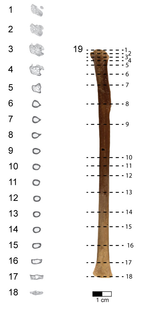
FIGURE 36. Left fibula of the holotype (UA 8656) of Rahonavis ostromi. 1, lateral view; 2, medial view; 3, proximal view (lateral surface facing up). Scale bar equals 1 cm. Morphosource link to mesh file: https://doi.org/10.17602/M2/M84062. Abbreviations: ft, fibular tubercle for m. iliofibularis.
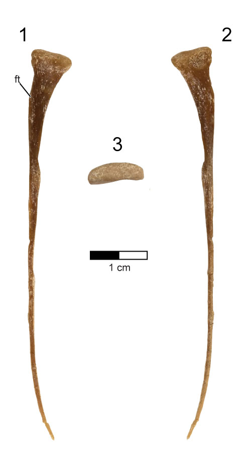
FIGURE 37. Right astragalus and calcaneum of the holotype (UA 8656) of Rahonavis ostromi. 1, cranial view; 2, caudal view (internal); 3, lateral view; 4, medial view; 5, distal view (lateral side to the left). Scale bar equals 1 cm. Morphosource link to mesh file: https://doi.org/10.17602/M2/M80697. Abbreviations: ap, ascending process; ast, astragalus; cal, calcaneum.
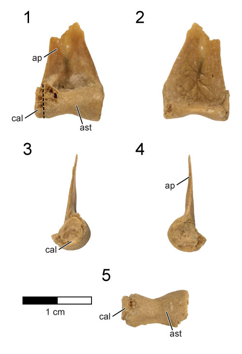
FIGURE 38. CT slices through the right astragalus and calcaneum of the holotype (UA 8656) of Rahonavis ostromi. CT slices (1) through (7) extend from the dorsal to the ventral (articular) end of the astragalus-calcaneum; in all slices cranial is up. The position of each individual CT slice is shown in the cranial view of the astragalus-calcaneum in (8). Scale bar equals 1 cm. Image in (8) is not to same scale. Morphosource link to stack data: https://doi.org/10.17602/M2/M80698. Abbreviations: ap, ascending process; cal, calcaneum.
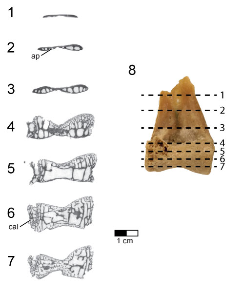
FIGURE 39. Right pes of the holotype (UA 8656) of Rahonavis ostromi. 1, lateral views; 2, dorsal views; 3, lateral views; 4, plantar views; 5, proximal views; 6, distal views. Roman numerals refer to the placement of the digit in the pes. Scale bar equals 2 cm. See Appendix for complete list of Morphosource links for mesh and stack data for right pedal elements. Abbreviations: dt, distal tarsal.
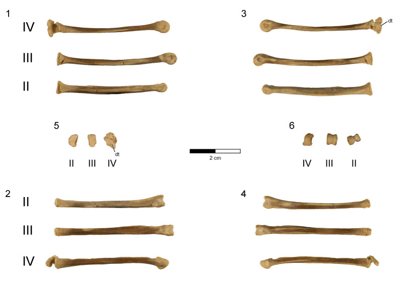
FIGURE 40. Digital surface reconstructions from CT scans of the left pes of the holotype (UA 8656) of Rahonavis ostromi. 1, medial views; 2, dorsal views; 3, lateral views; 4, plantar views. Proximal views are shown above each element in (1). Distal views are shown above each element in (2). Roman numerals refer to the placement of the digit in the pes. See Appendix for complete list of Morphosource links for mesh and stack data for left pedal elements. Scale bar equals 2 cm. Abbreviations: mpr, median plantar ridge; ph, proximoplantar heel.
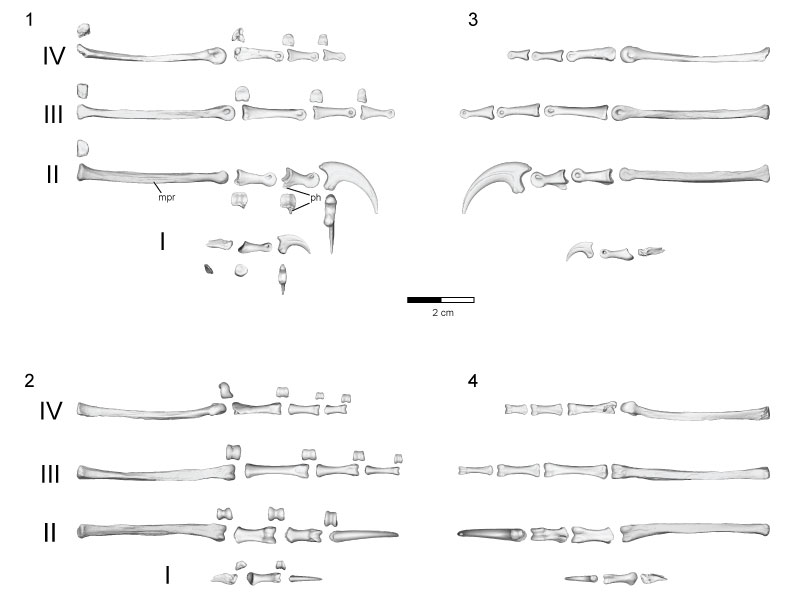
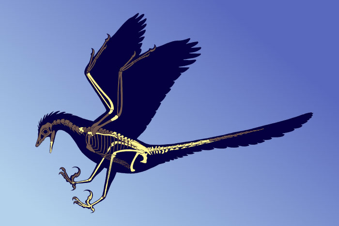
FIGURE 41. Life reconstruction of Rahonavis ostromi based on the holotype (UA 8656) and referred specimens (FMNH PA 740, FMNH PA 746, and UA 9604). Known elements are colored in yellow.