FIGURE 1. A, Regional map with early Paleocene outline of the San Jorge Basin (dark line; after Sylwan, 2001), with rectangular inset for study area (B); B, Salamanca Formation and Río Chico Group outcrops in the Sarmiento area and major geological structures. Fossil localities: OR, Ormachea Petrified Forest Park; PL, Palacio de los Loros; LF, Las Flores; RG, Rancho Grande.
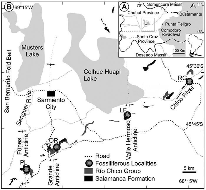
FIGURE 2. Paleocene fossil quarries at Palacio de los Loros (PL). A, General view of the Danian Salamanca (SA) and Peñas Coloradas (PC) formations, showing the distinctive butte at left that is home to large numbers of burrowing parrots (Cyanoliseus patagonus Vieillot, 1818); the locations of the PL1 quarry (white arrow) and the PL-1 tuff horizon (black arrow) dated in Clyde et al. (2014); and the Banco Negro Inferior (BNI) capping the Salamanca Formation. B, Quarry PL1 (assigned to Chron C29n, 65.58-64.86 Ma; Clyde et al., 2014; Comer et al., 2015). C, Quarry PL2 (Chron C28n, 64.67-63.49 Ma).
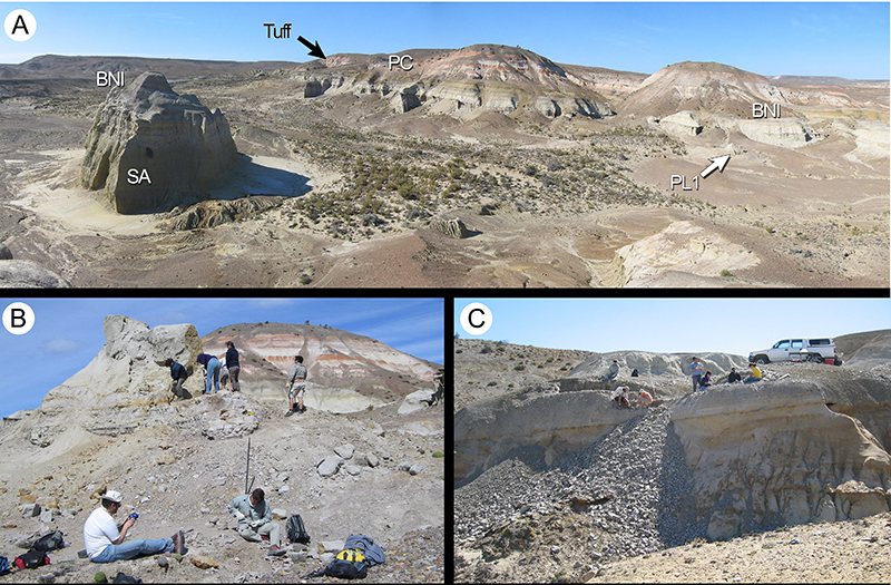
FIGURE 3. Strata exposed at Ormachea Petrified Forest Park. A, General view of the Danian Salamanca (SA) and Peñas Coloradas (PC) formations; white arrow marking OR2 quarry (Chron C29n), black arrow showing the white tuff horizon dated in Iglesias et al. (2007) and Clyde et al (2014). The prominent dark bed is the Banco Negro Inferior (BNI) at the top of the Salamanca Formation. Strata above the tuff include the Eocene Las Flores and Koluel Kaike formations. B, OR1 quarry (arrow, Chron C28n, 64.67-63.49 Ma) in heterolithic cross-bedded sandstones of the Salamanca Formation. C, Dromedary Hill, exposing part of the upper section of the Salamanca Formation (Chron C28n), arrow marking the DR fossil quarry. Scale ca. 7 m.
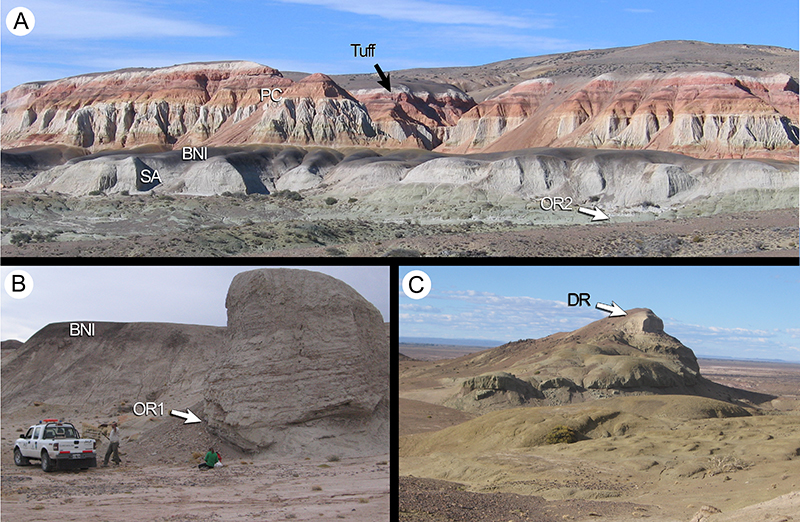
FIGURE 4. A-B, Las Flores oil field locality (La Campanita Ranch), Peñas Coloradas Formation. A, General view of coarse, continental fluvial channel deposits near the fossil locality, basal Peñas Coloradas Formation. B, Las Flores (LF) fossil quarry (Chron C27n, late Danian, 62.52-62.22 Ma). C, Palacio de los Loros PL4 quarry, Salamanca Formation (Chron C29n, early Danian, 65.58-64.86 Ma), arrow marking the fossiliferous level.
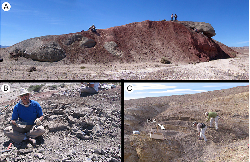
FIGURE 5. Morphotype SA001 (Akaniaceae?). A, MPEF-Pb-9000, note insect damage at the leaf margin (arrow; see also Labandeira et al., 2007 p.15, figure DT114); B, MPEF-Pb-2020 (exemplar, see also Figure 5H); C, MPEF-Pb-9001; D, MPEF-Pb-4000; E, detail of tooth venation in Figure 5B, note darkly preserved idioblasts (arrow); F, digital overlay drawing (DOD) of teeth and associated venation of specimen in Figure 5E; G, camera lucida drawing (CLD) of leaf venation of MPEF-Pb-9002; H, DOD of leaf venation of specimen in Figure 5B (exemplar); I, tooth detail of specimen in Figure 5C; J, CLD of tooth venation in Figure 5G; note the thick fimbrial vein (arrow). Single-color scale bars equal 10 mm; grid scales equal one millimeter (per rectangle).
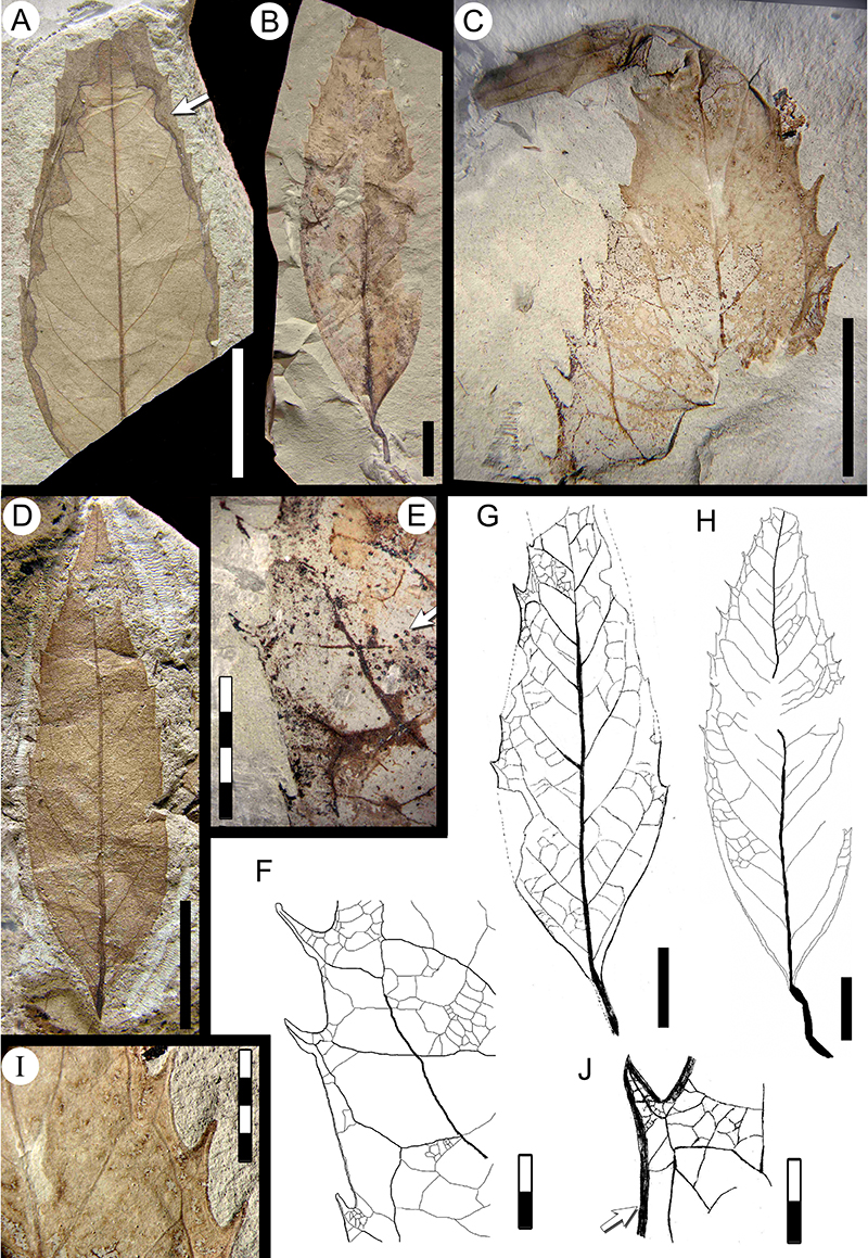
FIGURE 6. Morphotype SA002. A, MPEF-Pb-2021 (exemplar, see also Figure 6F); B, MPEF-Pb-4007; C, MPEF-Pb-4010 (white arrow, asymmetric base), note insect damage (black arrow, DT26; also see Donovan et al., 2018 figure 9.16); D, MPEF-Pb-9004; E, digital overlay drawing (DOD) of leaf venation in Figure 6C (arrow, swollen petiole base); F, DOD of leaf venation of specimen in Figure 6.A (exemplar); G, apex in MPEF-Pb-9005; H, tooth detail of specimen in Figure 6.A; I, DOD of tooth venation of specimen in Figure 6H; J, camera lucida drawing (CLD) of MPEF-Pb-9006; K, medial tooth venation of specimen in Figure 6J; L, basal tooth venation of specimen in Figure 6J; M, CLD of leaf venation of specimen in Figure 6D (arrow, chevroned epimedial tertiaries); N, tooth venation of specimen in Figure 6M. Single-color scale bars equal 10 mm; grid scales equal one millimeter (per rectangle).
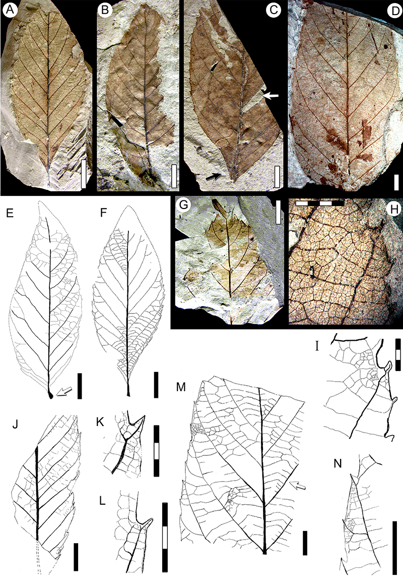
FIGURE 7. Morphotype SA004 “Dryophyllum” australis Berry 1937a (Nothofagaceae) and related material. A, “D. australis” syntype specimen USNM-208522 (illustrated in Berry 1937a, plate VI figure 4; specimen referred in this work to morphotype SA042 Cunoniaceae, see also Table 1, Table 2). B, “D.” australis syntype specimen USNM-208523a (illustrated in Berry 1937a, plate VI, figure 5; see also Table 1, Table 2) (arrow, primary vein sinuosity); C, MPEF-Pb-2022 (exemplar); D, MPEF-Pb-2053 (accessory exemplar; arrows, two basal branches from a secondary vein innervating teeth); E, camera lucida drawing (CLD) of tooth venation of specimen in Figure 7D (accessory exemplar); F, MPEF-Pb-9007 (arrow, primary vein sinuosity); G, MPEF-Pb-9008; H, digital overlay drawing (DOD) of specimen in Figure 7G (arrow, agrophic veins); I, CLD of tooth venation in MPEF-Pb-9009 (arrow, accessory vein on basal flank); J, DOD of MPEF-Pb-9010; K, MPEF-Pb-9011; L, MPEF-Pb-9012 (arrow, basal branches of agrophic veins); M, MPEF-Pb-4018. Single-color scale bars equal 10 mm; grid scales equal one millimeter (per rectangle).
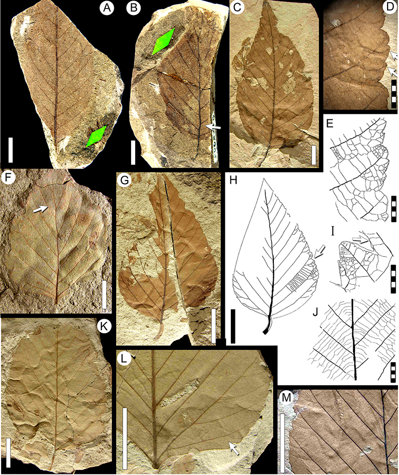
FIGURE 8. Morphotype SA005 (Cunoniaceae?). A, MPEF-Pb-4021; B, MPEF-Pb-4023 (arrow, basal portion of fimbrial vein); C, apical tooth detail of specimen in Figure 8D; D, MPEF-Pb-2023 (exemplar; arrow, basal portion of fimbrial vein); E, medial tooth detail of specimen in Figure 8D; F, camera lucida drawing (CLD) of leaf venation of specimen in MPEF-Pb-9017; G, CLD of MPEF-Pb-9013 (arrow, darkened apical gland); H, CLD of tooth venation of MPEF-Pb-9014 (arrow, darkened apical gland); I, CLD of leaf venation of MPEF-PB-9015, note margin-feeding insect damage (DT12, arrows); J, CLD of tooth venation of specimen in Figure 8I; K, CLD of tooth venation of specimen in Figure 8G; L, CLD of leaf venation of MPEF-Pb-9016; M, tooth venation detail from leaf in Figure 8F. Single-color scale bars equal 10 mm; grid scales equal one millimeter (per rectangle).
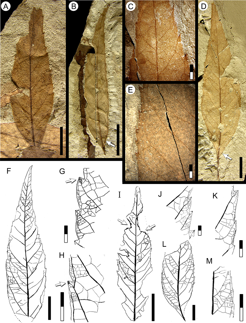
FIGURE 9. Morphotype SA008 (Rosaceae). A, MPEF-Pb-2024 (exemplar); B, MPEF-Pb-4028 (white arrow, concave base; black arrow, large polylobate hole feeding DT5, see also Donovan et al., 2018 figure 5.3); C, MPEF-Pb-4027 (white arrow, basal secondary veins), with Cochlichnus trace fossil (black arrow); D, MP¬¬EF-Pb-4030; E, MPEF-Pb-4031 (arrow, compound agrophic veins), with Cochlichnus; F, MPEF-Pb-9018; G, MPEF-Pb-4033; H, camera lucida drawing (CLD) of leaf venation of MPEF-Pb-9019; I, CLD of leaf base of MPEF-Pb-9020; J, CLD of leaf venation of MPEF-Pb-9021 (arrow, compound agrophic veins); K, CLD of leaf venation of MPEF-PB-9022; L, CLD of tooth venation of MPEF-Pb-9023; M, CLD of leaf venation MPEF-Pb-9024; N, CLD of apical venation of MPEF-Pb-9025. Scale bars equal 10 mm except for 2 millimeter scale in Figure 9L.
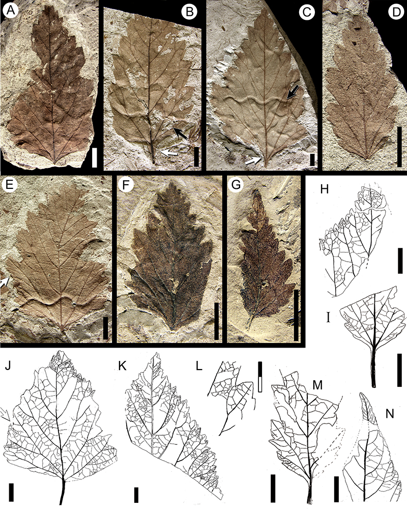
FIGURE 10. Morphotype SA009 “Cissites” patagonica Berry, 1937a (see also Figure 11). A, MPEF-Pb-2025 (exemplar); B, MPEF-PB-9026; C, MPEF-Pb-9027; D, MPEF-Pb-9028, note insect damage (black arrow, trenched margin feeding DT15; white arrow, hole feeding DT05; see also Donovan et al., 2018, figure 9.1); E, MPEF-Pb-4042; F, detail of glandular teeth of MPEF-Pb-9029; G, MPEF-Pb-4040; H, MPEF-Pb-4038; I, MPEF-Pb-4036; J, MPEF-Pb-2054 (accessory exemplar); K, MPEF-Pb-9031; L, MPEF-Pb-4041; M, detail of minor venation of MPEF-Pb-9032. N-Q, “C.” patagonica syntypes; N, USNM-201955 (illustrated in Berry 1937a, plate VII, figure 1; see also Table 2); O, USNM-201956a (illustrated in Berry 1937a, plate VII, figure 2; see also Table 2); P, tooth detail of specimen in Figure 10O (arrows, minute second-order teeth); Q, USNM-201957 (illustrated in Berry 1937a, plate VII, figure 3; see also Table 2), with Cochlichnus trace fossils. Scale bars equal 10 mm, except millimeter scales (per rectangle) in Figure 10F and 10P.
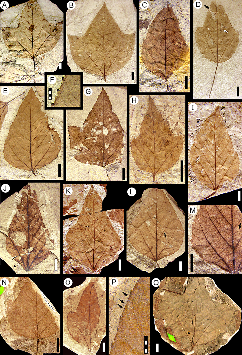
FIGURE 11. Morphotype SA009 “Cissites” patagonica Berry 1937a (continued from Figure 10), camera lucida drawings. A, MPEF-Pb-9020; B, MPEF-Pb-9152; C, tooth detail from Figure 11B; D, MPEF-Pb-9151 (arrow, compound agrophic veins); E, tooth detail from Figure 11D; F, MPEF-Pb-9145; G, tooth detail from Figure 11F; H, tooth detail from Figure 11A; I, tooth detail from Figure 11A (arrow, thin fimbrial vein); J, tooth venation detail from Figure 11A (arrow, looped marginal ultimate venation); K, MPEF-Pb-9160 (arrow, swollen petiole base); L, MPEF-Pb-9148; M, MPEF-Pb-9146; N, tooth detail from Figure 11B. Scale bars equal 10 mm (per rectangle).
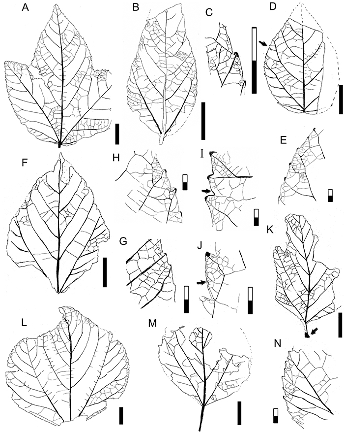
FIGURE 12. Morphotype SA010 (Lauraceae, see also Figure 13). A, Laurophyllum piatnitzkyi Berry, 1937a syntype specimen USNM-201966 (illustrated in Berry, 1937a, plate IX, figure 2; see also Table 1, Table 2); B, Cryptocaryoides maria-santisimensis Berry, 1937a syntype specimen USNM-208521 (illustrated in Berry, 1937a, plate VI, figure 3; see also Table 1, Table 2) (arrow, agrophic veins); C, MPEF-PB-9033; D, MPEF-PB-9034; E, MPEF-Pb-4048; F, MPEF-Pb-9035 (black arrow, agrophic veins), with skeletonization (DT79, white arrow; see also Donovan et al., 2018, figure 9.23); G, MPEF-Pb-3036; H-I, MPEF-Pb-2055 (accessory exemplar), note idioblasts (dark dots, arrow) preservation; J, detail of areoles and FEVs associated with idioblasts (arrow) from Figure 12F; K, counterpart of specimen in Figure 12E (arrow, agrophic veins); L, MPEF-Pb-9039; M, MPEF-Pb-2026 (exemplar; arrow, basal portion of fimbrial vein); N, MPEF-Pb-4049 (arrow, drip tip). Single-color scale bars equal 10 mm; grid scales equal one millimeter (per rectangle).
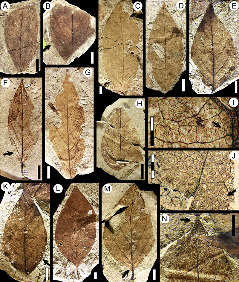
FIGURE 13. Morphotype SA010, Lauraceae (continue from Figure 12), camera lucida drawings. A, MPEF-Pb-9042; B, MPEF-Pb-9044; C, MPEF-PB-9049; D, MPEF-Pb-9051 (arrow, simple intersecondary vein); E, MPEF-Pb-9052; F, MPEF-Pb-9045 (arrow, compound agrophic veins); G, MPEF-Pb-9046; H, MPEF-Pb-9047 (arrow, simple intersecondary vein); I, MPEF-Pb-9048. Scale bars equal 10 mm.
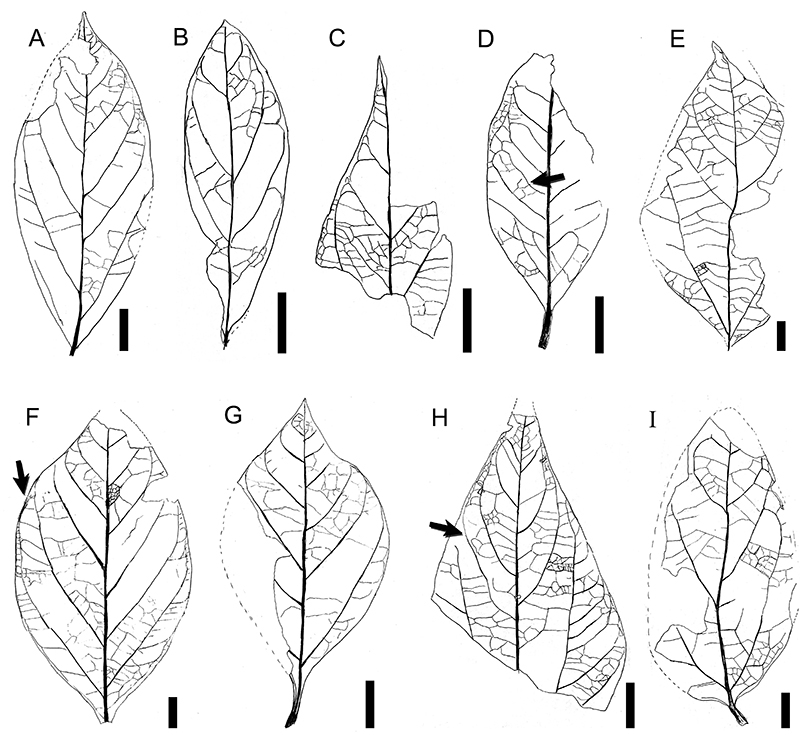
FIGURE 14. Morphotype SA014, “Sterculia” acuminataloba Berry 1937a (see also Figure 15). A, “S.” acuminataloba syntype USNM-208524 (illustrated in Berry, 1937a, plate VIII, figure 1; see also Table 2); B, “S.” acuminataloba syntype USNM-208526 (illustrated in Berry, 1937a, plate VIII, figure 3; see also Table 2); C, MPEF-Pb-2027 (exemplar; arrow, palinactinodromous venation); D, MPEF-Pb-4058; E, MPEF-Pb-9053 (arrow, weak brochidodromous secondary vein); F, MPEF-Pb-9054 (arrow, sinus-bracing vein); G, MPEF-Pb-4056; H, MPEF-Pb-4057; I, MPEF-Pb-4070, with margin feeding (DT14, arrow); J, lobe venation detail of MPEF-Pb-9056; K, MPEF-Pb-4053 (arrow, weak brochidodromous secondary vein); L, MPEF-Pb-9057; M, sinus-bracing veins, detail of MPEF-Pb-9058, and a fungal pycnidium or stroma leaf damage ring (arrow, DT334; see also Donovan et al., 2018 figure 6.13); N, MPEF-Pb-4059. Scale bars equal 10 mm, except 5 millimeter scale in Figure 14J.
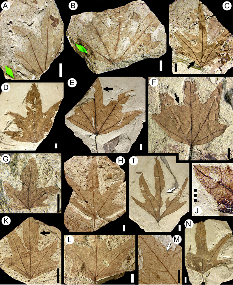
FIGURE 15. Morphotype SA014 “Sterculia” acuminataloba Berry 1937a (continued from Figure 14) and morphotype SA019 (see also Figure 17). A-D, camera lucida drawings (CLD) of morphotype SA014 “Sterculia” acuminataloba. A, MPEF-Pb-9059 (arrow, interior secondary vein); B, MPEF-Pb-9060 (arrow, sinus-bracing vein); C, MPEF-Pb-9061; D, MPEF-Pb-9062. E-I, CLD of morphotype SA019. E, MPEF-Pb-9063 (arrow, minor secondary vein); F, minor venation detail of specimen in Figure 15E; G, MPEF-Pb-9064 (arrow, sinus-bracing vein); H, MPEF-Pb-9065 (arrow, secondary lobe); I, MPEF-Pb-9066. Scale bars equal 10 mm, except 2 millimeter scale in Figure 15F.
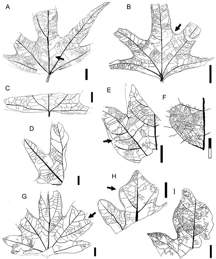
FIGURE 16. Morphotype SA016 (see also Figure 19F-I) and morphotype SA042 “Fagophyllum duseni” Berry 1937a (Cunoniaceae; see also Figure 25A-D). A-E, morphotype SA016. A, MPEF-Pb-2028 (exemplar). B, venation and skeletonization insect damage (DT19; see also Donovan et al., 2016) in apex detail of specimen in Figure 16A (arrow, intramarginal vein); C, MPEF-Pb-4062; D, MPEF-Pb-9067 (arrow, intramarginal vein); E, MPEF-Pb-9068. F-M, morphotype SA042, “F. duseni” (Cunoniaceae). F, MPEF-Pb-2035 (exemplar); G, articulated compound leaf MPEF-Pb-9156; H, tooth venation detail in MPEF-Pb-4093 (arrow, apically deflected vein from a secondary); I, MPEF-Pb-9070 (arrow, untoothed leaf base); J, MPEF-Pb-4098; K, MPEF-Pb-9069. L-M, “F. duseni” syntypes. L, USNM-201960 (illustrated in Berry 1937a, plate V, figure 1; see also Table 2) (arrow, swollen petiole base); M, USNM-201961 (illustrated in Berry 1937a, plate V, figure 2; see also Table 2). Single-color scale bars equal 10 mm; grid scales equal one millimeter (per rectangle).
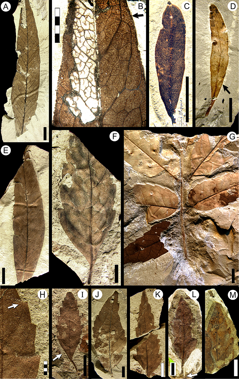
FIGURE 17. Morphotype SA019 (continued from Figure 15E-I). A, MPEF-Pb-2029 (exemplar, see also Figure 17D-E); B, MPEF-Pb-9149, with Cochlichnus trace fossils; C, venation detail of MPEF-Pb-4070 (arrow, sinus-bracing vein); D, minor venation detail from leaf in Figure 17A (exemplar); E, base detail of leaf in Figure 17A (exemplar; arrow, intramarginal vein); F, MPEF-Pb-9073 (arrow, secondary lobe; G, MPEF-Pb-9072; H, MPEF-Pb-4072, with several fungal pycnidia or stromata leaf-damage rings (DT334, arrow; see also Donovan et al., 2018, figure 10.13); I, “Sterculia” acuminataloba Berry, 1937a syntype USNM-208528 (illustrated in Berry 1937a, plate VIII, figure 5; see also Table 2); J, MPEF-Pb-4071; K, MPEF-Pb-9075; L, MPEF-Pb-9076 (arrow, cordate base). Single-color scale bars equal 10 mm; grid scales equal one millimeter (per rectangle).
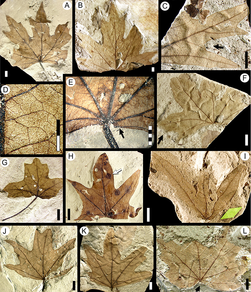
FIGURE 18. Morphotype SA020 (Cunoniaceae, see also Figure 19A-E). A, MPEF-Pb-2030 (exemplar, see also Figure 18F; arrow, extended acuminate apex); B, MPEF-Pb-9077; C, MPEF-PB-9078; D, MPEF-Pb-4076 (arrow, asymmetric decurrent base); E, MPEF-Pb-9079; F, perimarginal vein detail of specimen in Figure 18A (exemplar; arrow, an apical, short curved branch); G, tooth venation detail of specimen in Figure 18K (accessory exemplar; arrow, intramarginal vein); H, MPEF-Pb-9080; I, MPEF-Pb-9081; J, MPEF-Pb-9082; K, MPEF-Pb-3020 (accessory exemplar, see also Figure 18G); L, MPEF-Pb-9083 (arrow, asymmetric decurrent base); M, articulated compound leaf MPEF-Pb-9154. Single-color scale bars equal 10 mm; grid scales equal one millimeter (per rectangle).
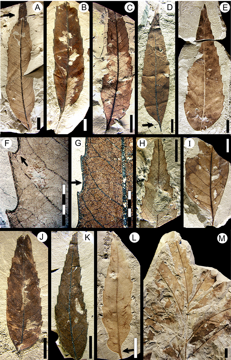
FIGURE 19. Morphotypes SA016 (continued, see also Figure 16A-E), SA035 (Bixaceae? see also Figure 20), SA020 (continued, see also Figure 18; Cunoniaceae), and SA041 (Rhamnaceae, see also Figure 23A-I). A-E, camera lucida drawings (CLD) of morphotype SA020 (Cunoniaceae). A, MPEF-Pb-9087 (arrow, bifurcation of a secondary vein); B, MPEF-Pb-9088 (arrow, intersecondary vein); C, MPEF-Pb-9089; D, MPEF-Pb-9090 (arrow, bifurcation of a secondary vein); E, MPEF-Pb-3020 (accessory exemplar, see also Figure 18K; arrow, intersecondary vein). F-I, CLD of morphotype SA016. F, MPEF-Pb-9091 (arrow, basal secondary vein); G, MPEF-Pb-9092 (arrow, intramarginal vein); H, MPEF-Pb-9093; I, MPEF-Pb-9094. J-K, digital overlay drawing (DOD) of morphotype SA035 (Bixaceae?). J, tooth venation of MPEF-Pb-2031 (exemplar, see also Figure 20A, C); K, MPEF-Pb-4078. L-M, morphotype SA041 (Rhamnaceae). L, hand drawing of MPEF-Pb-2034 (exemplar, see also Figure 23A); M, DOD of venation in MPEF-Pb-9095 (arrow, two small, scattered teeth). Scale bars equal 10 mm, except 4 millimeter scale in Figure 19F, J, M.
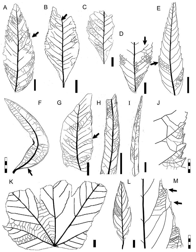
FIGURE 20. Morphotype SA035 (Bixaceae? continued, see also Figure 19J-K). A, MPEF-Pb-2031 (exemplar, see also Figure 19J, Figure 20C); B, MPEF-Pb-4078 (arrow, sinus-bracing vein); C, tooth detail of specimen in Figure 20A (exemplar); D, venation detail at leaf base, MPEF-Pb-9096; E, venation detail of MPEF-Pb-9097 (arrow, compound agrophic veins); F, basal venation of MPEF-Pb-9098; G, MPEF-Pb-4083 (arrow, sinus-bracing vein); H, MPEF-Pb-4082; I, MPEF-Pb-4079. Scale bars equal 10 mm, except 5 millimeter scale in Figure 20C.
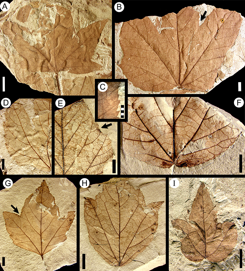
FIGURE 21. Morphotypes SA038 (see also Figure 22I), SA039 “Paranymphaea” aristolochiaformis Berry 1937a (see also Figure 22A-H), SA040 (see also Figure 22J-M), and SA055 (see also Figure 30K-L). A-B, drawings of morphotype SA039. A, redrawing of “Paranymphaea” aristolochiaformis syntype USNM-201964 (illustrated in Berry, 1937a, plate V, figure 5; see also Figure 22A and Table 2; arrow, primary vein distally forking); B, digital overlay drawing (DOD) of MPEF-PB-2032 (exemplar, see also Figure 22B, D; arrow, festooned brochidodromous agrophic veins). C, DOD of morphotype SA038, MPEF-Pb-3033 (exemplar, see also Figure 22I). D, DOD of morphotype SA040, MPEF-Pb-2033 (exemplar; see also Figure 22J; arrow, forked lateral primary vein). E-F, DOD of morphotype SA055, MPEF-PB-2047 (exemplar, part and counterpart; see also Figure 30K-L), counterpart reconstructed areas dotted (arrow, sinus-bracing and intramarginal veins). Scale bars equal 10 mm.
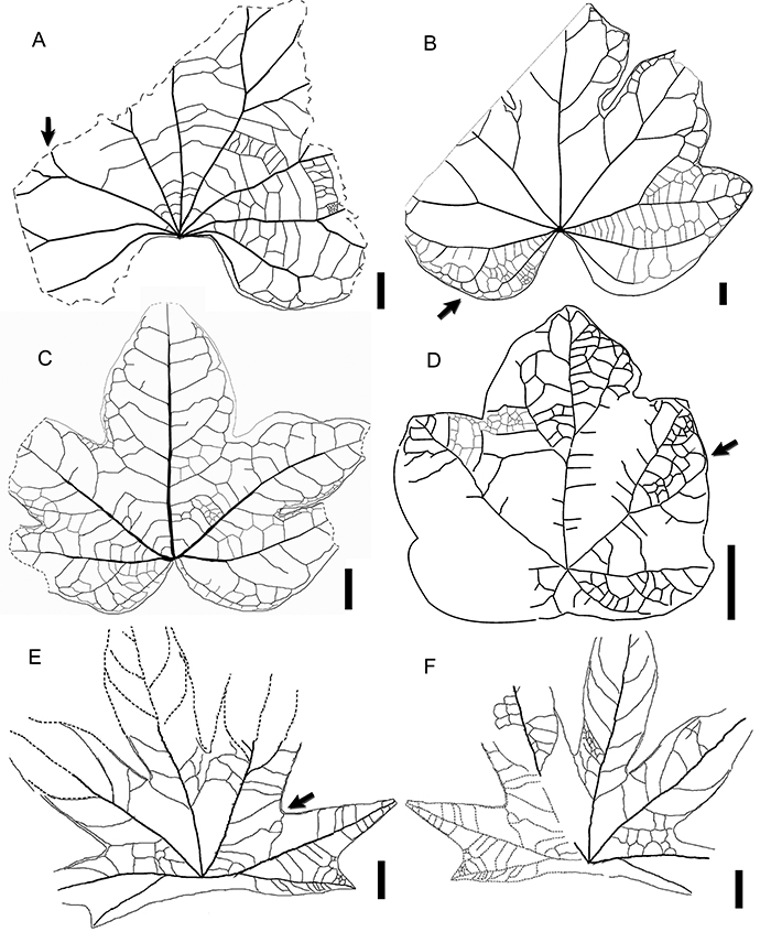
FIGURE 22. Morphotypes SA038 (continued, see also Figure 21C), SA039 “Paranymphaea” aristolochiaformis Berry 1937a (continued, see also Figure 21A-B), and SA040 (continued, see also Figure 21D). A-H, morphotype SA039. A, “P.” aristolochiaformis syntype USNM-201964 (illustrated in Berry, 1937a, plate V, figure 5; see also Figure 21A and Table 2); B, MPEF-Pb-2032 (exemplar, see also Figure 21B, Figure 22D); C, MPEF-Pb-9099; D, venation at leaf base of specimen in Figure 22B (exemplar; arrow, secondary vein); E, MPEF-Pb-9100; F, MPEF-Pb-4084 (arrow, primary vein); G, MPEF-Pb-9095; H, MPEF-Pb-9101. I, morphotype SA038, MPEF-Pb-3033 (exemplar, see also Figure 21C). J-M, morphotype SA040. J, MPEF-Pb-2033 (exemplar, see also Figure 21D); K, peltate base of MPEF-Pb-9102; L, peltate base of MPEF-Pb-9103; M, MPEF-Pb-9104. Scale bars equal 10 mm, except 5 millimeter scale in Figure 22F, L.
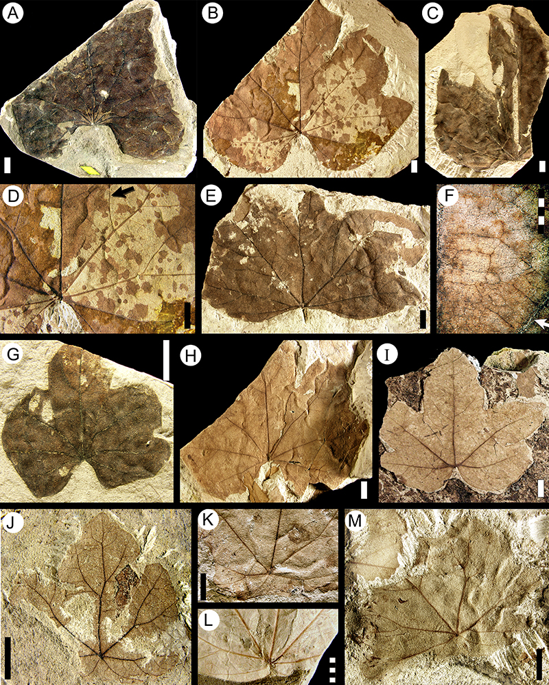
FIGURE 23. Morphotypes SA041 (Rhamnaceae; continued, see also Figure 19L-M) and SA044 (Cunoniaceae, see also Figure 25G-H). A-I, morphotype SA041 (Rhamnaceae). A, MPEF-Pb-2034 (exemplar, see also Figure 19L); B, MPEF-Pb-4085; C, MPEF-Pb-4089 (arrow, decurrent secondary veins); D, MPEF-Pb-4090; E, counterpart of MPEF-Pb-4090; F, MPEF-Pb-9105; G, MPEF-Pb-9106 (arrows, scattered teeth); H, MPEF-Pb-9107 (arrow, wavy margin); I, minor venation detail from leaf in Figure 23A (arrow, one-branched freely ending veinlets). J-O, morphotype SA044 (Cunoniaceae). J, MPEF-Pb-4105; K, MPEF-Pb-2037 (exemplar, see also Figure 25G-H); L, MPEF-Pb-3024 (accessory exemplar); M, MPEF-Pb-9108 (arrow, apical brochidodromous secondary vein); N, asymmetric base (arrow) of MPEF-Pb-9109; O, tooth venation of specimen in Figure 23K (exemplar; arrow, small triangular tooth). Scale bars equal 10 mm, except 4 millimeter scale in Figure 23I and 23O.
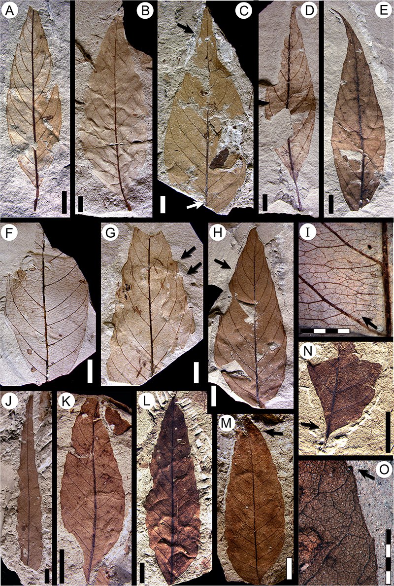
FIGURE 24. Morphotype SA043 (Cunoniaceae?/Sapindaceae? see also Figure 25E-F) and morphotype SA045 (Fabaceae; see also Figure 25J). A-G, morphotype SA043 (Cunoniaceae?/Sapindaceae?). A, MPEF-Pb-2036 (exemplar); B, MPEF-Pb-3023 (accessory exemplar 1; arrow, asymmetric base); C, articulated compound leaf, MPEF-Pb-9110 (accessory exemplar 2; arrow, petiolule subtending terminal leaflet); D, MPEF-Pb-4102; E, MPEF-Pb-6561, with hole-feeding damage (DT113; see also Labandeira et al., 2007 p.7, figure DT113; and Donovan et al., 2016, figure 1i); F, tooth venation detail from Figure 24E; G, MPEF-Pb-9159 with three attached leaflets. H-L, morphotype SA045 (Fabaceae). H, MPEF-Pb-4110; I-L, MPEF-Pb-2038 (exemplar, see also Brea et al., 2008 figure 6, and Figure 25J); I, counterpart preserving apical portion; J, detail of retuse apex; (arrow); K, base detail with a pulvinulate, horizontally striated petiolule (arrow); L, complete exemplar. Single-color scale bars equal 10 mm; grid scales equal one millimeter (per rectangle).
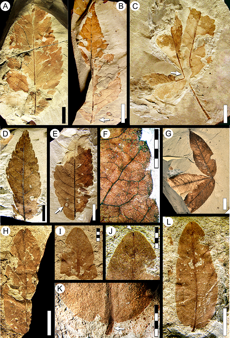
FIGURE 25. Morphotypes SA042 (Cunoniaceae; continued, see also Figure 16F-M), SA043 (Cunoniaceae?/Sapindaceae? continued, see also Figure 24A-G), SA044 (Cunoniaceae, see also Figure 23J-O), SA045 (Fabaceae; continued, see also Figure 24H-L), and SA046 Laurophyllum chubutensis Berry (Lauraceae, see also Figure 26A-F). A-D, camera lucida drawings (CLD) of morphotype SA042 (Cunoniaceae). A, MPEF-Pb-9069; B, MPEF-Pb-9113 (arrow, an intersecondary vein); C, tooth venation detail of specimen in Figure 25B (arrow, a forked secondary vein); D, tooth venation of specimen in Figure 25A (arrow, an intersecondary vein). E-F, morphotype SA043 (Cunoniaceae?/Sapindaceae?), CLD of MPEF-Pb-6561. G-H, morphotype SA044 (Cunoniaceae), MPEF-Pb-2037 (exemplar, see also Figure 23K, O). G, digital overlay drawing (DOD) of tooth venation; H, triangular tooth with glandular apex (arrow). I, morphotype SA046 Laurophyllum chubutensis Berry (Lauraceae), DOD of MPEF-Pb-2039 (exemplar, see also Figure 26A-B), note low rank venation (see also Figure 26A-B). J, morphotype SA045 (Fabaceae), CLD of MPEF-Pb-2038 (exemplar; see also Figure 24I-L), note pulvinulate petiolule (arrow). Single-color scale bars equal 10 mm; grid scales equal one millimeter (per rectangle).
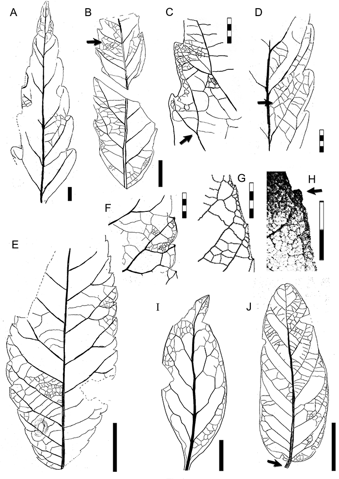
FIGURE 26. Morphotype SA046 Laurophyllum chubutensis Berry 1937a (Lauraceae; continued, see also Figure 25I) and morphotype SA047 (see also Figure 27A). A-F, morphotype SA046 (Lauraceae). A, MPEF-Pb-2039 (exemplar, see also Figure 25I), note patches of skeletonization (arrow, DT16; see also Donovan et a., 2018 figure 6.1); B, venation detail of specimen in Figure 26A (exemplar; arrow, fimbrial vein); C, MPEF-Pb-9114 (arrow, intersecondary vein); D, MPEF-Pb-4115; E, MPEF-Pb-3025 (accessory exemplar); F, Laurophyllum chubutensis syntype, USNM-208530 (illustrated in Berry, 1937a, plate IX, figure 3-4; see also Table 1, Table 2). G-L, morphotype SA047. G, MPEF-Pb-2040 (exemplar, see also Figure 27A); H, MPEF-Pb-9116, with hole feeding damage (DT03, arrow; see also Donovan et al., 2016); I, MPEF-Pb-4124 (arrow, second order teeth); J, detail of compound teeth, MPEF-Pb-9117 (arrow indicating glandular tooth apex); K, tooth and venation detail of specimen in Figure 26G (exemplar), note the fimbrial vein (arrow); L, MPEF-Pb-4121. Single-color scale bars equal 10 mm; grid scales equal one millimeter (per rectangle).
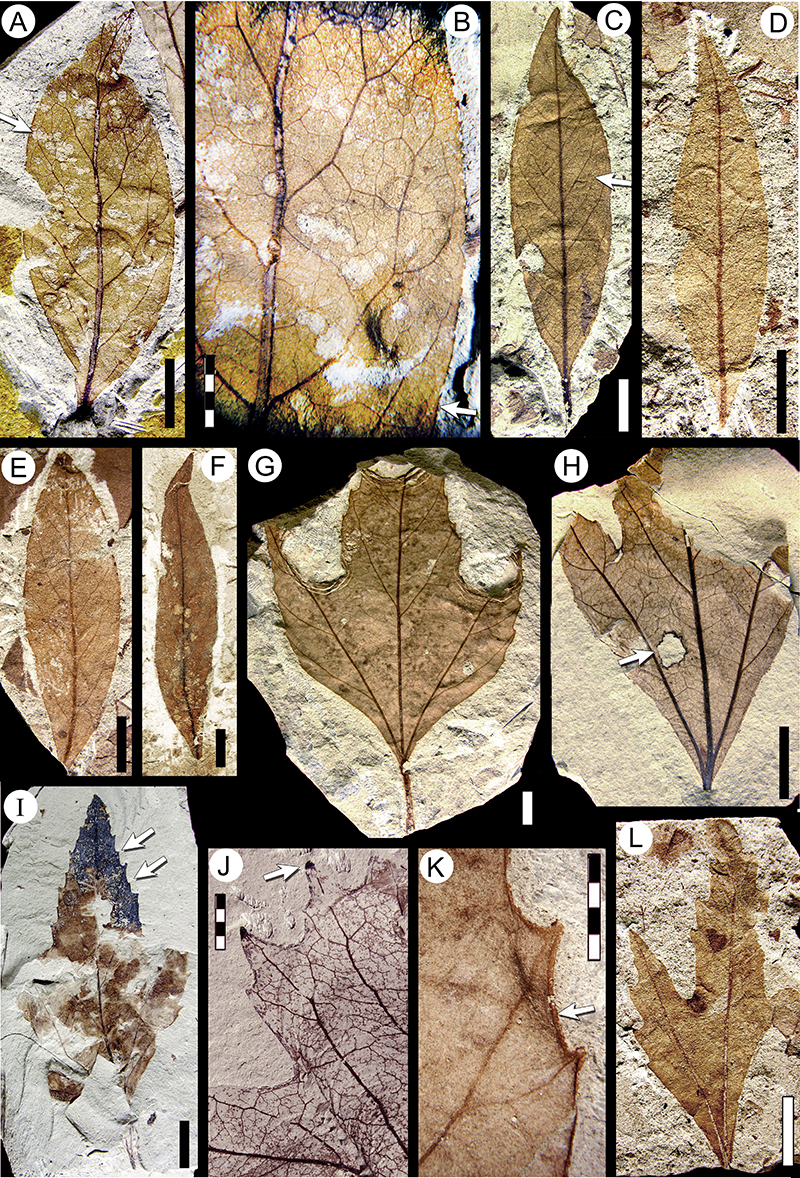
FIGURE 27. Morphotypes SA047 (continued, see also Figure 26G-L), SA048 (Rhamnaceae, see also Figure 28A-G), SA049 “Myrica” premira Berry 1937a (see also Figure 29A-K), SA051 (Urticaceae? see also Figure 30A-G), and SA052 (Sapindaceae? see also Figure 28H-I). A, morphotype SA047, digital overlay drawing (DOD) of MPEF-Pb-2040 (exemplar; arrow, agrophic veins; see also Figure 26G, K). B-D, morphotype SA048 (Rhamanceae). B, DOD of MPEF-Pb-2041 (exemplar, see also Figure 28D), note compound agrophic veins (arrow); C, DOD of Ziziphus chubutensis Berry, 1937a holotype USNM-208529 (see also Figure 28B and Table 2); D, DOD of “Banaraphyllum” ovatum Berry, 1937a holotype USNM-201953 (see also Figure 28A and Table 2). E-F, morphotype SA049 “Myrica” premira. E, DOD of tooth venation of MPEF-PB-2042 (exemplar; see also Figure 29D) (arrow, an independent tooth accessory vein); F, venation and margin of MPEF-Pb-9118. G-I, morphotype SA051 (Urticaceae?). G, camera lucida drawing (CLD) of MPEF-Pb-3028 (accessory exemplar; see also Figure 30C, E) (arrow, percurrent tertiaries); H, CLD of MPEF-Pb-2044 (exemplar; see also Figure 30A-B) (arrow, decurrent leaf base). I, DOD of teeth detail from Figure 27H (exemplar). J, morphotype SA052 (Sapindaceae?), DOD of MPEF-Pb-2045 (exemplar, see also Figure 28H-I); note secondary vein branches (arrow). Single-color scale bars equal 10 mm; grid scales equal one millimeter (per rectangle).
FIGURE 28. Morphotype SA048 (Rhamnaceae; continued, see also Figure 27B-D) and morphotype SA052 (Sapindaceae? continued, see also Figure 27J). A-G, morphotype SA048 (Rhamnaceae). A, “Banaraphyllum” ovatum Berry, 1937a holotype, USNM-201953 (illustrated in Berry, 1937a, plate IX, figure 1; see also Figure 27D and Table 1, Table 2); B, Ziziphus chubutensis Berry, 1937a holotype, USNM-208529 (illustrated in Berry, 1937a, plate IX, figure 2; see also Figure 27C and Table 1, Table 2); note the Cochlichnus trace fossils; C, MPEF-Pb-4129 (arrow, decurrent secondary veins); D, MPEF-Pb-2041 (exemplar, see also Figure 27B), note the naked basal vein (black arrow) and the margin feeding leaf damage (DT14, white arrow; see also Donovan et al., 2016); E, tooth detail from specimen in Figure 28D (arrow, an independent sinus vein); F, leaf base of MPEF-Pb-9119, note the naked basal vein (black arrow); G, MPEF-Pb-9120, with hole feeding leaf damage (DT05, arrow; see also Donovan et al., 2016). H-I, morphotype SA052 (Sapindaceae?), MPEF-Pb-2045 (exemplar, see also figure 27J). H, detail of the teeth and higher order venation (arrow, tooth venation forking at the sinus); I, complete (probable) leaflet. Single-color scale bars equal 10 mm; grid scales equal one millimeter (per rectangle).
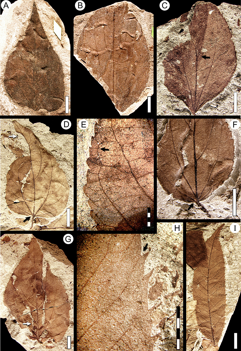
FIGURE 29. Morphotype SA049 “Myrica” premira Berry (continued, see also Figure 27E-F), morphotype SA050 (Anacardiaceae? see also Figure 31A-D), and related material. A-K, morphotype SA049 “Myrica” premira (except Figure 29C). A, “M.” premira syntype USNM-201963 (illustrated in Berry, 1937a, plate V, figure 4; see also Table 2); B, tooth detail (arrow) from specimen in Figure 29A; C, “M. premira” syntype USNM-201962 (illustrated in Berry, 1937a, plate V, figure 3; here referred to morphotype SA020 Cunoniaceae; see also Table 1, Table 2), note the thick fimbrial vein (arrow); D, MPEF-Pb-2042 (exemplar, see also Figure 27E), note swollen petiole base (arrow); E, MPEF-Pb-3027 (accessory exemplar 1) (arrow, a crenation); F, MPEF-Pb-3026 (accessory exemplar 2), with piercing and sucking leaf damage (DT 46, arrows; see also Donovan et al., 2016), often found on this leaf morphotype; G, venation detail from specimen in Figure 29F (arrow, a crene); H, higher order venation of specimen in Figure 29F, note dendritic branching of freely ending veinlets (arrow); I, MPEF-Pb-4135; J, MPEF-Pb-4134, with piercing and sucking leaf damage (DT 46; see also Donovan et al., 2016); K, MPEF-Pb-4136, with Cochlichnus trace fossils. L-O, morphotype SA050 (Anacardiaceae?). L, higher-order venation from Figure 29M, note admedially ramified tertiary venation (arrow); M, MPEF-Pb-2043 (exemplar, see also Figure 31A); N, MPEF-Pb-4143; O, leaf venation detail of MPEF-Pb-9150, showing admedially ramified venation (arrow). Single-color scale bars equal 10 mm; grid scales equal one millimeter (per rectangle).
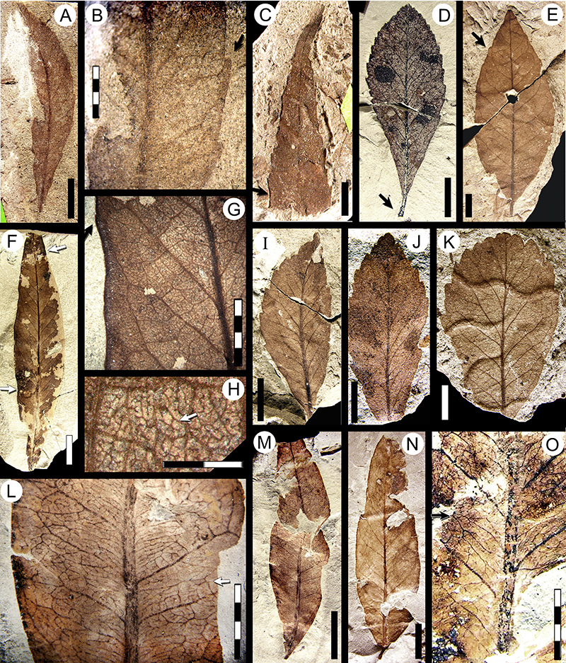
FIGURE 30. Morphotypes SA051 (Urticaceae? continued, see also Figure 27G-I), SA053 (see also Figure 31E-F), SA054 (Cunoniaceae; see also Figure 31G-H), and SA055 (continued, see also Figure 21E-F). A-G, morphotype SA051 (Urticaceae?). A, MPEF-Pb-2044 (exemplar, see also Figure 27H-I); B, tooth detail from specimen in Figure 30A (exemplar; arrow, spherulate apex); C, tooth detail from specimen in Figure 30E (accessory exemplar); D, MPEF-Pb-4145; E, MPEF-Pb-3028 (accessory exemplar; see also Figure 27G; arrows, spherulate teeth); F, venation detail from specimen in Figure 30E (accessory exemplar); G, leaf base from counterpart of specimen in Figure 30A. H-J, morphotype SA053. H, parallel venation detail from Figure 30I (exemplar); I, MPEF-Pb-3029 (exemplar; see also Figure 31E) (arrows, glandular teeth); J, MPEF-Pb-9121 (arrow, swollen petiole base; see also Figure 31F). K-L, morphotype SA055 MPEF-Pb-2047, part and counterpart (exemplar, see also Figure 21E-F). M-P, morphotype SA054 (Cunoniaceae). M, MPEF-Pb-2046 (exemplar, see also Figure 31G-H); N, MPEF-Pb-9122; O, MPEF-Pb-4147 (arrow, a glandular tooth apex); P, tooth venation detail from Figure 30M (exemplar; arrow, a second-order tooth). Single-color scale bars equal 10 mm; grid scales equal one millimeter (per rectangle).
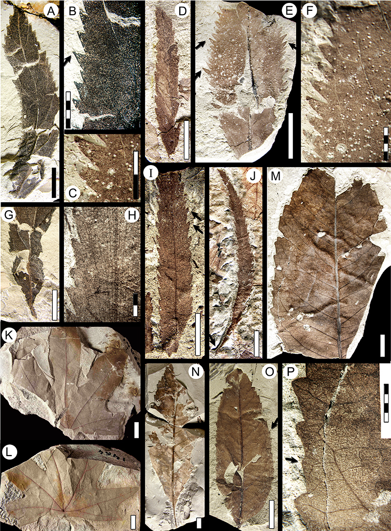
FIGURE 31. Morphotypes SA050 (Anacardiaceae? continued, see also Figure 29L-O), SA053 (continued, see also Figure 30H-J) SA054 (continued, see also Figure 30M-P), SA056 (see also Figure 32A-C), SA057 (see also Figure 32D-G), and SA058 (Malvaceae? see also Figure 32H-J). A-D, morphotype SA050 (Anacardiaceae?). A, digital overlay drawing (DOD) of MPEF-Pb-2043 (exemplar, see also Figure 29L-M); B, higher order venation detail from Figure 30A (arrow, intramarginal vein); C, camera lucida drawing (CLD) of MPEF-Pb-9123; D, higher order venation detail from Figure 31C (arrow, a dichotomizing fourth-order vein). E-F, morphotype SA053. E, CLD of MPEF-Pb-3029 (exemplar, see also Figure 30H-I; arrow, a secondary vein anastomosis); F, DOD of MPEF-Pb-9121 (from Figure 30J). G-H, morphotype SA054, MPEF-Pb-2046 (exemplar, see also Figure 30M, P). G, DOD of complete leaf; H, CLD of tooth venation, note vein junction (arrow) before entering the tooth. I, morphotype SA057, DOD of MPEF-Pb-2049 (exemplar, see also Figure 32D-G), note proximal secondary veins extended for over half the length of the blade (arrow). J-K, morphotype SA058 (Malvaceae?). J, DOD of MPEF-Pb-2050 (exemplar, see also Figure 32H, J); K, CLD of higher order venation from Figure 30J (exemplar; arrow, intersecondary vein). L, morphotype SA056, DOD of MPEF-Pb-2048 (exemplar, see also Figure 32A, C). Single-color scale bars equal 10 mm; grid scales equal one millimeter (per rectangle).
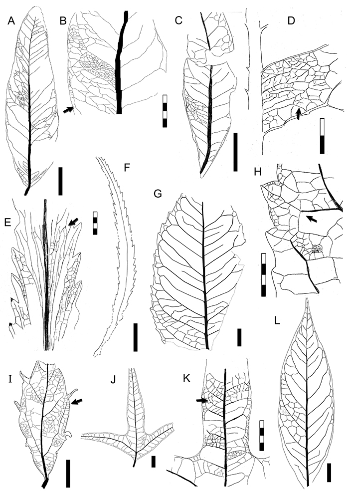
FIGURE 32. Morphotype SA056 (continued, see also Figure 31L), SA057 (continued, see also Figure 31I), SA058 (Malvaceae? continued, see also Figure 31J-K), and SA059 (see also Figure 33A-B). A-C, morphotype SA056. A, MPEF-Pb-2048 (exemplar; see also Figure 31L; arrow, drip tip); B, MPEF-Pb-4150; C, higher order venation from specimen in Figure 32A (exemplar; arrow, an intersecondary secondary vein). D-G, morphotype SA057, MPEF-Pb-2049 (exemplar, see also Figure 31I). D, spinose tooth; E, tooth venation detail, with margin-feeding leaf damage (DT12, arrow; see also Donovan et al., 2016); F, higher order venation and idioblasts (dark dots, arrow); G, complete leaf of exemplar. H-J, morphotype SA058 (Malvaceae?). H, MPEF-Pb-2050 (exemplar, see also Figure 31J-K); I, “Sterculia” acuminataloba Berry, 1937a syntype USNM-208525 (illustrated in Berry 1937a, plate VIII, figure 2; see also Table 2); J, venation detail from specimen in Figure 32H (exemplar). K-M, morphotype SA059, MPEF-Pb-2051 (exemplar, see also Figure 33A-B). K, complete leaf of exemplar, note mucronate apex (arrow); L, basal teeth (arrow) and venation detail; M, detail of apical compound teeth and venation (arrow, a small, opaque, spherulate tooth apex). Single-color scale bars equal 10 mm; grid scales equal one millimeter (per rectangle).
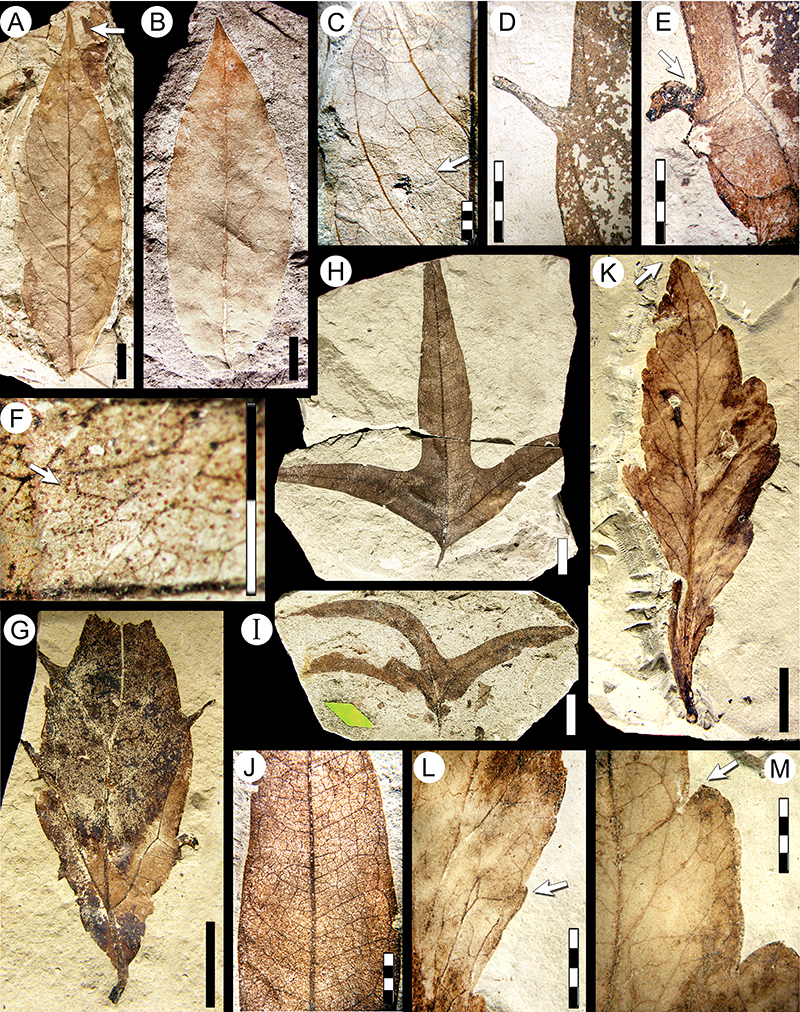
FIGURE 33. Morphotypes SA059 (continued, see also Figure 32K-M), SA060 Wilkinsoniphyllum menispermoides Jud, Gandolfo, Iglesias, and Wilf, 2018 (Menispermaceae, see also Jud et al., 2018 figure 4; and Figure 34A-C), SA063 (see also Figure 34G-I), SA066 (see also Figure 34L-M), SA067 (see also Figure 34D-F), SA068 (see also Figure 35G-K), and SA069 (Rhamnaceae; see also Figure 34J-K). A-B, morphotype SA059, MPEF-Pb-2051 (exemplar, see also Figure 32K-M). A, digital overlay drawing (DOD), note decurrent base shape and swollen petiole base (arrow); B, DOD of tooth venation detail (arrow, anastomosis in venation). C-D, morphotype SA060 W. menispermoides (Menispermaceae), MPEF-Pb-2052 (holotype, see also Figure 34A-C). C, DOD of the entire leaf (arrow, tapering fimbrial vein); D, DOD of mucronate apex and detail of regular, orthogonal higher order venation (arrow). E, morphotype SA063, camera lucida drawing (CLD) of MPEF-Pb-3002 (exemplar, see also Figure 34G, I). F, morphotype SA066, CLD of MPEF-Pb-3004 (exemplar, see also Figure 34L-M). G, morphotype SA067, CLD of MPEF-Pb-3006 (exemplar, see also Figure 34D-F). H-I, morphotype SA069, MPEF-Pb-3010 (exemplar, see also Figure 34J-K). H, CLD of tooth venation; I, CLD of the complete leaf (arrow, naked basal veins). J, morphotype SA068, CLD of MPEF-PB-3008 (accessory exemplar, see also Figure 35G-H). Single-color scale bars equal 10 mm; grid scales equal one millimeter (per rectangle).
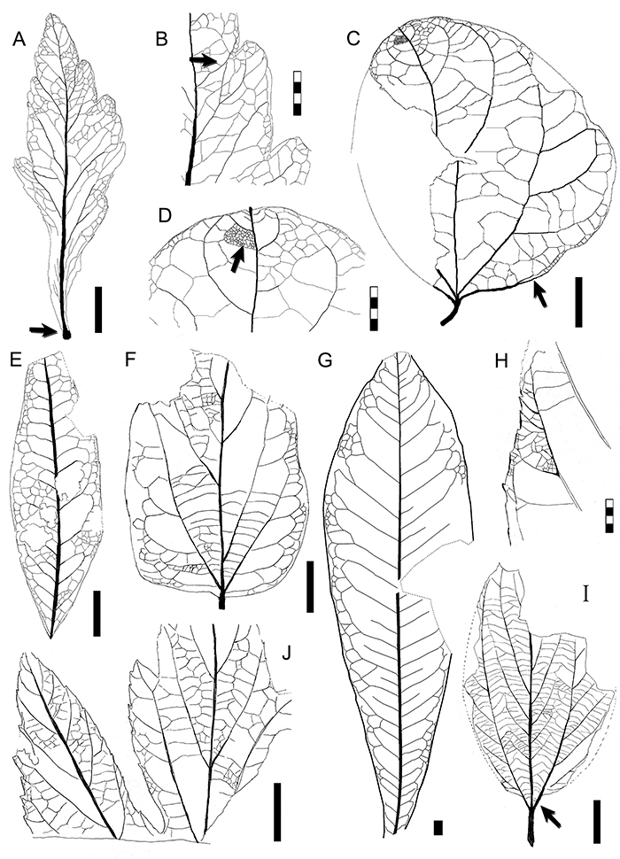
FIGURE 34. Morphotypes SA060 Wilkinsoniphyllum menispermoides Jud Gandolfo, Iglesias, and Wilf, 2018 (Menispermaceae; continued, see also Jud et al., 2018 figure 4, and Figure 33C-D), SA063 (continued, see also Figure 33E), SA066 (continued, see also Figure 33F), SA067 (continued, see also Figure 33G), and SA069 (Rhamnaceae; continued, see also Figure 33H-I). A-C, morphotype SA060 W. menispermoides (Menispermaceae), MPEF-Pb-2052 (holotype, see also Figure 33C-D). A, complete leaf; B, detail of mucronate apex (arrow) and higher order venation; C, counterpart. D-F, morphotype SA067, MPEF-Pb-3006 (exemplar, see also Figure 33G). D, complete leaf; E, leaf base detail; F, apex venation detail. G-I, morphotype SA063. G, venation detail from specimen in Figure 34I; H, MPEF-Pb-9124; I, MPEF-Pb-3002 (exemplar, see also Figure 33E), with galling leaf damage (arrow, DT112, also Labandeira et al., 2007 p.14). J-K, morphotype SA069 (Rhamnacaeae), MPEF-Pb-3010 (exemplar, see also Figure 33H-I). J, complete leaf preserving petiole and naked basal veins (arrow); K, counterpart (arrow, chevroned interior veins). L-M, morphotype SA066, MPEF-Pb-3004 (exemplar, see also Figure 33F). L, complete leaf; M, venation detail along part of the basal margin. Single-color scale bars equal 10 mm; grid scales equal one millimeter (per rectangle).
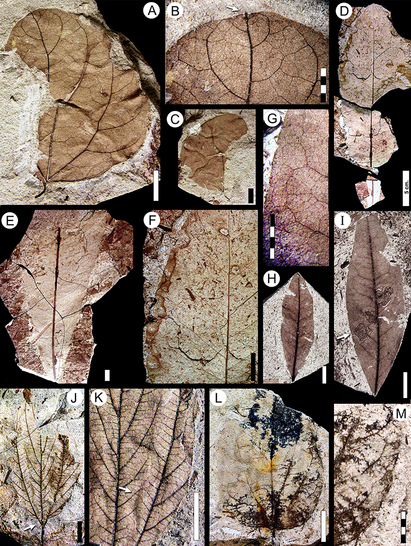
FIGURE 35. Morphotypes SA070 (Juglandaceae? see also Figure 39A-B), SA068 (continued, see also Figure 33J). A-F, morphotype SA070 (Juglandaceae?). A, MPEF-Pb-3012 (accessory exemplar, see also Figure 39A-B), note asymmetric base and basally crowded secondary veins (arrow); B, MPEF-Pb-9125; C, MPEF-Pb-3011 (exemplar); D, MPEF-Pb-3071; E, MPEF-Pb-9126; F, tooth detail of MPEF-Pb-9127 (arrows, second order teeth). G-K, morphotype SA068. G, MPEF-Pb-3008 (accessory exemplar, see also Figure 33J); H, tooth detail around a second-order lobe sinus from specimen in Figure 35G (accessory exemplar), with hole-feeding leaf damage (DT01, arrow); I, MPEF-Pb-9128; J, MPEF-Pb-3007 (exemplar), with Cochlichnus trace fossils (arrow); K, tooth detail from specimen in Figure 35J (exemplar). Single-color scale bars equal 10 mm; grid scales equal one millimeter (per rectangle).
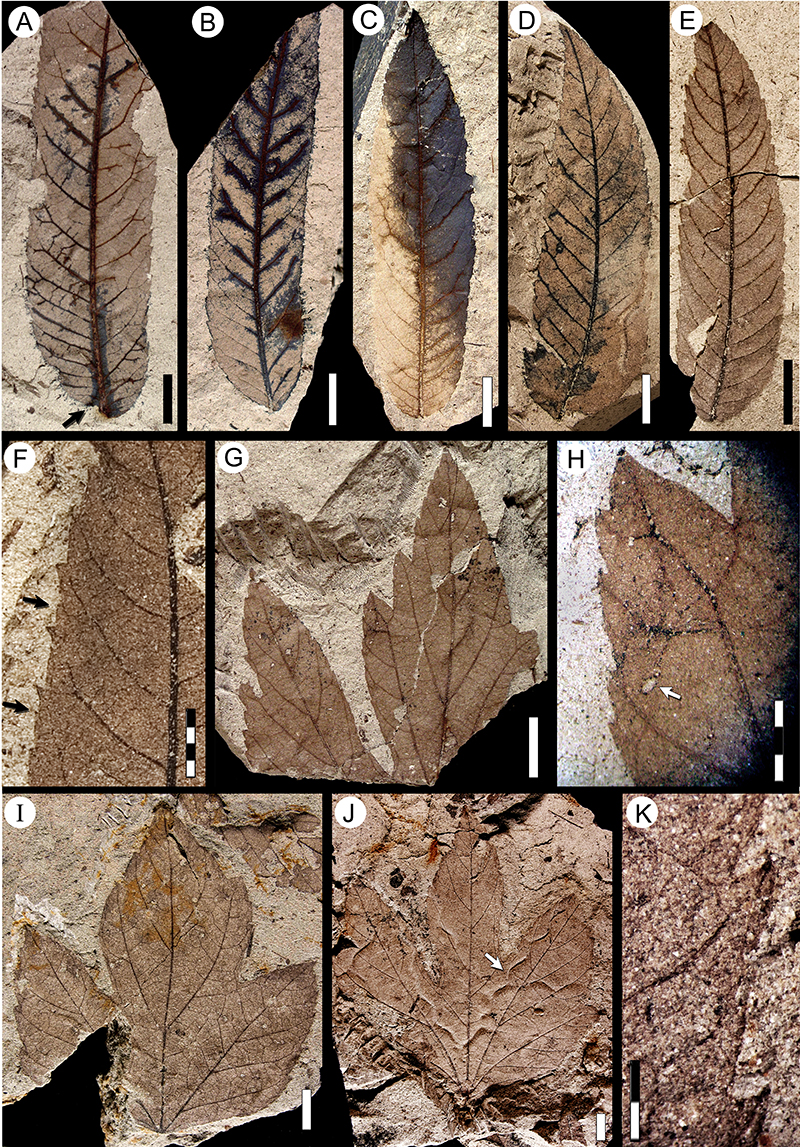
FIGURE 36. Morphotypes SA073 (see also Figure 39E) and SA074 (see also Figure 39F-G). A-E, morphotype SA073. A, MPEF-Pb-9129; B, MPEF-Pb-3031 (exemplar); C, “Cissites patagonica” Berry, 1937a syntype specimen USNM-201959 (illustrated in Berry, 1937a, plate IX, figure 5; see also Table 2), note the extended lateral primary veins (arrow); D, MPEF-Pb-9131; E, higher order venation detail of MPEF-Pb-3032 (accessory exemplar; arrow, fimbrial vein). F-H, morphotype SA074. F, MPEF-Pb-3015 (exemplar, see also Figure 36H, Figure 39F-G); G, venation detail at base of MPEF-Pb-9132, note looped ultimate venation (arrow); H, detail of tooth venation from specimen in Figure 36F (exemplar), note glandular tooth apex (arrow). Single-color scale bars equal 10 mm; grid scales equal one millimeter (per rectangle).

FIGURE 37. Morphotypes SA076 (see also Figure 40A-B) and SA075 (see also Figure 39C-D). A-D, morphotype SA076. A, MPEF-PB-3017 (exemplar, see also Figure 37B, Figure 40A); B, base detail from counterpart of specimen in Figure 37A (arrow, swollen petiole base); C, MPEF-Pb-9135; D, higher order venation detail of MPEF-Pb-9136 (arrow, one-branched freely ending veinlets). E-H, morphotype SA075. E, MPEF-PB-3016 (exemplar, see also Figure 39C); F, higher order venation in MPEF-Pb-9137 (arrow, regular polygonal reticulate higher-order veins); G, MPEF-Pb-9138; H, tooth venation of MPEF-Pb-9139 (arrow, sinus-bracing vein, see also Figure 39D). Single-color scale bars equal 10 mm; grid scales equal one millimeter (per rectangle).
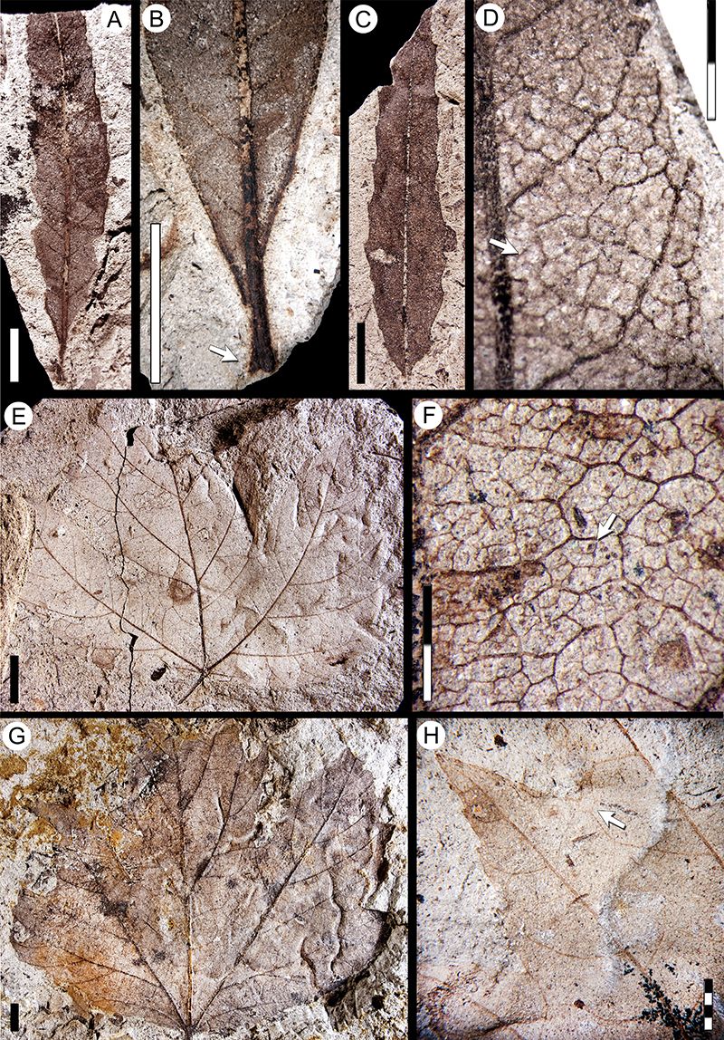
FIGURE 38. Morphotypes SA077 (see also Figure 40C), and SA078 (see also Figure 40E). A-D, morphotype SA077. A, MPEF-Pb-3018 (exemplar, see also Figure 40C); B, MPEF-Pb-9140 (arrow, forked secondary vein); C, MPEF-Pb-9141; D, venation detail from Figure 38A (exemplar; arrow, percurrent tertiary vein). E-H, morphotype SA078. E, base venation from counterpart of specimen in Figure 38F (exemplar); F, MPEF-Pb-3019 (exemplar, see also Figure 38E, Figure 40E), note asymmetrical blade and base (black arrow) and large polylobate hole damage (white arrow DT5; see also Donovan et al., 2018 figure 13.4); G, MPEF-Pb-9142; H, venation detail from Figure 38G (arrow, alternate percurrent tertiary veins). Single-color scale bars equal 10 mm; grid scales equal one millimeter (per rectangle).
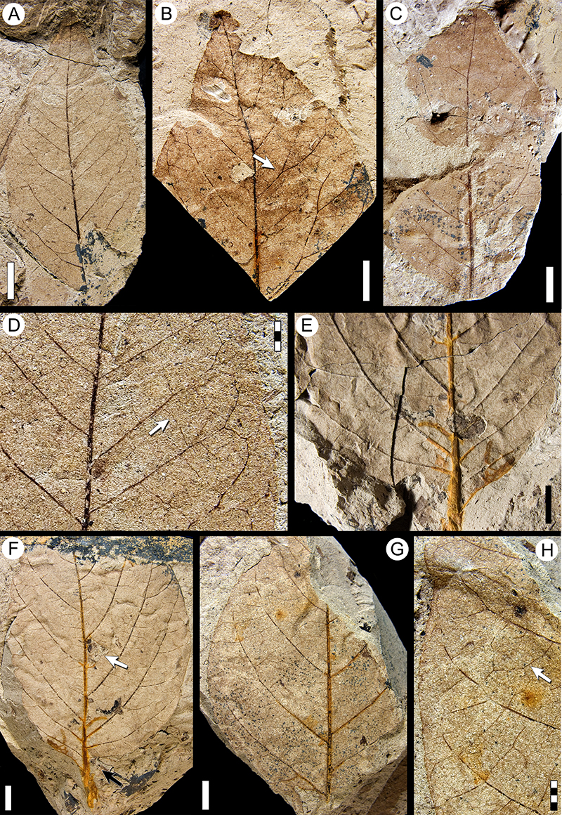
FIGURE 39. Morphotypes SA070 (Juglandaceae? continued, see also Figure 35A-F), SA073 (continued, see also Figure 36A-E), SA074 (continued, see also Figure 36F-H), and SA075 (continued, see also Figure 37E-H). A-B, morphotype SA070, MPEF-Pb-3012 (accessory exemplar, see also Figure 35A). A, camera lucida drawing (CLD) of complete leaf, note asymmetric base and basally crowded secondary veins (arrow); B, CLD of tooth venation detail (arrow, regular minor venation). C-D, morphotype SA075. C, CLD of MPEF-Pb-3016 (exemplar; see also Figure 37E; arrow, sinus-bracing vein); D, CLD of venation and teeth in MPEF-Pb-9139 (see also Figure 37H). E, morphotype SA073, CLD of MPEF-Pb-9143 (arrow, decurrent base). F-G, morphotype SA074, MPEF-Pb-3015 (exemplar, see also Figure 36F, H). F, CLD of complete leaf, note epimedial venation with concentric tertiary veins (arrow); G, CLD of tooth venation. Single-color scale bars equal 10 mm; grid scales equal one millimeter (per rectangle).
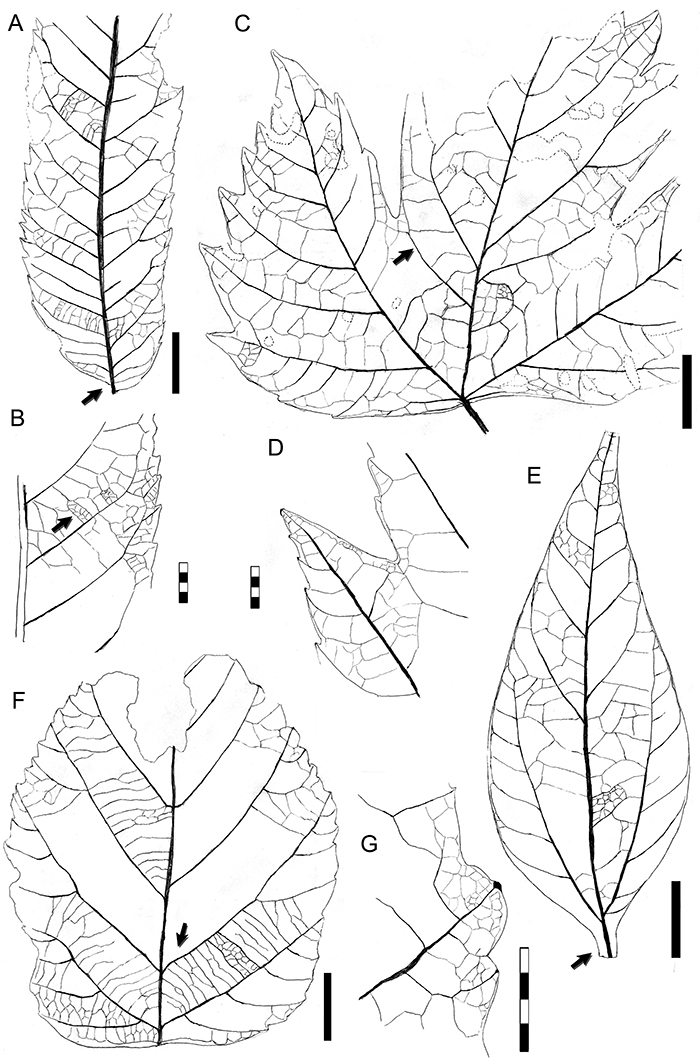
FIGURE 40. Morphotypes SA061 (Araceae? see also Figure 41H-K), SA065 (Arecaceae, see also Figure 41F-G), SA072 (Arecaceae, see also Figure 41D-E), SA076 (continued, see also Figure 37A-D), SA077 (continued, see also Figure 38A-D), SA078 (continued, see also Figure 38E-H). A-B, morphotype SA076. A, digital overlay drawing (DOD) of MPEF-Pb-3017 (exemplar, see also Figure 37A-B); B, DOD of MPEF-Pb-9153. C, morphotype SA077, DOD of MPEF-Pb-3018 (exemplar, see also Figure 38A, D). D, morphotype SA065 (Arecaecae), camera lucida drawing (CLD) of MPEF-Pb-3003 (exemplar, see also Figure 41F-G). E, morphotype SA078, DOD of MPEF-Pb-3019 (exemplar; see also Figure 38E-F) (arrow, reticulate higher-order veins). F, reconstruction of morphotype SA072 (Arecaceae) based on three leaf samples (MPEF-Pb-3013, -3014, and -4155). G, morphotype SA061 (Araceae?), CLD of MPEF-Pb-3000 (exemplar; see also Figure 41H-K; arrow, transverse veins). Scale bars equal 10 mm, except 3 millimeter scale in Figure 40G.
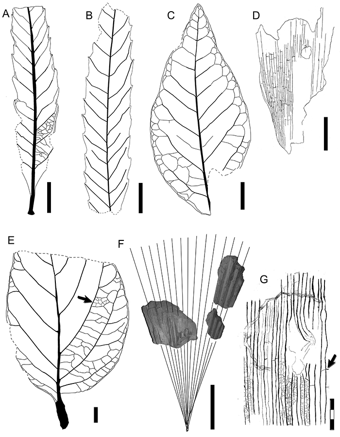
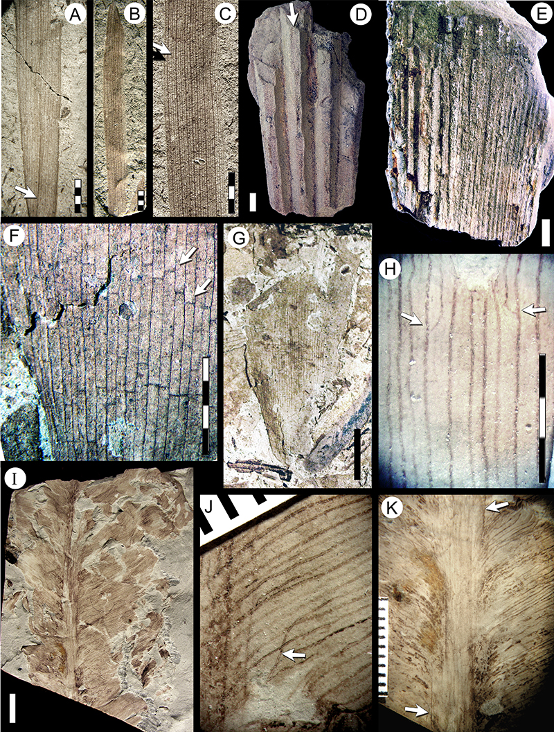
FIGURE 41. Monocot leaf morphotypes of Araceae? (SA061 continued, see also Figure 40G) and Arecaceae (SA032; SA065, continued, see also Figure 40D; SA072, continued, see also Figure 40F). A-C, morphotype SA032 (Arecaceae). A, MPEF-Pb-3021 (exemplar; arrow, segment plication); B, MPEF-Pb-3022 (accessory exemplar); C, MPEF-Pb-9144 (black arrow, A veins; white arrow B veins). D-E, morphotype SA072 (Arecaceae). D, MPEF-Pb-3013 (exemplar), note the flattened carena (arrow); E, MPEF-Pb-3014 (accessory exemplar). F-G, morphotype SA065 (Arecaceae), MPEF-Pb-3003 (exemplar, see also Figure 40D). F, detail of the segment venation (arrows indicating straight, sinuous or oblique transverse veins); G, detail of a segment. H-K, morphotype SA061 (Araceae?), MPEF-Pb-3000 (exemplar, see also Figure 40G). H, detail showing vein anastomoses (arrows) and higher order venation; I, complete view of fossil; J, detail of fibers (arrow) accompanying lateral veins; K, detail of massive midvein, with thick surrounding tissue (arrows). Single-color scale bars equal 10 mm; grid scales equal one millimeter (per rectangle).