APPENDIX 1.
Dimensions of specimens of Chuandianella ovata investigated in this study.
| Specimen | Site | Carapace length/height (mm) |
Body length (mm)♠ |
| YKLP 13967 | HM | 8.84/6.30 | 24.51 |
| YKLP 16215 | HE | 7.06/4.85 | >13.75 |
| YKLP 16216 | HE | 5.15/3.09 | 18.07- |
| YKLP 16217 | HE | 13.09/8.22 | - - |
| YKLP 16218 | HM | 8.15/5.13 | 17.89 |
| YKLP 16238 | HJ | 12.49/8.30 | - - |
| YKLP 16239 | HE | 11.32/8.10 | - - |
| Ch5 | HE | 7.50/4.96 | - - |
| Ch6 | HM | 13.06/7.63 | - - |
| Ch11a | HM | 9.77/6.95 | - - |
| Ch12 | HE | 8.97/7.40 | - - |
| RCCBYU 10272 | HM | 7.69/5.95 | 21.53♦ |
| He-f-6-4-294 | HE | 8.45/6.05 | - - |
| Hz-f-4-777 | HE | 7.51/4.14 | - - |
“♠”: The sagittal body length is measured from the anterior end of the carapace to the posterior end of the caudal processes. “-“: Slightly underestimated value due to incomplete preservation. “- -”: The corresponding value is not available. “♦”: This body-length value is calculated by summing up the carapace length and the length of the body protruding from the valve, in view of the flexed appearance of the body. Abbreviations: HE, Ercaicun of Haikou, Kunming; HJ, Jianshan of Haikou, Kunming; HM, Mafang of Haikou, Kunming.
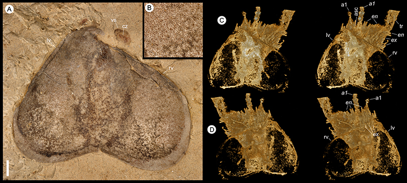
APPENDIX 2.
Chuandianella ovata, YKLP 16256. (A) Microscope image, dorsal vieww. Scale bar equals 0.9 mm. (B) Microscope image, showing pits on left valve. Scale bar equals 0.4 mm. (C-D) Stereo-pairs of micro-CT images. The posterior part of the body is reflexed so that it emerges from the anterior end of the carapace. Scale bar equals 2.0 mm. (C) Dorsal view. (D) Ventral view. Figure 1, Figure 2, Figure 3, Figure 4. Italics indicate a right-side appendage. Scale bar in (A) applies to all panels.
APPENDIX 3.
Chuandianella ovata, YKLP 16218, microscope images (for micro-CT images of this specimen see Figure 1A, C-E,G). Scale bar equals 1.0 mm. (A) YKLP 16218a, dorsal view of anterior part. (B) YKLP 16218b, ventral view of anterior part; posterior part is buried in sediment. Abbreviations as for Figure 1, Figure 2, Figure 3, Figure 4. Italics indicate a right-side appendage. Scale bar in (A) equals 1.0 mm and applies to both panels.
APPENDIX 4.
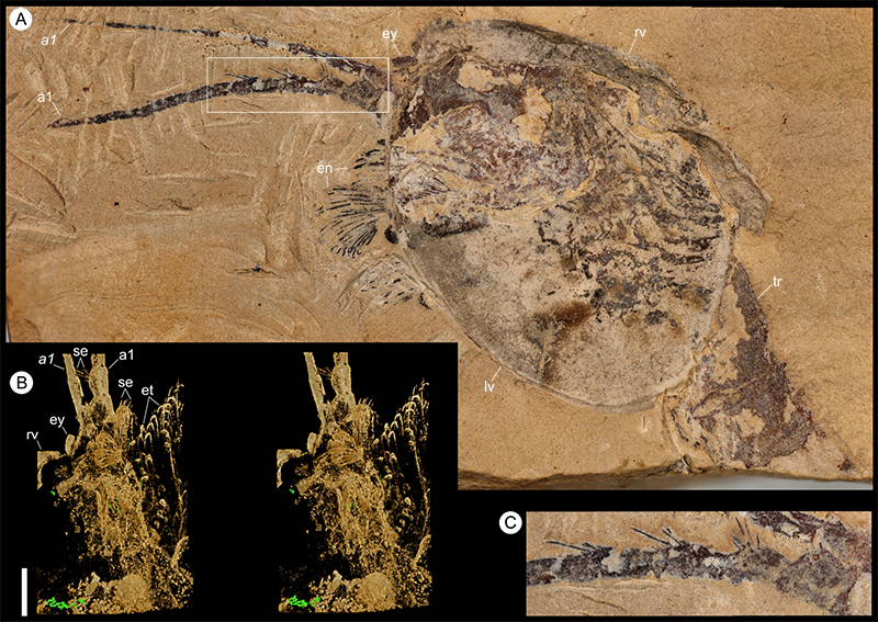
Chuandianella ovata, Hz-f-4-777. (A) Microscope image, dorsal view; the left and posterior parts of the specimen are missing. Scale bar equals 1.8 mm. (B) Details of left eye. Scale bar equals 0.7 mm. (C) Stereo-pair of micro-CT image, ventral view of anterior part. Scale bar equals 1.0 mm. Abbreviations as for Figure 1, Figure 2, Figure 3, Figure 4. Italics indicate a right-side appendage. Scale bar in (C) applies to all panels.
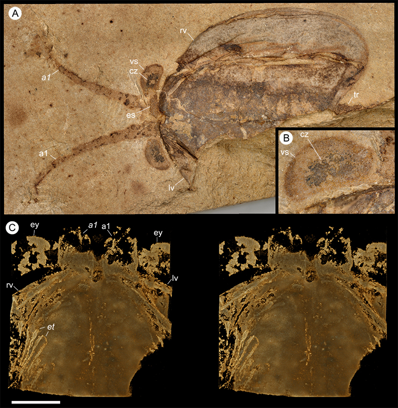
APPENDIX 5.
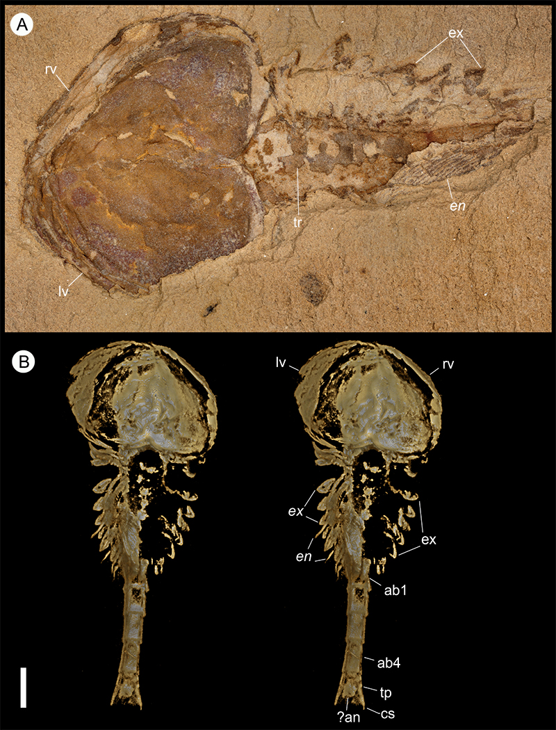
Chuandianella ovata, He-f-6-4-294. (A) Microscope image, left view; the posterior part of the trunk is missing. Scale bar equals 1.0 mm. (B) Stereo-pair of micro-CT image, details of anterior part of specimen, oblique-right view (viewed from underside of slab). Scale bar equals 1.2 mm. (C) Details of setae on left a1 (white rectangle in A). Scale bar equals 0.5 mm. Abbreviations as for Figure 1, Figure 2, Figure 3, Figure 4. Italics indicate a right-side appendage. Scale bar in (B) applies to all panels.
APPENDIX 6.
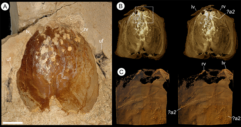
Chuandianella ovata, YKLP 16257. (A) Microscope image, dorsal view. Scale bar equals 2.3 mm. (B-C) Stereo-pairs of micro-CT images. (B) Dorsal view. Scale bar equals 5.0 mm. (C) Ventral view of the anterior part (white rectangle in B), showing a2. Scale bar equals 1.5 mm. Abbreviation additional to Figure 1, Figure 2, Figure 3, Figure 4: uf, unidentified fossil. Italics indicate a right-side appendage. Scale bar in (A) applies to all panels.
APPENDIX 7.
Chuandianella ovata, YKLP 16216. (A) Microscope image of specimen on rock slab. Scale bar equals 0.8 mm. (B) Micro-CT stereopair image. Scale bar equals 2.0 mm. Abbreviations as for Figure 1, Figure 2, Figure 3, Figure 4. Note: The soft parts of this specimen are taphonomically dislocated and are flipped vertically, so that this figure shows the dorsal views of the carapace and the ventral views of the trunk and appendages, with the left appendages associated with the right valve while the right appendages are associated with the left valve (cf. Figure 1B, G). Italics indicate a right-side appendage. Scale bar in (B) applies to both panels.
APPENDIX 8.
Chuandianella ovata, YKLP 16215a. (A) Microscope image of specimen on rock slab, dorsal view. Note that the posterior part of the trunk is missing. Scale bar equals 3.3 mm. (B-C) Micro-CT images of anterior part, stereo-pairs (anterior end to the left). Scale bar equals 3.1 mm. (B) Dorsal view. (C) Ventral view. Abbreviations as for Figure 1, Figure 2, Figure 3, Figure 4. Italics indicate a right-side appendage. Scale bar in (A) applies to all panels.
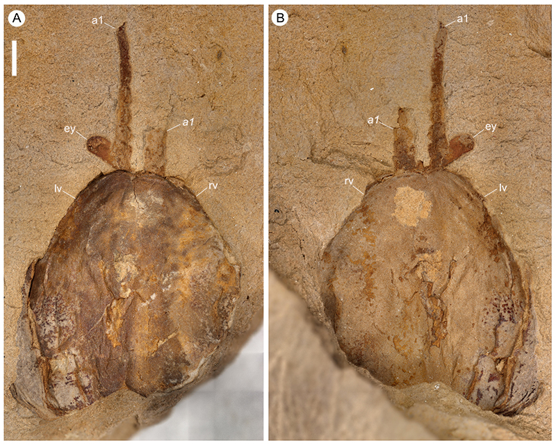
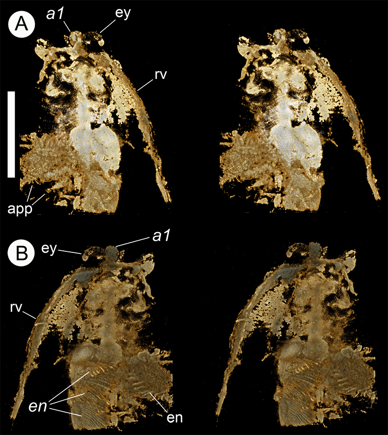
APPENDIX 9.
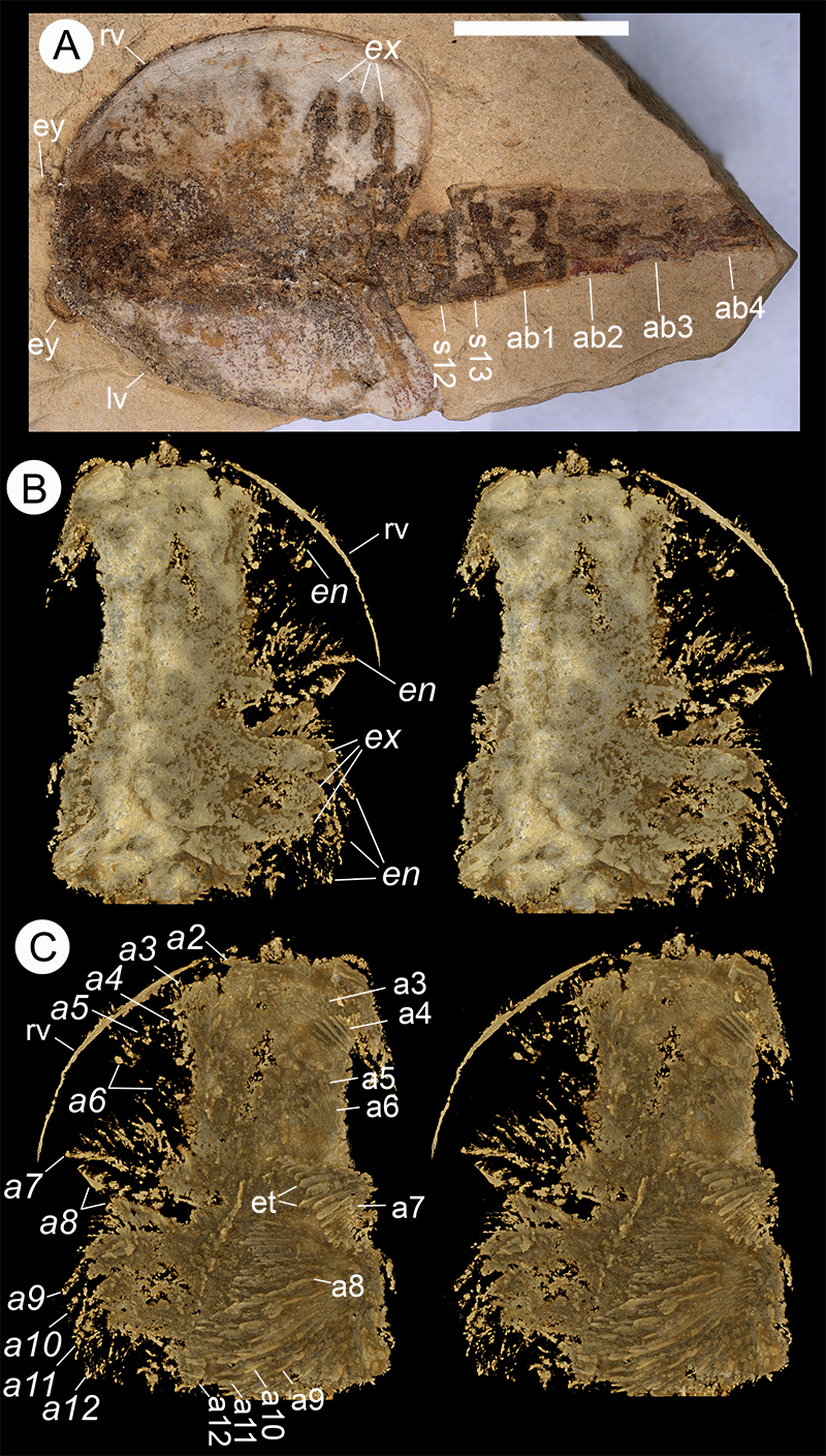
Chuandianella ovata, YKLP 16217 (for microscope image of this specimen see Figure 4A-E), stereo-pairs of micro-CT images. Scale bar equals 5.0 mm. (A) Dorsal view of anterior part. (B) Ventral view of anterior part. Italics indicate a right-side appendage. Scale bar in (A) equals 5.0 mm and applies to both panels.
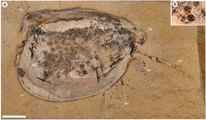
APPENDIX 10.
Chuandianella ovata, YKLP 16258, an egg-bearing specimen, microscope images. (A) Overview, oblique-right view. Scale bar equals 2.0 mm. (B) Details of eggs (white rectangle in A). Scale bar equals 1.4 mm. Abbreviations as for Figure 1, Figure 2, Figure 3, Figure 4. Italics indicate a right-side appendage. Scale bar in (A) applies to both panels.