

FIGURE 1. A-C: Maps showing the geographic situation of the Kaltensundheim sinkhole north of the farm in the village Kaltensundheim (Thuringia) in Germany. D: Plan of the excavation pits in the former sinkhole (Böhme, 1992). (Fig 1C modified from GoogleEarth).
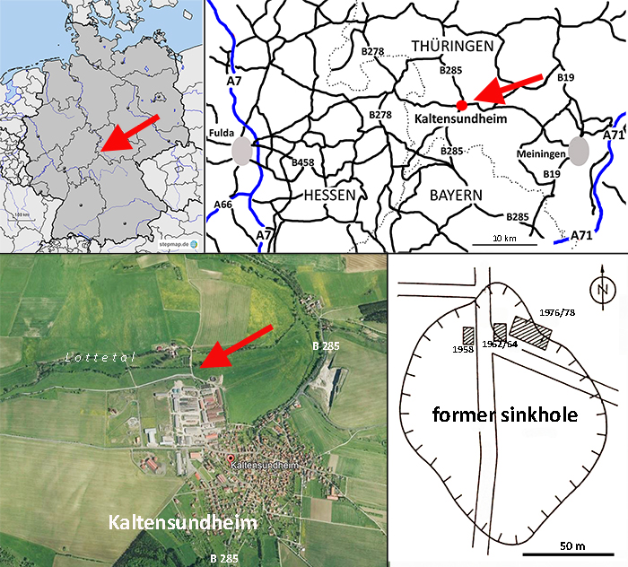
FIGURE 2. Excavation sketches of two “Mammut” borsoni individuals from the Kaltensundheim sinkhole. A: The specimen excavated in 1958 is discussed in this paper. Yellow-shaded bones are housed in the NHMS (MTe 810) and are described below. The whereabouts of the other bones is unknown. B: The second individual excavated in 1976-1978 is housed at the SFQW (modified from Kahlke, 1985) and was not available for this study.
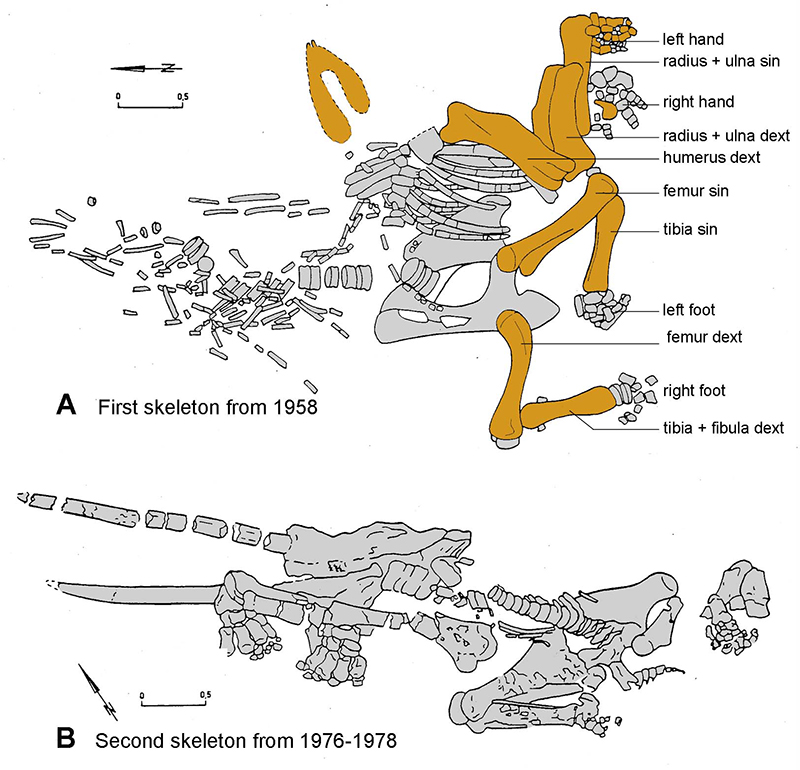
FIGURE 3. Stratigraphic position of important European localities with Zygolophodon and “Mammut”, and the expansion of Zygolophodon from Africa to Eurasia and to North America. A second immigration of Mammutidae to North America cannot be proven. NALMA - North American Land Mammal Ages; MN - European Neogene mammals Zones and stratigraphic scale in million years according to (Hilgen et al., 2012).
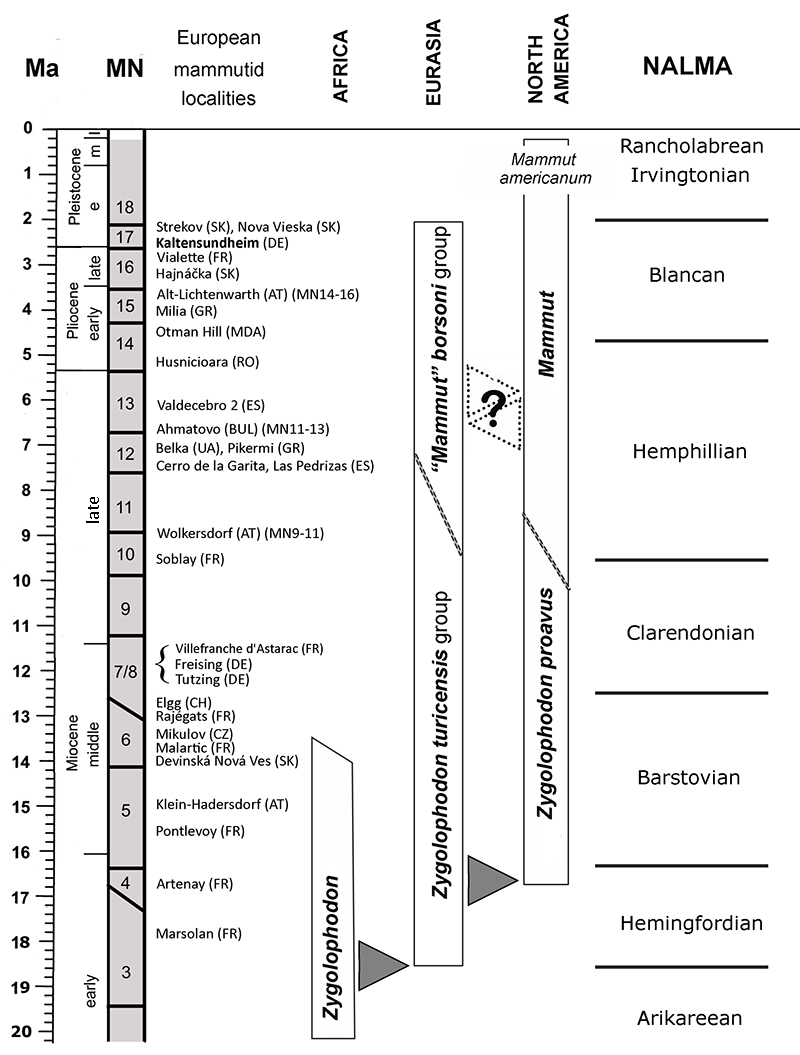
FIGURE 4. “Mammut” borsoni from Kaltensundheim. A: Historical photo (prior to 1988) of the mounted left arm and the mandible of the first “Mammut” borsoni at a temporary exposition in Meiningen with Dr. Böhme. In the unrestored mandible the two original mandibular tusks are obvious. B: The actual state of the mandible with reconstructed tusks in the exhibition of the NHMS in 2017.
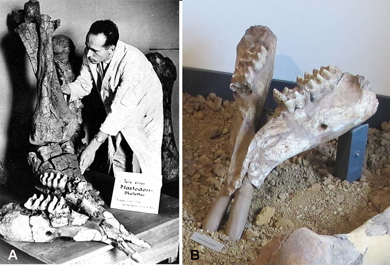
FIGURE 5. Preservation and restoration of “Mammut” borsoni long bones from Kaltensundheim: 3D models based on CT scans of the left femur (NHMS-MTe 810/4) (A: caudal, C: cranial) and left humerus (NHMS-MTe 810/2) from caudal (D). After subtracting the (low density) glue and added artificial material, the fragmentation of the bones and the state of preservation are evident (A, C, and D). B: The x-ray shows the internal stabilization with metallic rods.
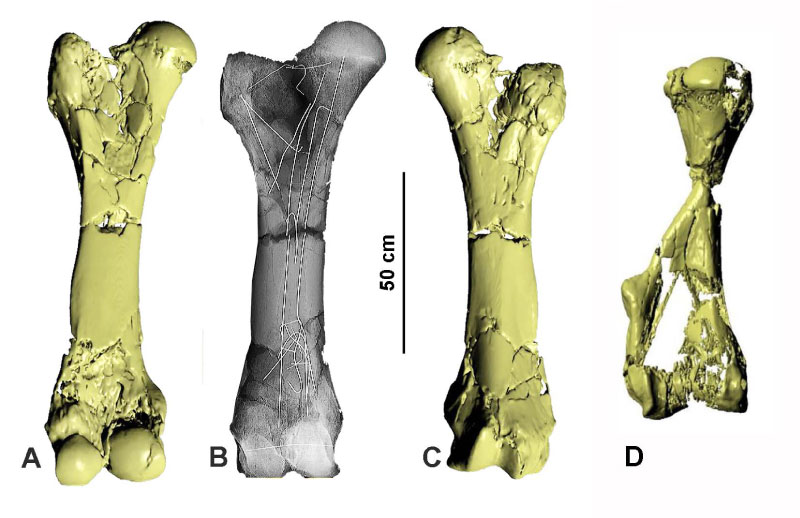
FIGURE 6. Mandible of “Mammut” borsoni from Kaltensundheim (NHMS-MTe 810/1). A: Anterior aspect of the symphysis, note that the rami are slightly distorted (not to scale due to perspective view). B: Left ramus from buccal. C: Right ramus from buccal. D: Occlusal aspect of the dentition and the mandible. Abbreviations: cm - corpus mandibulae, fm - foramen mandibulae, i1 - mandibular tusk, s - symphysis.
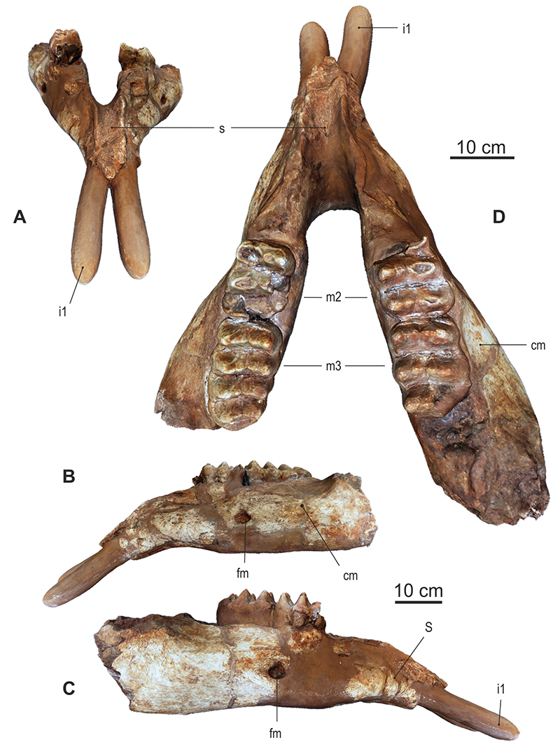
FIGURE 7. Left m3 of “Mammut” borsoni from Kaltensundheim. A: buccal (pretrite), B: occlusal, C: lingual (posttrite); B photo, A and C from 3D reconstruction.
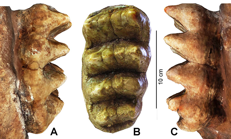
FIGURE 8. Long bones of the forelimb of “Mammut” borsoni from Kaltensundheim. A-F: humerus dext (NHMS-MTe 810/6), G-L: radius and ulna sin in articulation (NHMS-MTe 810/7). A - caudal, B -cranial, C and L - lateral, D and K - medial, F and G - proximal, E and H - distal, I - palmar, J - dorsal. Abbreviations: Humerus: ch - caput humeri, csh - crista humeri, csl - crista supracondylaris lateralis, el - epicondylus lateralis, em - epicondylus medialis, fo - fossa olecrani, fr - fossa radialis, mf - median furrow, si - sulcusintertubercularis, smb - sulcus musculus brachialis, td - tuberositas deltioidea, th - trochlea humeri, tmj - tuberculum majus, tmn - tuberculum minus, ttm - tuberositas teres minor. Radius: cr - corpus radii, faCi - facies articularis carpea for Ci, faCr - facies articularis carpea for Cr, fcr - fovea capitis radii. Ulna: faCu - facies articularis carpea for Cu, it - incisura trochlearis, o - olecranon, pa - processus anconeus, pcl - processus coronoideus lateralis, pcm - processus coronoideus medialis, to - tuber olecrani.
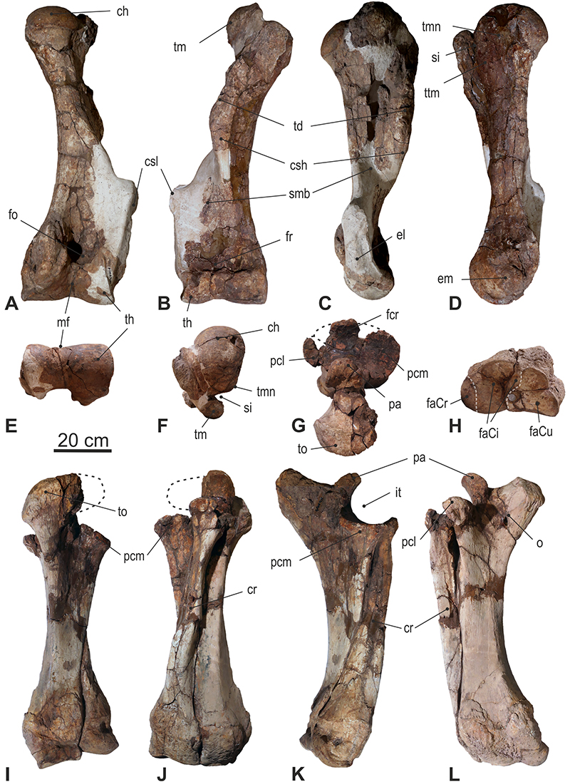
FIGURE 9. Left manus of “Mammut” borsoni from Kaltensundheim. A: Dorsal aspect of the left articulated hand skeleton. B-D: Comparison of proportions in mammutid forelimbs. B: Z. turicensis group (Turgai KAS, Mikulov-Czujan’s sandpit CZ); C: “Mammut” borsoni (individual from Kaltensundheim DE); and D: Mammut americanum (individual from Fort Wayne-Bueshing, from UMORF). Abbreviations: Cr - radiale, Ci - intermedium, CI-CIV - carpalia I-IV; McI-McIV - metacarpalia I-IV; Ph 1-Ph 2 - proximal and medial phalanges of digits I-IV.
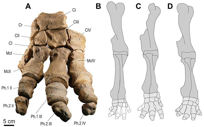
FIGURE 10. Articulation of carpus of “Mammut” borsoni from Kaltensundheim. The colors indicate to which bone each specific facet of the carpals and metacarpals articulates.

FIGURE 11. Proximal row of carpalia of “ Mammut” borsoni from Kaltensundheim. A-F: Cr = Os carpi radiale sin (scaphoideum) (NHMS-MTe 810/21), G-L: Ci =Os carpi intermedium sin (lunatum) (NHMS-MTe 810/20), M-R: Cu = Os carpi ulnare dext (cuneiforme) (mirrored) (NHMS-MTe 810/24), S-X: Ca = Os carpi accessorium sin (pisiforme) (NHM-MTe 810/23). Abbreviations: dfCI - distal facet for CI, dfCII - distal facet for CII, dfCIII - distal facet forCIII, dfCIV - distal facet for CIV, dfMcIV - distal facet for McIV, lfCi - lateral facet for Ci, lfCr - lateral facet for Cr, lfu - lateral facet for ulna, mfCa - medial facet for Ca, mfCi - medial facet for Ci, pfCu - proximal facet for Cu, plfCa - palmar facet for Ca, pfr - proximal facet for radius, pfu - proximal facet for ulna, plt - palmar tuberosity.
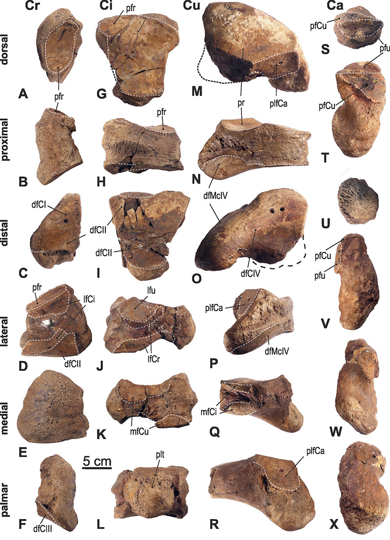
FIGURE 12. Distal row of left carpalia of “Mammut” borsoni from Kaltensundheim. A: distal row of left carpalia in articulation in dorsal view, B-F: CI = trapezium (NHMS-MTe 810/33), G-J: CII = trapezoideum (NHMS-MTe 810/32), K-O: CIII = magnum (NHMS-MTe 810/22), P-T: CIV = hamatum (NHMS-MTte 810/25). Abbreviations: dfMcI - distal facet for McI, dfMcII - distal facet for McII, dfMcIII - distal facet for McIII, dfMcIV - distal facet for McIV, dfMcV - distal facet for McV, lfCII - lateral facet for CII, lfCIII - lateral facet for CIII, lfCIV -lateral facet for CIV, lfMcII - lateral facet for Mc-II, mfCI - medial facet for CI, mfCII - medial facet for CII, mfCIII - medial facet for CIII, pfCII - proximal facet for CII, pfCr -proximal facet for Cr, pfCu - proximal facet for Cu, pfCi - proximal facet for Ci, pfMcI - proximal facet for McI, plt - palmar tuberosity.
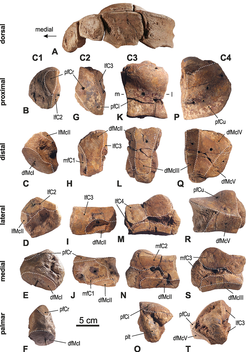
FIGURE 13. Left metacarpal bones of “Mammut” borsoni from Kaltensundheim: A-E: McIV (NHMS-MTe 810/28), F-J: McIII (NHMS-MTe 810/29), K-O: McII (NHMS-MTe 810/35), P-T: McI (NHMS-MTe 810/34). Abbreviations: lfMcIII - lateral facet for McIII, lfMcIV - lateral facet for McIV, lfMcV - lateral facet for McV, mfMcII - medial facet for McII, mfMcIII - medial facet for McIII, mfMcI - medial facet for McI, pfCI - proximal facet for CI, pfCII - proximal facet for CII, pfCIII - proximal facet for CIII, pfCIV - proximal facet for CIV, tMc - trochlea metacarpalis.
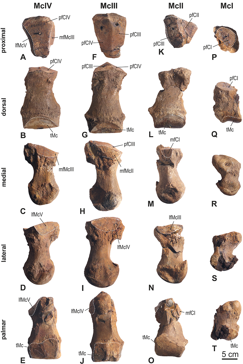
FIGURE 14. Proximal and medial phalages of the left hand of “Mammut” borsoni from Kaltensundheim. A-D: phalanx proximalis II (NHMS-MTe 810/36), E-H: phalanx proximalis III (NHMS-MTe 810/30), I-L: phalanx proximalis IV (NHMS-MTe 810/26), M-P: phalanx medialis II (NHMS-MTe 810/37), Q-T: phalanx medialis III (NHMS-MTe 810/21), U-X: phalanx medialis IV (NHMS-MTe 810/27).
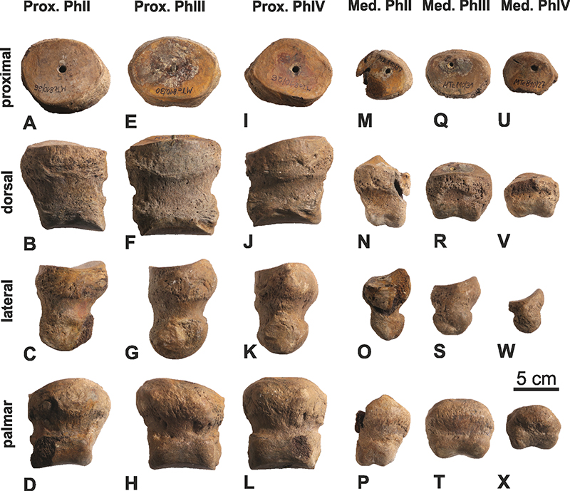
FIGURE 15. Long bones of the hindlimb of “Mammut” borsoni from Kaltensundheim. A-F: femur sin (NHMS-MTe 810/8), G-L: Tibia and fibula sin in articulation (NHMS-MTe 810/5). Views: A - cranial, B - caudal, C and K - medial, D and L- lateral, E and H - distal, F and G - proximal, I - dorsal, J - plantar. Abbreviations: aicn - area intercondylare centralis, aicr - area intercondylare cranialis, cf - caput femoris, cl - condylus lateralis, cm - condylus medialis, ct -cochlea tibiae, dfTc - distal facet for Tc, dfTt - distal facet forTt, el - epicondylus lateralis, em - epicondylus medialis, fa - facies aspera, fc - fovea capitis, fe - fossa extensoria, fi - fossa intercondylaris, fp - facies poplitea, ft - fossa trochanterica, mc - medial crest, ml - malleolus lateralis, mm - malleolus medialis, sm - sulcus malleolaris, til - tuberculum intercondylare lateralis, tim - tuberculum intercondylare medialis, tof - trochlea ossis femoris, trmj - trochanter major, tsll - tuberositas supracondylaris lateralis, tt - tuberositas tibiae.
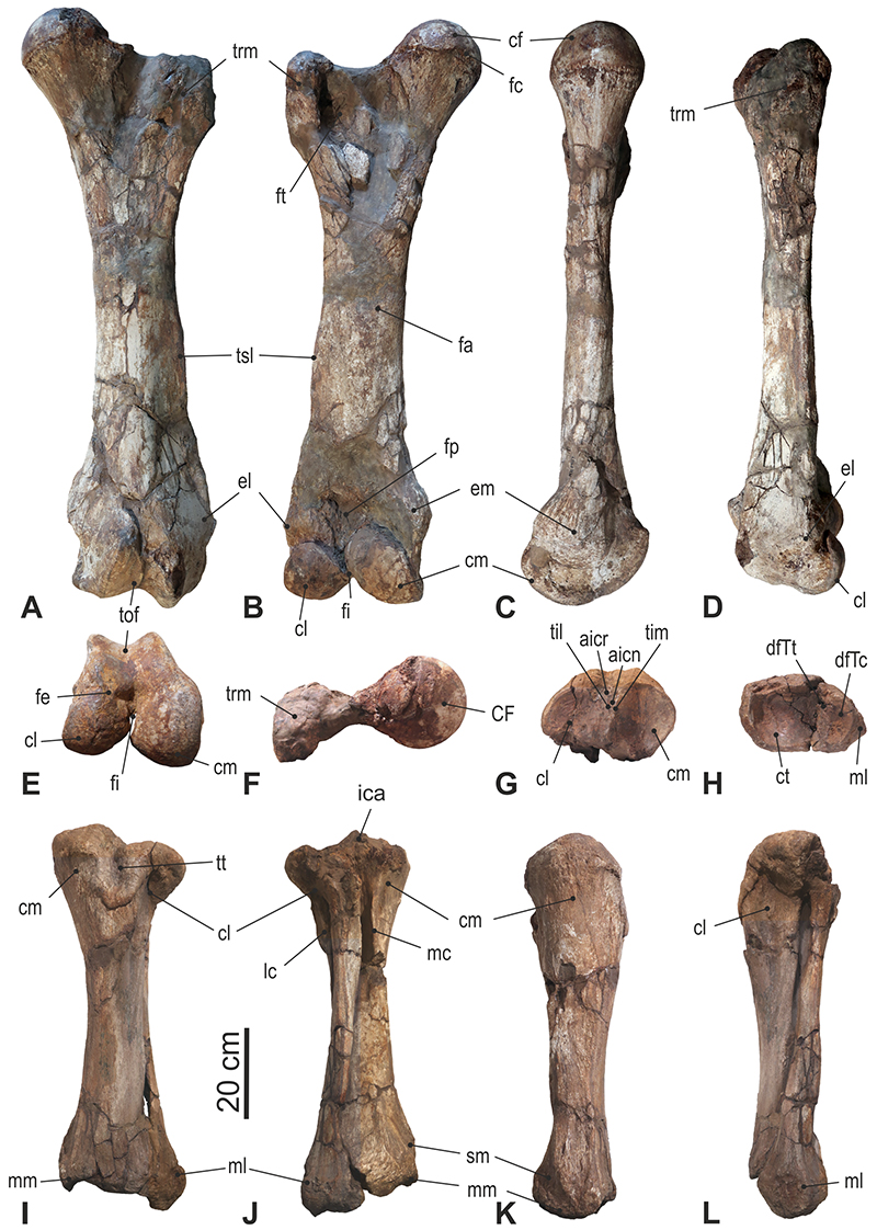
FIGURE 16. Metrical variability of the m3 in Zygolophodon turicensis (blue) and “Mammut” borsoni (red, Table 7), compared to the approximate variability of the m3 in Mammut americanum (green) (data for M. americanum from Dooley et al., 2019, figure 32). The m3 from Kaltensundheim (K) is relatively small compared to other specimens from Central Europe, especially Milia (M).
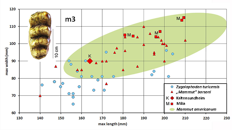
FIGURE 17. Comparison of the body size in various proboscideans. The silhouettes are from Larramendi, 2016. The one for Kaltensundheim (gray) is adjusted to the predicted shoulder height in flesh 320 cm. "Mammut" borsoni was larger than most individuals of Mammut americanum. Abbreviations: BM - predicted body mass, SH - shoulder height.

