

 Techniques for digital restoration of cranial elements on three-dimensional surface models
Article number: 28.2.a26
https://doi.org/10.26879/1506
Copyright Paleontological Society, June 2025
Author biographies
Plain-language and multi-lingual abstracts
PDF version
Submission: 13 November 2024. Acceptance: 14 June 2025.
ABSTRACT
Digital restoration is the process by which preservational or other types of damage are removed to recover the putative original morphology of fossil specimens. In the present work, three-dimensional (3D) surface models of skulls and mandibles of 32 cynognathians (Synapsida, Cynodontia) specimens from South America were digitally restored. The main restoration techniques used come from the literature and are adapted to the particular preservational problem. A classification into three restoration grades (low, medium and high) is proposed, taking into account the number of techniques used and the percentage of damage of the fossil. The higher the degree of damage, the greater the complexity of the restoration and the higher the level of interpretation added. Of all the specimens, 36.5% presented a low grade of restoration, 15% presented a medium grade, and 7.3% a high grade. Among the major damages recorded in the cynognathians specimens the following stand out: partial or total fragmentation of one of the zygomatic arches, total loss of the postdentary bones, fragmentation of the coronoid processes, and lithostatic deformation. The unidirectional deformation could not be completely eliminated in the fossils that presented it, being the only unresolved restorative problem. Detailing and documenting the totality of the restorative procedures allows establishing a traceability of the changes applied to the specimens, being extremely useful for researchers confronted with similar problems.
Florencia S. Filippini. Consejo Nacional de Investigaciones Científicas y Técnicas (CONICET). Museo Argentino de Ciencias Naturales “Bernardino Rivadavia” (MACN). Av. Angel Gallardo 470, C1405DJR Ciudad Autónoma de Buenos Aires, Argentina. flor.s.filippini@gmail.com
Fernando Abdala. Unidad Ejecutora Lillo, Consejo Nacional de Investigaciones Científicas y Técnicas (CONICET)-Fundación Miguel Lillo, Miguel Lillo 251, 4000, San Miguel de Tucumán, Tucumán, Argentina, and Evolutionary Studies Institute, University of the Witwatersrand, Johannesburg, South Africa. nestor.abdala@wits.ac.za
Guillermo H. Cassini. Consejo Nacional de Investigaciones Científicas y Técnicas (CONICET). División Mastozoología, Museo Argentino de Ciencias Naturales “Bernardino Rivadavia” (MACN), Av. Ángel Gallardo 470, C1405DJR Ciudad Autónoma de Buenos Aires, Argentina and Departamento de Ciencias Básicas, Universidad Nacional de Luján (UNLu). Ruta 5 y Av. Constitución s/n, 6700 Luján, Buenos Aires, Argentina. gcassini@macn.gov.ar
Keywords: restoration; retrodeformation; 3D models; digital techniques; fossil vertebrates; cynodonts
Final citation: Filippini, Florencia S., Abdala, Fernando, and Cassini, Guillermo H. 2025. Techniques for digital restoration of cranial elements on three-dimensional surface models. Palaeontologia Electronica, 28(2):a26.
https://doi.org/10.26879/1506
palaeo-electronica.org/content/2025/5560-3d-surface-models-restoration-techniques
Copyright: June 2025 Paleontological Society
This is an open access article distributed under the terms of Attribution-NonCommercial-ShareAlike 4.0 International (CC BY-NC-SA 4.0), which permits users to copy and redistribute the material in any medium or format, provided it is not used for commercial purposes and the original author and source are credited, with indications if any changes are made.
creativecommons.org/licenses/by-nc-sa/4.0/
INTRODUCTION
Digitization of fossil material has advanced significantly over the past few decades and has proven to be very useful in enabling new approaches in the studies of extinct organisms (Cunningham et al., 2014), as well as generating digital specimens accessible to researchers as they conform digital collections (Digital Atlas of Ancient Life, Morphosource, MorphoMuseuM, Sketchfab). The preservation of fossil material of vertebrates remains a problematic issue as the specimens are often disarticulated, fractured, incomplete, and/or deformed (Shipman, 1981; Lyman, 1994). Many studies require the analysis of specimens in which morphological distortions were previously corrected (Mallison, 2010; Molnar et al., 2012; Tschopp et al., 2013; Cuff and Rayfield, 2015). With the enormous progress in digital techniques, emphasis has been placed in recent years on the restoration of fossil material. This area has not been extensively explored in vertebrate paleontology as it has been in archeology and paleoanthropology, where restoration and reconstruction, especially of hominid crania, is a widespread practice (Davis and Napier, 1963; Walker et al., 1983; Ponce de León and Zollikofer, 1999; Zollikofer and Ponce de León, 2005; Gunz et al., 2009; Schlager et al., 2018).
Digital restoration involves the process of removing preservational and other artifacts to restore the morphology of a fossil specimen as close as possible to its assumed natural condition prior to fossilization, employing a combination of different software packages and techniques (Lautenschlager, 2016a). Although digital modeling offers multiple tools to perform this task, it is frequently the case in which the protocols and criteria used to restore fossils specimens are not described in detailed for other researchers to follow the same protocol, being Lautenschlager (2016b) one of the few guiding works for a first approach to the subject. It is important to note that the majority of publications usually describes the restoration of a single specimen or of several elements of different unrelated specimens (Díez Díaz et al., 2020; Demuth et al., 2022; DeVries et al., 2022; Ruella et al., 2024). Restoration of a large sample of fossil material from the same taxon is not commonly documented. Principally, due to the fact that a large sample of collected specimens is needed and is not common for fossil vertebrates, an exceptional case would be the cynognathians.
Cynognathians are a group of non-mammalian cynodonts that are well represented particularly in southern Pangaea and are the most abundant component in the Triassic faunas of Argentina (Abdala et al., 2020). Thus, many species of the group are represented by several specimens. However, like usually happens with most fossils, diagenesis and taphonomic processes affect their preservation. Cynognathians are characterized by broad skulls with a triangular appearance in dorsal view due to the contrast between the snout and the wide temporal fossae that produce a much broader posterior portion of the skull. They have broad zygomatic arches, a generally robust snout, and a jaw composed of a large dentary and more delicate postdentary elements forming a postdentary bar that loosely articulate with the dentary (Kielan-Jaworowska et al., 2004, Kemp, 2005; Abdala et al., 2020; Rougier et al., 2021).
In this contribution, we describe modified restoration techniques from Lautenschlager (2016b) to apply specifically to 3D surface models. We record the damage and deformations present in combination with the restoration techniques used for each cranial element in our fossil sample. In addition, a classification based on the level of restoration is proposed that qualitatively considers the degree of uncertainty added and the amount of work performed. In this way, a more complete record of the restoration process is reported and thus is more clearly defined for future use.
MATERIALS AND METHODS
Institutional Abbreviations
MACN-PV, Colección de Paleovertebrados, Museo Argentino de Ciencias Naturales “Bernardino Rivadavia”, Buenos Aires, Argentina; MLP, Museo de La Plata, La Plata, Argentina; PULR, Universidad Nacional de La Rioja, Argentina; PVL, Colección de Paleontología de Vertebrados, Instituto Miguel Lillo, Universidad Nacional de Tucumán, Argentina.
Specimens
The digitally restored material corresponds to a series of skulls and mandibles of 32 specimens of Cynognathia from Argentina, housed in MACN, MLP, PULR, and PVL paleontological collections. The species included are:
Cynognathus crateronotus Seeley, 1895. Horizon: Rio Seco de la Quebrada Formation (Puesto Viejo Group); age: ?lower Carnian (Upper Triassic). Represented by PVL 3859.
Pascualgnathus polanskii Bonaparte, 1966; horizon: Rio Seco de la Quebrada Formation (Puesto Viejo Group); age: ?lower Carnian (Upper Triassic). Represented by tree specimens: PVL 3466, PVL 4416 and MLP 65-VI-18-1.
Andescynodon mendozensis Bonaparte, 1969; horizon: Cerro de las Cabras Formation: age: upper Anisian (Middle Triassic). Represented by 11 specimens: PVL 4390, PVL 3092, PVL 3899, PVL 3900, PVL 3833, PVL 3834, PVL 3840, PVL 3835, PVL 3891, PVL 3898, PVL 4072, PVL 3890 and PVL 3894.
Massetognathus pascuali Romer, 1967; horizon: Chañares Formation; age: upper Ladinian-lower Carnian (Upper Triassic). Represented by 13 specimens. PULR v02, PVL 3901, PVL 3902, PVL 3904, PULR v10, PULR v13, PVL 3903, PVL 4728, PULR v11, PVL 4727, PVL 4729, PVL 5441, PVL 4726 and PVL 4613.
Exaeretodon argentinus Cabrera, 1943; horizon: Ischigualasto Formation; age: upper Carnian (Upper Triassic). Represented by five specimens: MACN-PV 18125, PVL 2564, PVL 2473, PVL 2056, PVL 2066, PVL 2467, PVL 2554 and PVL 2565.
Restoration
To proceed with the restoration, the workflow proposed by Lautenschlager (2016) for the digital restoration of surface models was used as baseline. Likewise, the techniques proposed in that work were adapted to be performed on surface models since they are mostly explained and applied to CT scanned models. The restoration process was performed by the same person to ensure consistency.
Digitization. Fossils were initially digitalized to obtain three-dimensional (3D) morphology as originally preserved. Photogrammetry was utilized to create surface-based 3D models. A DSLR (Digital Single Lens Reflex) camera with a fixed focal range of 55 mm was used to capture between 300 and 400 photos per cranial element, with a resolution of 24 Mpx (6000 x 4000 pixels) per photo. To achieve maximum coverage and to maintain necessary overlap, multiple photo cycles were taken at different planes, with photos taken every 7° (48 photos per cycle). The photographs were then processed using Agisoft® Metashape v.1.6.5 software to obtain the 3D models in PLY format.
Software. Various programs were used to achieve the restorations on the 3D surface models, depending on the techniques employed. Agisoft® Metashape v.1.6.5 was used to visualize, edit, and isolate elements of the 3D models. Most of the restoration techniques were carried out in Blender v. 2.93 (www.blender.org); retrodeformation was carried out using Landmark v.3.0.0.6 (Institute for Data Analysis and Visualization, University of California; Wiley et al., 2005). Finally, the restored models were obtained using MeshLab (https://www.meshlab.net/; Cignoni et al., 2008).
Restoration techniques. The following sections describe the techniques applied, detailing the necessary steps for restoration using Blender and Landmark programs on 3D models obtained by photogrammetry in Metashape.
Reflection of elements: This method is commonly used when the fossil is incomplete due to fragmentation. It exploits the bilateral symmetry of vertebrates to complete the missing element by mirroring its counterpart, as long as the other half of the fossil is well-preserved.
Superimposition: Fossils often exhibit varying degrees of fragmentation, even within the same specimen. In some cases, neither side of the specimen is complete, nor are larger areas of the fossil compromised. Therefore, the problem cannot be resolved by simply mirroring one side, or if the affected area has no bilaterally symmetrical counterpart (e.g., sagittal crest). To restore incomplete areas, the counterparts of the elements must be superimposed with each other, especially when each side of the fossil has a different state of preservation. In the case of non-symmetrical elements, the area can be restored by superimposing the element from other specimens of similar size.
Retrodeformation: Plastic deformation alters the original form of the fossil, modifying its morphology while preserving its elements. Retrodeformation is the process of restoring the original shape by applying the same amount of deformation but in the opposite direction. Most three-dimensional retrodeformation methods use the principle of bilateral symmetry, assuming that a symmetric counterpart has undergone shear or deformation with respect to the other part.
Restoration grades. A simple classification is proposed to better categorize the fossil sample. This is necessary due to the varying degrees of damage and deformation that can be observed, which resulted in different levels of complexity in the restoration process. As a result, there is a certain level of uncertainty. The number and extension of structures to be restored from a specimen varies based on its preservation and deformation, and could be expressed as a percentage. Therefore, based on the number of techniques used and the original condition of the specimen, we establish three levels of restoration:
Low restoration level: A restoration defined as simple, where the material is complete but deformed or only slightly damaged, with less than 15% of the fossil missing. It is therefore necessary to use only one restoration technique.
Medium restoration level: A moderately complex restoration that is solved by applying at least two techniques, for example, the specimen is deformed and broken; or only one technique is applied, but large sections of the fossil (20 - 50%) are missing.
High restoration level: Refers to a complex restoration process that involves multiple techniques to address the problem. It includes specimens that have a high degree of deformation and a significant amount of damage or missing elements (over 50%).
RESULTS
Damage and Deformation Observed
Table 1 lists the specimens detailing the damage recorded, the restoration techniques used, the percentage restored, and the level of restoration according to the proposed classification. Regarding the degree of damage, in the entire sample, the zygomatic arches of the skull and the coronoid processes of the dentary are usually partially preserved throughout the sample. A delicate skeletal part, which tends to be completely missing, are the postdentary bones. These elements are loosely articulated to the dentary and in most cases are not preserved even when the skull and mandible (dentary plus splenial) are articulated. Within each species, different degrees of damage or deformation are found. The Cynognathus skull and mandible are complete with a very slight degree of deformation which allowed a rapid restoration. The skulls of Andescynodon are, in general, poorly preserved, presenting often crushing, and, less frequently compression. In several specimens of this taxon, large parts of the zygomatic arch and the postorbital bar are missing, whereas, the coronoid processes of the mandible are usually incomplete, even when both hemimandibles are preserved. As for Pascualgnathus it presents two particular cases, on the one hand the specimen MLP 65-VI-18-1 it seems has the zygomatic arch removed by preparation action. On the other hand, the PVL 4416 specimen presents the highest degree of deformation of the sample. Massetognathus shows a high level of preservation, with low deformation and some fragmentation of parts of the zygomatic arch or the tips of the coronoid processes of the mandible.
Exaeretodon specimens are by far the largest fossils in the sample, and in the skulls the main damage is observed in the zygomatic arches (in which one of them is usually incomplete). In addition, presents parietal and occipital ridges with different degrees of damage, partially or completely missing. Mandibles often have missing parts of the coronoid processes, and some specimens show a high degree of compression (dorsoventrally or mediolaterally).
Adaptation of Techniques to Surfaces 3D Models
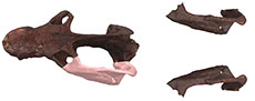
 Reflection of elements. Many specimens in our sample have only one complete zygomatic arch in the cranium (e.g., Massetognathus specimens PVL 3901, PVL 4613 PULR v02, Exaeretodon PVL 2056, Andescynodon PVL 3890, and Pascualgnathus MLP 65-VI-18-1, Table 1) or have missing fragments of the coronoid processes of the mandible (e.g., Massetognathus PVL 3903 and PVL 4613, Exaeretodon PVL 2564, Table 1). In these cases, incomplete elements can be repaired by mirroring the bilaterally symmetrical counterpart from the same specimen (Figure 1).
To perform these techniques, the element to be mirrored must first be isolated using Metashape, selecting and isolating the area of interest by editing the dense point cloud. Once the element is isolated, a model of the edited dense cloud is generated that can be exported to a 3D model editing program, such as Blender. Two models are imported to Blender, the specimen to restore and the edited fragment; and through the option Object → Mirror (in the x, y, or z axis, or manual) the isolated fragment is mirrored on the side to repair. Then using the option transformation manipulator → translating and rotating, both structures are manually aligned in the exact position. Once the missing piece has been accommodated in the original model, both models are merged with the option Object → Join (Figure 1).
 Superimposition. To carry out this type of restorative process it is necessary to work with two specimens. Specimen A would correspond to the specimen to be restored, while specimen B is used to extract the missing piece needed to restore specimen A. First, specimen B is taken in the Metashape program, and the area or piece necessary to complete specimen A is selected and isolated in the same way as described in the previous procedure. Then, both models, the specimen A and the isolated piece of specimen B, are imported in the same project in Blender. In this program, the restoration is continued by scaling the isolated piece of specimen B to have the same size as specimen A. This part of the process is done manually with a transformation manipulator for scaling: manual transformation → translation/rotation/scaling.
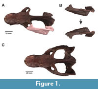
Transformation manipulators allow greater control of the process, especially if the morphologies are not exactly the same. Once the correct scaling and superimposition is achieved, both models are joined. (Object → Join). This technique was performed in Massetognathus: on the mandibles of specimens PVL 3902, PVL 3903, PVL 4613 with PVL 3901 being used as specimen B, and on the skull of PVL 3903 using PULR v10 as specimen B. The skull PVL 2564 and the mandibles PVL 2066 of Exaeretodon , were restored utilizing as specimen B the skull and mandibles of MACN-PV 18125; and the snout of MACN-PV 12125 was restored using PVL 2564 as specimen B. In Pascualgnathus, the skull and mandibles of PVL 4416 (previously dismantled, see below) and mandibles of PVL 3466 were restored utilizing as specimen B the skull and mandible of MLP 65-VI-18-1 (Figure 2).
When no specimen of the same species is available, a specimen of another closely related species may be used for restoration and missing parts are superimposed. This procedure assumes that there is no morphological variability between the taxonomic units selected and therefore implies a certain degree of interpretation. This method was only applied to the dentaries of Andescynodon specimens (PVL 3092, PVL 3835, PVL 3891, PVL 3894, PVL 3898, PVL 3900, PVL 4072), which none of them had a complete coronoid process. In this case, to restore the missing parts of the mandible we superimposed sections of the dentary dentary bone from the closely related Pascualgnathus, using MLP 65-VI-18-1.
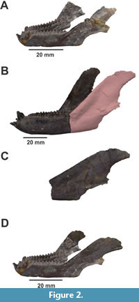
 Retrodeformation. As mentioned above, retrodeformation methods use the principle of bilateral symmetry, assuming that one symmetric counterpart has undergone deformation with respect to the other. A common method uses paired landmarks placed on both sides of the sagittal plane. Based on this set of landmarks, the object is then deformed so that each landmark occupies the same plane as its counterpart, which are orthogonal with respect to the sagittal plane of symmetry. This was done using the Landmark software. Once the set of paired landmarks are arranged, go to the Project → Retrodeform Pairs → Single axis and the software performs the retrodeformation automatically (Figure 3). The resulting model is then exported as a PLY file. This method was useful for shear deformed fossils, like in Andescynodon PVL 3835, PVL 3894, PVL 3898, and PVL 4072; Exaeretodon PVL 2468; Massetognathus PVL 3901 and PVL 4726; and Pascualgnathus PVL 4416. Nevertheless, this procedure was not very effective in cases where the specimens were crushed or compressed, as in specimens PVL 3092 of Andescynodon , PVL 2056 and MACN-PV 18125 of Exaeretodon , PULR v11 and PULR v13 of Massetognathus (PULR v 13 is shear deformed but the landmark editor could not correct completely). In cases where the deformation is strongly directional (PVL 3092, PVL 2056, PULR v11) or one part of the fossil is deformed (the snout of MACN-PV 18125), manual retrodeformation can be performed. However, because of its high level of subjectivity, it is necessary to proceed with caution and in a gradual manner so as to avoid distorting the anatomy of the specimen. Manual retrodeformation was realized using Blender’s manual transformers. It was not possible to completely retrodeform specimens highly compressed as in Andescynodon PVL 3092, PVL 3891, Exaeretodon PVL 2056 y PVL 2066.
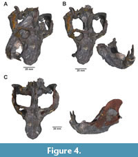 Dismantling of elements. Pascualgnathus PVL 4416, consists of an articulated skull and mandible with a high degree of shear deformation. This type of damage caused the mandible to be misaligned with respect to the cranial sagittal plane, displacing the anterior part of the dentary mostly on the right side of the snout (Figure 4A). Initially we tried to use the retrodeformation method following the steps explained above. However, the resulting model did not recover the symmetry completely of the specimen and distorted the anatomy. Therefore, we had to recur to another way of recovering the original shape of the specimen without modifying its anatomy.
Since the skull and the mandible appeared to present different degrees of deformation, the simplest solution was to separate both elements and then restore them separately. This was achieved by editing the dense point cloud in Metashape, resulting in two separate models for the skull and the mandible. Subsequently, the retrodeformation and restoration was performed independently in skull and mandible as it was done with other specimens. The retrodeformation was realized with the Landmark editor as explained above. Fragmented sections of the skull and the coronoid processes of the mandible of PVL 4416 were restored using superimposition and MLP 65-VI-18-1 as specimen B. This procedure was created to solve this specific complex case.
Percentage and Grade of Restoration
The majority of the specimens, 56% of the sample, present a medium level of restoration, followed by 36.5% requiring low level of restoration and 7.3% (three specimens) that required high level of restoration.
The Cynognathus skull and mandible are nearly complete and required about 5% of restoration (i.e., low level). Of 11 Andescynodon specimens that underwent restoration, eight were restored by superimposition using a specimen from the close related species ( Pascualgnathus ), being the species in which this method was more often used. One of the three specimens of Pascualgnathus , PVL 4416 showed the highest-degree of deformation in the sample, especially crushing and shearing of the skull and mandible, which led to a longer restoration process (Figure 4). The other two Pascualgnathus specimens were restored using one technique, reflection of elements or superimposition (i.e., medium to low level). The specimens of Massetognathus presented a low degree of damage and deformation in general (53.3% low level, 46.6% medium level restoration). In general, these restorations were easily resolved, with 80% of the specimens needing to undergo only one of the techniques to be restored. The method most commonly used was retrodeformation applied in 60 % of the specimens, while the superimposition was less used (26.6%). For Exaeretodon , the most used techniques were reflection and superimposition of elements to resolve the most severe damage (i.e., medium level restoration), which was applied in all the specimens. Additionally, manual retrodeformation was applied in 57% of the samples.
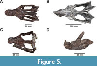 In general, the deformation produced by compression in the cynognathian specimens was corrected manually. However, in the most extreme cases as observed in PVL 3092, PVL 3891, PVL 2056 y PVL 2066 it was not possible to perform a total retrodeformation (Figure 5, A and B), being the 12.5% of the total sample that could not be fully restored. In addition, specimens were left out from the restoration process due to the impossibility of applying any of the techniques, which are not listed in this work and had to be left out entirely of the posterior analysis made. The left out specimens are two from Andescynodon species, the skull of PVL 3892 and mandibles of PVL 3890, and one Exaeretodon specimen, the skull of PVL 2554. These specimens present such a degree of damage that it is not possible to avoid a high degree of uncertainty at the moment of attempting to restore them. In the case of the skulls PVL 3892 and PVL 2554, have large missing areas which makes certain important anatomical features such as the postcanine dental rows or the height of the skull very uncertain (Figure 5C). While the mandibles of PVL 3890 present an incomplete preparation having a block of sediment within the hemimandibles (Figure 5D).
In summary, the total sample consisted of 32 specimens, 62.5%, (20) of the specimens were deformed and 90.6% (29) were fragmented and represented missing areas. All the specimens that have missing areas were restored completely. Of the specimens that were affected by deformation, 80% were successfully retrodeformed and only 20% were not able to remove the deformation completely. Additionally, three specimens were completely left out of the restorative process due to the level of damage presented.
DISCUSSION
Among the damages observed in our sample, partial fragmentation or total loss of one of the zygomatic arches, total loss of the postdentary bones and deformation due to lithostatic load stand out. The cranial elements of cynognathians are mostly dorsoventrally compressed, and to a lesser extent, mediolaterally compressed or sheared.
Massetognathus shows the best grade of preservation of its specimens, with little deformation and fragmentation regardless of the specimen size. The only specimen of Cynognathus is very well preserved, probably for their characteristic anteroposteriorly short zygomatic areas, broad occipital plate, and low coronoid process, which gives a greater robustness to the skull and jaws compared to other cynognathians. Thus, a more robust construction of the skull allows a greater resistance against fragmentation. Andescynodon small specimens commonly show deformation and fragmentation. Only Pascualgnathus PVL 4416 shows the highest degree of multidirectional deformation, which does not occur in other specimens. The large Exaeretodon specimens show as distinction the fragmentation in the parietal and occipital crests that could be a size effect. In general, the most fragile part in cynognathians skull are zygomatic arches, due to both brittle and plastic deformation. This is the area most affected by compressive forces, often leaving the zygomatic arches closer to the cranial walls.
As for the mandible, most of the damaged areas correspond to the detachment (an absence) of the postdentary bones and the broken tips at different levels of the coronoid processes. The postdentary bones are small and loosely attached to the dentary, and they are frequently lost in fossils of non-mammaliaform cynodonts before fossilization. The loss of these bones occurs in all species to an equal extent, regardless of size. In Andescynodon specimen’s large areas or coronoid processes are usually missing. Sometimes the complete processes are missing, preserving only the alveolar ramus. As for deformation, it is common for the dentaries to present shear deformation.
The grade of damage and deformation recorded in cynognathian fossils is variable also depending on the different deposits in which they were found. Most specimens come from siltstone or sandstone deposits intercalated with sediments of pyroclastic origin such as tuffs deposits (Abdala, 1996; Bonaparte, 1962, 1966a, 1966b, 1969, 1997; Martinelli and de la Fuente, 2008; Martinelli et al., 2009; Martínez et al., 2012; Mancuso et al., 2021). Specimens of Pascualgnathus extracted from the Rio Seco de la Quebrada Formation show different states of deformation, or in Andescynodon from the Cerro de las Cabras Formation the specimens have the same degree of deformation, but different fragmentation, Exaeretodon from the Ischigualasto Formation features different degree of fragmentation and deformation. Most of the fossils of the Chañares Formation were found in facies rich in calcareous concretions (Romer et al., 1966; Bonaparte, 1997; Rogers et al., 2001; Fiorelli et al., 2013; Mancuso et al., 2014, 2021), mostly within them, so they were protected and therefore less affected by diagenetic processes. This could explain why Massetognathus skulls and mandibles had the best preservation in the whole sample, regardless of their size. Cynognathus and Massetognathus specimens required a low level of restoration, while the remaining cynodonts specimens required a medium to high level of restoration.
Most of the damages and deformations were solved by means of one of the three chosen techniques or a combination of them (reflection and/or superimposition of elements, and retrodeformation). In Massetognathus , Pascualgnathus , and Cynognathus reflection of elements and retrodeformation were the most used techniques, whereas in Andescynodon and Exaeretodon superimposition and retrodeformation were the most used. Other kinds of damages such as, unidirectional deformation in dorsoventral and mediolateral compressions of greater magnitude, were the only restorative problem that could not be solved. This is due to the fact that the landmark software uses bilateral symmetry to calculate the degree and direction of compression to perform retrodeformation, which is useful when the symmetrical deformation occurs differentially on each side of the specimen. Although manual retrodeformation could be used instead, it requires a higher level of speculation about the original morphology of the fossil. Therefore, we avoided its overuse.
Digital restoration techniques are a very useful tool to remove damage from fossil specimens (Lautenschlager, 2016), and help to increase the sample in studies of morphological variation and paleobiological analysis. Usually, studies that requires linear measurements or landmark data for morphometric analyses select specimens with little to no apparent deformation or with completeness of parts of interest (Foth et al., 2012; Cassini, 2013; Ordonez et al., 2019; Ercoli et al., 2021; Knapp et al., 2021), so it is necessary to correct morphological distortion as much as possible (Herbst et al., 2022). This is the case with the restored cynognathians specimens, which were subsequently subjected to morpho-functional analysis (Filippini, 2023). Considering that in the sample predominate medium (56%) and low (36.5%) restoration level we consider the results in these cases to be reliable (i.e., the restorations would not introduce significant biases in morpho-functional analyses). At the same time, this process allowed us to eliminate the errors that could introduce the deformation of most of the specimens and to incorporate ten specimens of Andescynodon , six of Exaeretodon , five of Massetognathus , two of Pascualgnathus , and one Cynognathus to the total sample of cynodonts that were part of the subsequent morpho-functional analysis (see Filippini et al., 2022).
Several studies used similar digital restoration techniques in 3D models to those described in the present work. Mirroring and superimposition techniques to complete the missing part of the fossil specimen, either with the same specimen or using another specimen as reference (Zollikofer et al., 2005; Gunz et al., 2009; Grine et al., 2010; Attard et al., 2014), performing restoration using manual manipulators of the programs (Lautenschlager, 2013; Cuff and Rayfield, 2015; Porro et al., 2015) and retrodeformation using landmarks (Ogihara et al., 2006; Molnar et al., 2012; Tallman et al., 2014). These works are commonly focused on performing a restoration in a single or multiple elements of one single specimen or a couple of specimens. Hence, studies involving large numbers of restored specimens from different species with different degrees of damage are not commonly found in the literature, being this work, in our knowledge, one of the first recorded. In our particular case, the goal was to organize and adapt a restoration protocol that could be applied sequentially throughout the entire fossil sample which contained numerous specimens. And more importantly, to remove as much damage and deformation from the fossils as possible, in order to increase the sample per species in a posterior morpho-functional analysis.
Detailed description of the type of damage in the fossil sample is important to identify the most common damage present in each taxon, and accordingly establish a procedure to be applied consistently throughout the sample. In addition, it is advisable to identify the best-preserved specimens of the sample of the same taxon to use them as reference models to complete other specimens or as a comparative model to use as an anatomical guide. All types of reference are also recommended to minimize user bias, e.g., photographs, drawings, descriptions, and any other source of anatomical information from fossils or extant taxa (Lautenschlager, 2016a; Herbst et al., 2022). Recording the levels of restoration can provide practical information for other researches of the type of 3D model being dealt with. The restoration process does not always recover the original anatomy completely (Angielczyk and Sheets, 2007; Tschopp et al., 2013; Schlager et al., 2018), models with high restoration levels or unsolved deformed specimens may influence the results of geometric morphometric analysis of shape (Foth and Rauhut, 2013; Kammerer et al., 2020). The recording of restoration levels would allow easy identification of specimens that could cause subsequent problems in analyses, especially when dealing with large samples. As these specimens have the highest level of uncertainty it is necessary to pay special attention to the effect they may have in shape variation analyses. Distorted specimens can be misplaced in morphospace respect other specimens of the same species, however when included to calculate mean morphologies in multispecies analysis the error of disparity is minimal (Kammerer et al., 2020).
Digital tools have advanced in recent years and have been applied to different groups of fossil vertebrates (Lautenschlager 2016a, b), and studies describing new detailed restoration methodologies on 3D models are more frequent in paleontology (Lautenschlager, 2016b; Demuth et al., 2022; Herbst et al., 2022; Rowe and Rayfield, 2022; Ruella et al., 2024). As previous works had established, it is recommended that a detailed description of the entire restoration process is made in order to inform with clarity the decision-making process. We proposed a detailed description and categorization of the percentage of the damage that the specimen presents, which allows establishing a concise and clear protocol to minimize user bias, additionally permitting traceability of the modifications made to the model with the level of restoration classification. This work will be very useful for other researchers, for ensuring transparency at the moment to inform about the process, in addition to providing a guide tool for the techniques and procedures for restoration of surface 3D models and set as an example of case study for digital restoration with large samples.
CONCLUSIONS
In the last few years, works describing the development of techniques and protocols to be followed for the restoration of 3D models in vertebrate paleontology have started to become more frequent. We present a guide to performing restorations specifically on surface models of cranial elements, as one of the few examples of restorations with a large sample of specimens from the same group. At the same time, we present a scale of restoration levels that allows us to give an estimate of the level of uncertainty that is added to the sample and to recognize which taxa could introduce biases in subsequent analyses.
The main damages registered in the cynognathian specimens are fragmentation of the zygomatic arches, the coronoid processes, and loss of postdentary bones, in addition to lithostatic deformation, predominantly compression. For the restoration process, three techniques previously described by other authors and adapted to 3D surface models were used: reflections of elements, superimposition, and retrodeformation. Additionally, a particular technique was created for the case of the specimen of Pascualgnathus PVL 4416, the disarticulation of elements. For Cynognathus , Andescynodon , and Massetognathus specimens the most used technique was the reflection of elements, while for Andescynodon and Exaeretodon , the superimposition was most used. Retrodeformation was used equally for all species in general, either using landmarks or manual transformers in Blender.
The majority of the cynognathians specimens present a medium and low restoration level (56% and 36.5% of the sample, respectively), being the specimens with high restoration level a low percentage (7.3%). Of the total number of specimens showing deformation (90.6%), only 20% could not be fully retrodeformed due to a strong unidirectional deformation. Despite the tools available, there were three specimens that could not be properly restored due to a high degree of damage and have been eliminated from the sample for future analysis.
This work contributes to the record of restoration processes and computational tool sets available to other researchers facing similar problems with fossil vertebrates. In addition to proposing a simple way to inform about the type of processes and ensure traceability of the restorative processes.
ACKNOWLEDGMENTS
The access to the specimens was possible thanks to the assistance of Dr. G. Cisterna (Colección de La Universidad Nacional de la Rioja), M. Reguero (Museo de La Plata, Universidad Nacional de La Plata, Argentina), Dr. M. Ezcurra (Colección de Paleovertebrados, Museo Argentino de Ciencias Naturales “Bernardino Rivadavia”, Buenos Aires, Argentina), R. González and P. Ortiz (Colección Paleontología de Vertebrados Lillo, Universidad Nacional de Tucumán, Argentina). The authors wish to express their sincere thanks to the reviewer Dr. A. Rowe for his valuable comments and suggestions. For financial assistance FSF acknowledges Consejo Nacional de Investigaciones Científicas y Técnicas (CONICET - doctoral grant); GC acknowledges Agencia Nacional de Promoción Científica y Tecnológica (ANPCyT, PICT 2016-2665 and PICT 2021-I-A-00271), Universidad Nacional de Luján (CDD-CB 014/19) and Museo Argentino de Ciencias Naturales “Bernardino Rivadavia” (PUE 0098). FA acknowledges the National Research Foundation of South Africa, CONICET and Agencia Nacional de Promoción Científica y Tecnológica (PICT-2020-SERIEA-01498).
REFERENCES
Abdala, F. 1996. Redescripción del cráneo y reconsideración de la validez de Cynognathus minor (Eucynodontia- Cynognathidae) del Triásico inferior de Mendoza. Ameghiniana, 33(2):115-26.
Abdala, F., Gaetano, L.C., Martinelli, A.G., Soares, M.B., Hancox, P.J., and Rubidge, B.S. 2020. Non-mammaliaform cynodonts from western Gondwana and the significance of Argentinean forms in enhancing understanding of the group. Journal of South American Earth Sciences, 104: 102884.
https://doi.org/10.1016/j.jsames.2020.102884
Angielczyk, K. and Sheets, H.D. 2007. Investigation of simulated tectonic deformation in fossils using geometric morphometrics. Paleobiology, 33 (1):125-48.
https://doi.org/10.1666/06007.1
Attard, M.R.G., Parr, W.C.H., Wilson, L.A.B., Archer, M., Hand, S.J., Rogers, T.L., and Wroe, S. 2014. Virtual Reconstruction and Prey Size Preference in the Mid Cenozoic Thylacinid, Nimbacinus dicksoni (Thylacinidae, Marsupialia). PLOS ONE, 9(4): e93088.
https://doi.org/10.1371/journal.pone.0093088
Bonaparte, J.F. 1962. Descripción del cráneo y mandíbula de Exaeretodon frenguellii Cabrera y su comparación con Diademodontidae, Tritylodontidae y los Cinodontes sudamericanos, vol. 1. Publicaciones del Museo Municipal de Ciencias Naturales y Tradición, Mar del Plata, p. 135-202.
Bonaparte, J.F. 1966a. Una nueva “fauna” Triásica de Argentina. (Therapsida: Cynodontia - Dicynodontia). Ameghiniana, 4(8):243-96.
Bonaparte, J.F. 1966b. Sobre nuevos terápsidos triásicos hallados en el centro de la provincia de Mendoza, Argentina (Therapsida: Dicynodontia y Cynodontia). Acta Geológica Lilloana, 91-100.
Bonaparte, J.F. 1969. Dos Nuevas ‘Faunas’ de Reptiles Triásicos de Argentina. Gondwana Stratigraphy vol. 1967. I. U. G. S. Coloquio Mar del Plata p. 283-302.
Bonaparte, J.F. 1997. El triásico de San Juan, La Rioja : Argentina y sus dinosaurios, Buenos Aires, Museo Argentino de Ciencias Naturales Bernardino Rivadavia.
Cabrera, A. 1943. El primer hallazgo de terápsidos en la Argentina. Notas del Museo de La Plata. Sección Paleontología, 8(55):317-31.
Cassini, G.H. 2013. Skull geometric morphometrics and paleoecology of Santacrucian (late early miocene; Patagonia) native Ungulates (Astrapotheria, Litopterna, and Notoungulata). Ameghiniana, 50(2):193-216.
https://doi.org/10.5710/AMGH.7.04.2013.606
Cignoni, P., Callieri, M., Corsini, M., Dellepiane, M., Ganovelli, F., and Ranzuglia, G. 2008. MeshLab: an Open-Source Mesh Processing Tool., p. 129-36, in Computing - 10.2312/LocalChapterEvents/ItalChap/ItalianChapConf2008/129-136
Cuff, A.R. and Rayfield, E.J. 2015. Retrodeformation and muscular reconstruction of ornithomimosaurian dinosaur crania. PeerJ, 3: e1093.
https://doi.org/10.7717/peerj.1093
Cunningham, J.A., Rahman, I.A., Lautenschlager, S., Rayfield, E.J., and Donoghue, P. C.J. 2014. A virtual world of paleontology. Trends in Ecology & Evolution, 29(6):347-57.
https://doi.org/10.1016/j.tree.2014.04.004
Davis, P.R. and Napier, J. 1963. A Reconstruction of the Skull of Proconsul africanus (R.S.51). Folia Primatologica, 1(1):20-28.
https://doi.org/10.1159/000164878
Demuth, O.E., Benito, J., Tschopp, E., Lautenschlager, S., Mallison, H., Heeb, N., and Field, D.J. 2022. Topology-Based Three-Dimensional Reconstruction of Delicate Skeletal Fossil Remains and the Quantification of Their Taphonomic Deformation. Frontiers in Ecology and Evolution, 10:828006.
https://doi.org/10.3389/fevo.2022.828006
DeVries, R.P., Sereno, P.C., Vidal, D., and Baumgart, S.L. 2022. Reproducible Digital Restoration of Fossils Using Blender. Frontiers in Earth Science, 10: 833379.
https://doi.org/10.3389/feart.2022.833379
Díez Díaz, V., Demuth, O., Schwarz, D., and Mallison, H. 2020. The Tail of the Late Jurassic Sauropod Giraffatitan brancai : Digital Reconstruction of Its Epaxial and Hypaxial Musculature, and Implications for Tail Biomechanics. Frontiers in Earth Science, 8: 160.
https://doi.org/10.3389/feart.2020.00160
Ercoli, M.D., Álvarez, A., Moyano, S.R., Youlatos, D., and Candela, A.M. 2021. Tracing the Paleobiology of Paedotherium and Tremacyllus (Pachyrukhinae, Notoungulata), the Latest Sciuromorph South American Native Ungulates. Journal of Mammalian Evolution, 28(2): 377-409.
https://doi.org/10.1007/s10914-020-09516-7
Filippini, F.S. 2023. Paleobiología de los cinodontes gonfodontes (Therapsida: Cynodontia): una aparoximación morfométrica y morfofuncional al estudio del aparato masticatorio. Unpublished PhD Thesis. Universidad Nacional de Luján, Buenos Aires, Argentina.
Filippini, F., Abdala, F., and Cassini, G. 2022. Body mass estimation in Triassic cynodonts from Argentina based on limb variables. Acta Palaeontologica Polonica, 67(2): 543-557.
https://doi.org/10.4202/app.00919.2021
Fiorelli, L.E., Ezcurra, M.D., Hechenleitner, E.M., Argañaraz, E., Taborda, J.R. A., Trotteyn, M.J., Von Baczko, M.B., and Desojo, J.B. 2013. The oldest known communal latrines provide evidence of gregarism in Triassic megaherbivores. Scientific Reports, 3(1):3348.
https://doi.org/10.1038/srep03348
Foth, C., Brusatte, S.L., and Butler, R.J. 2012. Do different disparity proxies converge on a common signal? Insights from the cranial morphometrics and evolutionary history of Pterosauria (Diapsida: Archosauria). Journal of Evolutionary Biology, 25(5): 904-915.
https://doi.org/10.1111/j.1420-9101.2012.02479.x
Foth, C. and Rauhut, O. 2013. The Good, the Bad, and the Ugly: The Influence of Skull Reconstructions and Intraspecific Variability in Studies of Cranial Morphometrics in Theropods and Basal Saurischians. PLoS ONE, (8) e72007.
https://doi.org/10.1371/journal.pone.0072007
Grine, F., Gunz, P., Betti-Nash, L., Neubauer, S., and Morris, A. 2010. Reconstruction of the Late Pleistocene human skull from Hofmeyr, South Africa. Journal of Human Evolution, 59:1-15.
https://doi.org/10.1016/j.jhevol.2010.02.007
Gunz, P., Mitteroecker, P., Neubauer, S., Weber, G., and Bookstein, F. 2009. Principles for the Virtual Reconstruction of Hominin Crania. Journal of Human Evolution, 57: 48-62.
https://doi.org/10.1016/j.jhevol.2009.04.004
Herbst, E., Meade, L., Lautenschlager, S., Fioritti, N., and Scheyer, T. 2022. A toolbox for the retrodeformation and muscle reconstruction of fossil specimens in Blender. Royal Society Open Science, 9:220519.
https://doi.org/10.1098/rsos.220519
Kammerer, C.F., Deutsch, M., Lungmus, J.K., and Angielczyk, K.D. 2020. Effects of taphonomic deformation on geometric morphometric analysis of fossils: a study using the dicynodont Diictodon feliceps (Therapsida, Anomodontia). PeerJ, 8: e9925.
https://doi.org/10.7717/peerj.9925
Kemp, T.S. 2005. The Origin and Evolution of Mammals Oxford University Press, Oxford , p. 331.
Kielan-Jaworowska, Z., Cifelli, R.L., and Luo, Z-X. 2004. Mammals from the age of dinosaurs. Origins, evolution, and structure. Columbia University Press, New York.
https://doi.org/10.7312/kiel11918
Knapp, A., Knell, R., and Hone, D. 2021. Three-dimensional geometric morphometric analysis of the skull of Protoceratops andrewsi supports a socio-sexual signalling role for the ceratopsian frill. Proceedings Royal Society B, 288: 20202938.
https://doi.org/10.1098/rspb.2020.2938
Lautenschlager, S. 2013. Cranial myology and bite force performance of Erlikosaurus andrewsi : a novel approach for digital muscle reconstructions. Journal of Anatomy, 222(2):260-272.
https://doi.org/10.1111/joa.12000
Lautenschlager, S. 2016a. Digital reconstruction of soft-tissue structures in fossils. The Paleontological Society Papers, 22: 101-17.
https://doi.org/10.1017/scs.2017.10
Lautenschlager, S. 2016b. Reconstructing the past: methods and techniques for the digital restoration of fossils. Royal Society Open Science, 3(10):160342.
https://doi.org/10.1098/rsos.160342
Lyman, R.L. 1994. Vertebrate Taphonomy. Cambridge Manuals in Archaeology, Cambridge, Cambridge University Press.
Mallison, H. 2010. CAD assessment of the posture and range of motion of Kentrosaurus aethiopicus Hennig 1915. Swiss Journal of Geosciences, 103(2):211-233.
https://doi.org/10.1007/s00015-010-0024-2
Mancuso, A.C., Gaetano, L.C., Leardi, J.M., Abdala, F., and Arcucci, A.B. 2014. The Chañares Formation: a window to a Middle Triassic tetrapod community. Lethaia, 47(2):244-265.
https://doi.org/10.1111/let.12055
Mancuso, A.C., Horn, B.L.D., Benavente, C.A., Schultz, C.L., and Irmis, R.B. 2021. The paleoclimatic context for South American Triassic vertebrate evolution. Journal of South American Earth Sciences, 110: 103321.
https://doi.org/10.1016/j.jsames.2021.103321
Martinelli, A.G. and de la Fuente, M. 2008. Los cinodontes no-mamaliaformes de la Formación Puesto Viejo, San Rafael, Mendoza. Actas Tercer Encuentro Científico ICES, Malargüe, p. 67-74.
Martinelli, A.G., Fuente, M.D.L., and Abdala, F. 2009. Diademodon tetragonus Seeley, 1894 (Therapsida: Cynodontia) in the Triassic of South America and its biostratigraphic implications. Journal of Vertebrate Paleontology, 29(3):852-862.
https://www.jstor.org/stable/20627095
Martínez, R.N., Apaldetti, C., Alcober, OA., Colombi, C. E., Sereno, P.C., Fernandez, E., Malnis, P.S., Correa, G.A., and Abelin, D. 2012. Vertebrate succession in the Ischigualasto Formation. Journal of Vertebrate Paleontology, 32(1):10-30.
https://doi.org/10.1080/02724634.2013.818546
Molnar, J., Pierce, S., Clack, J., and Hutchinson, J. 2012. Idealized landmark-based geometric reconstructions of poorly preserved fossil material: A case study of an early tetrapod vertebra. Palaeontologia Electronica, 15:18.
https://doi.org/10.26879/274
Ogihara, N., Nakatsukasa, M., Nakano, Y., and Ishida, H. 2006. Computerized Restoration of Nonhomogeneous Deformation of a Fossil Cranium Based on Bilateral Symmetry. American journal of physical anthropology, 130:1-9.
https://doi.org/10.1002/ajpa.20332
Ordonez, M. de L.A., Cassini, G.H., Vizcaíno, S.F., and Marsicano, C.A. 2019. A geometric morphometric approach to the analysis of skull shape in Triassic dicynodonts (Therapsida, Anomodontia) from South America. Journal of Morphology, 280(12):1808-1820.
https://doi.org/10.1002/jmor.21066
Ponce de León, M.S. and Zollikofer, C.P. 1999. New evidence from Le Moustier 1: computer-assisted reconstruction and morphometry of the skull. The Anatomical Record, 254(4): 474-489.
https://doi.org/10.1002/(SICI)1097-0185(19990401)254:4<474::AID-AR3>3.0.CO;2-3
Porro, L., Rayfield, E., and Clack, J. 2015. Descriptive Anatomy and Three-Dimensional Reconstruction of the Skull of the Early Tetrapod Acanthostega gunnari Jarvik, 1952. PloS one, 10: e0118882.
https://doi.org/10.1371/journal.pone.0118882
Rogers, RR., Arcucci, A. B., Abdala, F., Sereno, P.C., Forster, C. A., and May, C.L. 2001. Paleoenvironment and Taphonomy of the Chañares Formation Tetrapod Assemblage (Middle Triassic), Northwestern Argentina: Spectacular Preservation in Volcanogenic Concretions. Palaios, 16(5):461-481.
https://doi.org/10.1669/0883-1351(2001)016<0461:PATOTC>2.0.CO;2
Romer, A.S. 1967. The Chanares (Argentina) Triassic reptile fauna. III. Two new gomphodonts, Massetognathus pascuali and M. teruggii . Breviora, 264:1-25.
Romer, A.S., Romer, A.S., and Jensen, J.A. 1966. The Chanares (Argentina) Triassic reptile fauna. II. Sketch of the geology of the Rio Chanares-Rio Gualo region. Breviora, 252:1--20.
Rougier, G.W., Martinelli, A.G., and Forasiepi, A.M. 2021. The Origin and the Radiation of Early Mammals: A Southern Perspective. In Mesozoic Mammals from South America and Their Forerunners. Springer Earth System Sciences. Springer, Cham.
https://doi.org/10.1007/978-3-030-63862-7_3
Rowe, A.J. and Rayfield, E.J. 2022. The efficacy of computed tomography scanning versus surface scanning in 3D finite element analysis. PeerJ 10:e13760.
https://doi.org/10.7717/peerj.13760
Ruella, A., Pérez Moreno, A., and Herrera, Y. 2024. A guide to the reconstruction of the autopodia of Tetrapoda through 3D technology: the case of Neuquensaurus australis (Sauropoda: Titanosauria). Publicación Electrónica de la Asociación Paleontológica Argentina, 24:26-43.
https://doi.org/10.5710/PEAPA.04.10.2023.483
Schlager, S., Profico, A., Di Vincenzo, F., and Manzi, G. 2018. Retrodeformation of fossil specimens based on 3D bilateral semi-landmarks: Implementation in the R package “Morpho”, (A. R. Evans, Ed.). PLOS ONE, 13(3): e0194073.
https://doi.org/10.1371/journal.pone.0194073
Seeley, H.G. 1895. XV. Researches on the structure, organisation, and classification of the fossil reptilia. Part IX. Section 4. On the Gomphodontia. Proceedings of the Royal Society of London, B 186:1-57.
Shipman, P. 1981. Life History of a Fossil: an Introduction to Taphonomy and Paleoecology. Cambridge, Mass., London, Harvard University Press.
Tallman, M., Amenta, N., Delson, E., Frost, S.R., Ghosh, D., Klukkert, Z.S., Morrow, A., and Sawyer, G.J. 2014. Evaluation of a new method of fossil retrodeformation by algorithmic symmetrization: crania of papionins (Primates, Cercopithecidae) as a test case, (K. Kupczik, Ed.). PLoS ONE, 9(7): e100833.
https://doi.org/10.1371/journal.pone.0100833
Tschopp, E., Russo, J., and Dzemski, G. 2013. Retrodeformation as a test for the validity of phylogenetic characters: An example from diplodocid sauropod vertebrae. Palaeontologia electronica, 16:1-23.
https://doi.org/10.26879/312
Walker, A., Falk, D., Smith, R., and Pickford, M. 1983. The skull of Proconsul africanus : reconstruction and cranial capacity. Nature, 305(5934):525-27.
Wiley, D.F., Amenta, N., Alcantara, D.A., Ghosh, D., Kil, Y.J., Delson, E., Harcourt-Smith, W., Rohlf, F.J., St. John, K., and Hamann, B. 2005. Evolutionary Morphing, in Visualization Conference (VIS 05), Minneapolis, Piscataway: IEEE. p. 431-32.
https://doi.org/10.1109/VISUAL.2005.1532826
Zollikofer, C.P. and Ponce de León, M.S. 2005. Virtual Reconstruction: A Primer in Computer-Assisted Paleontology and Biomedicine, New York, NY, Wiley-Interscience.
Zollikofer, C.P.E., Ponce de León, M.S., Lieberman, D.E., Guy, F., Pilbeam, D., Likius, A., Mackaye, H.T., Vignaud, P., and Brunet, M. 2005. Virtual cranial reconstruction of Sahelanthropus tchadensis . Nature, 434(7034):755-59.
https://doi.org/10.1038/nature03397
