FIGURE 1. LF1370 Makrodactylus oligodontus under A) daylight and B) under UV light. Scale bar is 100 mm with 10 mm divisions.
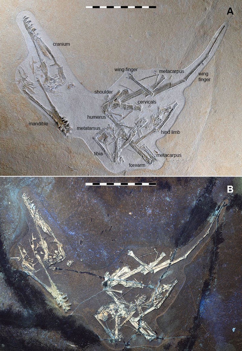
FIGURE 2. Line drawing of LF 1370. Abbreviations as follows and in later figures: car, carpal; co, coracoid; cv, cervical vertebra; dr, dorsal rib; dv, dorsal vertebra; fe, femur; fr, frontal; hu, humerus; il, ilium; is, ischium; j, jugal; lc, lacrimal; mc, metacarpals; mn, mandible; mt, metatarsal; mx, maxilla; na, nasal; pal, palate; pm, premaxilla; po, postobital; pp, prepubis; pt, pterygoid; pu, pubis; px, phalanx; q, qudarate; qj, quadratojugal; rd, radius; sa, sacrum; sc, scapula; st, sternum; ta, tarsal; ti, tibiotarsus; ul, ulna; wmc, wing metacarpal; wpx 1-4, wing phalanges. Scale bar is 100 mm with 10 mm divisions.
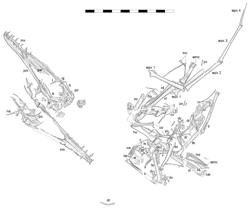
FIGURE 3. X-ray image of specimen LF1370 Makrodactylus oligodontus. Note that the end of the further wing phalanx is missing and the breaks in the slab that have been restored. Scale bar is 100 mm with 10 mm divisions.
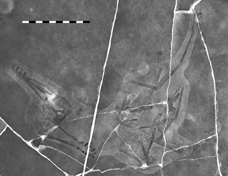
FIGURE 4. Close up of the skull of LF 1370. Abbreviations as in Figure 2. Scale bar is 50 mm with 10 mm divisions.
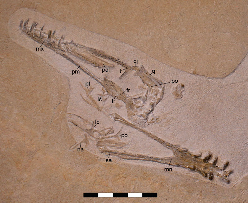
FIGURE 5. Close up photo of the body of LF 1370. Abbreviations as in Figure 2. Scale bar is 50 mm with 10 mm divisions.
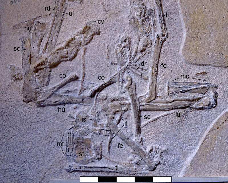
FIGURE 6. Close up photos of the body of LF1370 under UV light. A) Cervical series, shoulder and part of the wing, B) sternum, shoulder, pelvis and hindlimb. Abbreviations as in Figure 2. Scale bars are 20 mm with 10 mm divisions.
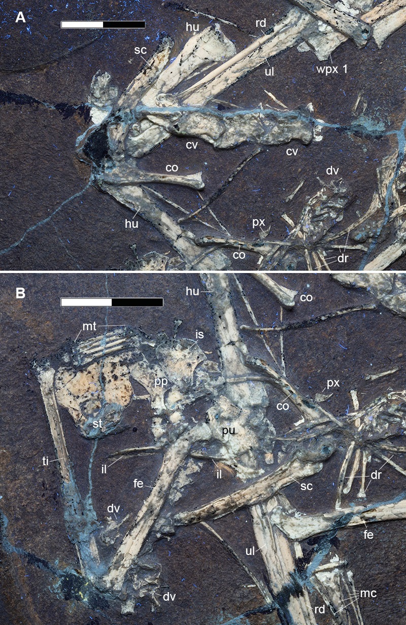
FIGURE 7. Restored skull and comparisons of Solnhofen region early monofenestratans and an early pterodactyloid seen in lateral view and with the mandible in dorsal view for some: Skiphosaura (Hone et al., 2024); Makrodactylus (this study); Propterodactylus (based on Spindler, 2024) and Diopecephalus (based on Vidovic & Martill, 2014 - their figure 3 G and H). Scale bars are all 10 mm.
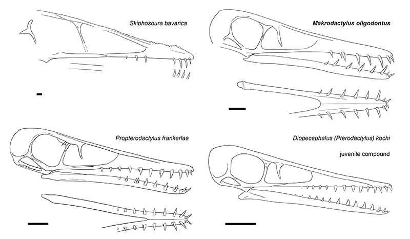
FIGURE 8. Nopsca curves of the Solnhofen region early monofenestratans showing the sizes of various key elements. Data from Table 2.
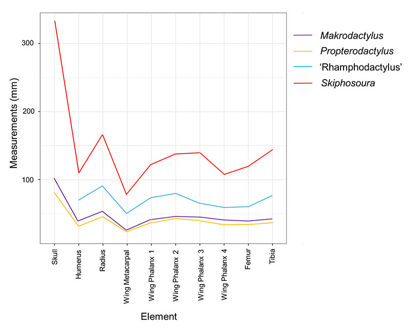
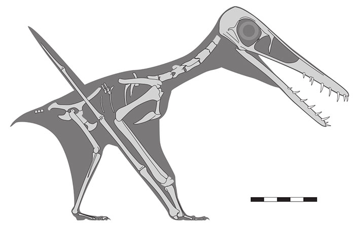
FIGURE 9. Restored skeleton of Makrodactylus oligodontus in right lateral view is a standing posture showing the preserved elements (pale grey) and inferred body and soft tissues (dark grey). Dashed outlines to elements indicates uncertain margins of bones that are not well preserved. Scale bar in 50 mm with 10 mm divisions.