|
|
|
SYSTEMATIC PALEONTOLOGYOrder SQUAMATA
Oppel, 1811 Generic Diagnosis. Dentary small (preserved length 9.20 – 9.60 mm); three pleurodont teeth, recurved third pleurodont tooth followed by tricuspid, acrodont and pleuroacrodont teeth with incipient development of lateral cuspules that are smaller in size than the central cusp as compared to those of Tinosaurus; relatively deep anterior part of the dentary; differs from all known taxa of agamid lizards in having a subspherical, vertically oriented, ventrally sloping symphyseal facet covering almost the entire anterolingual face of the dentary bone; broad and slightly convex to flat, platform-like subdental ridge between the alveolar margin and the dorsal margin of the Meckelian fossa; and close spacing of teeth. Etymology. The genus is named after the Vastan Lignite Mine from where the specimens were recovered. Type Species. Vastanagama susani sp. nov. Holotype. IITR/ SB/ VLM 1050, left dentary. Referred Specimens. IITR/SB/VLM/793, left dentary, IITR//SB/VLM/ 886 right dentary. Horizon & Locality. Lower Eocene Cambay Shale of Vastan Lignite Mine, District Surat, Gujarat state, India. Vastanagama susani sp. nov. Specific Diagnosis. Same as for the genus. Etymology. The species is named in honor of Dr. Susan E. Evans.
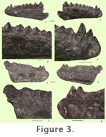
IITR/SB/VLM/886 (Figures 3.5-8) is very similar to IITR/SB/VLM/793 and 1050 in the morphology of the dentary, except for the presence of a strong vertical groove separating the symphyseal facet from the anterior border and a ridge of eroded teeth posterior to the pleurodont teeth. Only three highly eroded pleuroacrodont teeth are distinguished posteriorly. Vertical grooves are present between the three posteriormost teeth. The posterior part of the jaw is not preserved (see Table 1). Comparisons. The dentaries described here are referred to the family Agamidae because they exhibit a distinctive combination of characters, such as a subdental ridge, an acrodont dentition with some pleuroacrodont teeth posterior in the jaws, pleurodont teeth in the symphyseal region (Moody and Rocek 1980), and the absence of wear facets on the lingual surfaces of the dentary teeth (Evans et al. 2002). Augé and Smith (1997) also considered the presence of a subdental ridge and acrodont teeth followed by pleuroacrodont teeth posterior to the anterior pleurodont series to be primitive characters for agamids. Teeth with such an implantation were designated as "subacrodont"(pleuroacrodont), a condition intermediate in morphology between the large majority of lizards and typical agamids. Fossil lizards with acrodont teeth are known in the form of Tikiguania (Datta and Ray, 2006), Bharatagama (Evans et al. 2002), Priscagama, Pleurodontagama (Borsuk-Bialynicka and Moody 1984, Borsuk-Bialynicka 1996), Mimeosaurus (Gilmore 1943, Gao and Hou 1995) and Flaviagama (Alifanov 1989) (Priscagaminae), Tinosaurus (Marsh 1872) and Quercyagama (Augé and Smith 1997). The dentaries referred here to Vastanagama have three anterior pleurodont teeth as in Tikiguania and Quercyagama, but unlike in Bharatagama (five), Priscagama (three to five), Flaviagama (two) and Tinosaurus (four). The posterior pleuroacrodont teeth are labiolingually compressed and tricuspid in Vastanagama, as in Tikiguania and Tinosaurus. In contrast, the posterior pleuroacrodont teeth are labiolingually compressed, blade-like and not tricuspid in Bharatagama and all priscagamids. However, the tricuspid teeth of Vastanagama and Tikiguania differ from those of Tinosaurus in having a relatively high central cusp and weakly defined lateral cuspules. Vastanagama has a comparatively short tooth row, as in Bharatagama and priscagamids. Tikiguania and Tinosaurus europeocaenus (Augé and Smith 1997), on the other hand, have a long tooth row. (Augé and Smith 1997). Dorsoventral wear facets are characteristic of Tikiguania, Bharatagama, priscagamids, Tinosaurus, Quercyagama, and living acrodonts, and they are also present in the dentaries of Vastanagama. In acrodont iguanians, the dentary symphyseal facet lies medial in position, restricted to the dorsal margin of the Meckelian fossa. A strong, largely horizontally oriented symphyseal facet restricted to the dorsal margin of the Meckelian fossa is characteristic of Bharatagama, priscagamids, Tinosaurus and living acrodonts. In Tikiguania, this facet is comparatively small and slightly obliquely oriented to the long axis of the dentary. Vastanagama differs from all these taxa in possessing a large, subcircular, vertically oriented, ventrally sloping symphyseal facet. In Tikiguania, Bharatagama, priscagamids, Tinosaurus, and Quercyagama, the dentaries taper anteriorly and have shallow anterior regions with the bases of the teeth closer to the ventral dentary margin. In Vastanagama, the anterior part of the dentary is relatively deep, does not taper and the bases of the teeth are more distant to the ventral dentary margin. As in Bharatagama, Tinosaurus and Tikiguania, no splenial is present in Vastanagama, however, this bone is known to occur in the dentaries of priscagamids. Although Vastanagama compares well with Tinosaurus sp. in its short jaws and tricuspid posterior pleuroacrodont teeth (Augé 1990), the lateral cuspules of the teeth are well separated from the central cusp by deep vertical grooves, and the height difference between the central cusp and lateral cusps is much lower in Tinosaurus sp. On the whole, the three characters: subspherical, vertically oriented, large symphyseal facet; deep anterior part of the dentary; and broad, flat to convex, platform-like subdental ridge, are absent in any other known acrodont iguanian and strongly favor placement of the dentaries in the new genus Vastanagama. Among all known acrodont agamids, the Vastanagama dentaries are morphologically closer to Tikiguania in the development of a broad, convex or flat, platform-like subdental ridge, incipient lateral cuspules and a high median cusp on the teeth. Genus TINOSAURUS
Marsh, 1872 Emended Generic Diagnosis. Dentary generally elongated; several pleurodont teeth in the symphyseal region, one of which could be caniniform; acrodont teeth behind the anterior pleurodont teeth; posterior teeth pleuroacrodont; acrodont and pleuroacrodont teeth tricuspid; size of teeth reduced regularly anteroposteriorly; presence of a subdental ridge between the tooth row and the dorsal margin of Meckelian fossa; Meckelian fossa at times open anteriorly and ventrally at least to the front of the dentary. Differs from Tikiguania and Vastanagama in having a horizontal symphyseal facet; anterior part of premaxillary process of maxilla not raised; and well-developed palatine process of maxilla. Type Species.Tinosaurus stenodon Marsh, 1872 Tinosaurus indicus sp. nov. Holotype. IITR/SB/VLM/ 904, left dentary. Referred Specimens. IITR/SB/VLM 1051, maxilla, IITR/SB/VLM 748, right dentary; IITR/SB/VLM 820, right dentary; IITR/SB/VLM 1040, right dentary. Horizon & Locality. Lower Eocene Cambay Shale of Vastan Lignite Mine, District, Surat, Gujarat state, India. Specific Diagnosis. Maxilla and dentary large in size (maximum preserved length 23 mm), anteroposteriorly elongated jaws with shallow anterior regions, teeth with high median cusp and poorly differentiated lateral cuspules; narrow, cylindrical subdental ridge between Meckelian fossa and alveolar border; symphyseal facet elliptical in outline, anteroposteriorly elongated and obliquely or horizontally oriented with respect to the long axis of the dentary; anteriorly closely spaced acrodont and posteriorly widely spaced pleuroacrodont teeth. Etymology. Species is named after India, the country of its origin.
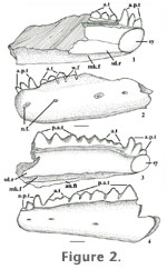
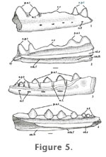
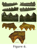
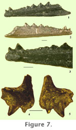
IITR/SB/VLM 820 (Figures 5.1-2, Figures 6.1-2) is a right dentary in which the anterior and middle parts are preserved. This specimen is heavily eroded with rounded ends. It has a straight and rounded ventral border and several small, shallow depressions on its lateral face, which may represent nutrient foramina. On the medial surface of the dentary, the subdental ridge is a rounded convex horizontal bar that extends anteroposteriorly (Figure 5.2, Figure 6.2). This bar overhangs the Meckelian fossa, which is restricted to the ventral border and widens posteriorly. It has two pleurodont teeth anteriorly and there is a socket in front of the first preserved tooth, possibly for the first pleurodont tooth. The two preserved pleurodont teeth are not closely spaced but are rather separated by a broad U- shaped groove. The preserved first pleurodont tooth is larger than the second one and mesially inclined, whereas the second one is slightly recurved distally. The symphyseal facet of the dentary is elongated anteroposteriorly, but slightly obliquely oriented to the long axis of the dentary and subventral in position (Figure 5.2). Posterior to the pleurodont teeth, the dental series is highly eroded in the middle part and only two large, triangular teeth are present at the posterior portion of the dentary (Figures 5.1-2, Figures 6.1-2). These teeth can be regarded as pleuroacrodont as their bases are more strongly developed lingually. A short diastema is present between the pleurodont anterior teeth and the hatchling teeth of the eroded ridge (Figures 6.1-2). The lateral cuspules are not discernible. At the posterior end of the dentary, a narrow angular facet is present medially (Figure 5.2). IITR/SB/VLM 748 appears to be similar to IITR/SB/VLM 904 and IITR/SB/VLM/820 in morphology and size, but it is heavily eroded. As in IITR/SB/VLM 904, a shallow longitudinal depression occurs between the rounded ventral margin and the area ventral to the vertical grooves between the teeth.
IITR/SB/VLM 1051 (Figures 6.7-9) is a left maxillary fragment broken on both its anterior and posterior ends. The size of this maxilla matches the dentaries described above, and hence it is described as belonging to the same taxon. In lateral view, the dorsal margin of the maxilla is broken anteriorly above the first and second preserved teeth. The margin is intact posteriorly and dorsal to the remaining two teeth. The lateral face of the maxilla is generally flat, but slightly convex dorsal to the anteriormost three teeth. Dorsal to the fourth tooth, there is a slightly depressed area between the tooth-bearing surface and the dorsal margin of the maxilla. In lateral view, the four teeth have an acrodont implantation, and in medial view the teeth have a pleuroacrodont implantation. In medial view, the teeth overhang a rounded horizontal bar, which rises regularly from anterior to posterior. The teeth appear to be tricuspid with minute lateral cuspules at their mid-height on the posterior face. The central cusp of these teeth is very high, and the lateral cuspules are located at mid-height or close to the base. The absence of anterior lateral cuspules can be explained by lingual wear facets, which are located in this position. The presence of wear facets on the lingual face of the teeth suggests that this bone is a maxilla. The teeth increase in size gradually from anterior to posterior. The horizontal bar is expanded posteriorly dorsal to the third and fourth teeth and this expansion is possibly for the palatine process, which is broken in the present specimen. IITR/SB/VLM 1052 (Figures 7.4-5) is a right premaxillary bone bearing a broad alveolar margin with two teeth. The premaxilla is broken both anteriorly and posteriorly. The nasal process is relatively thin, and its anterior face and tip are broken. The lateral face of the bone is convex. Posterior to the nasal process, the dorsal margin of this bone is straight. The nasal process is almost vertically orientated. See Table 2. Comparisons. The dentary bones represented by IITR/SB/VLM/904 possess three pleurodont teeth anteriorly, but they are comparatively larger than those referred to Vastanagama. Furthermore, these dentaries differ from those of Vastanagama in having an elliptical and anteroposteriorly elongated symphyseal facet oriented obliquely or horizontally to the long axis of the dentary, and in a narrow and subcylindrical subdental ridge. Within the family Agamidae, the morphology of IITR/SB/VLM/904 and other Vastan specimens described here is reminiscent of the Late Triassic genus Tikiguania and the Paleocene-Eocene genus Tinosaurus. Although in most of the specimens the dentary is broken posterior to the posterior teeth, in IITR/SB/VLM/904 an unworn additional tooth occurs posterior to the adult permanent teeth. This is considered as the posteriormost tooth of the dentary because it is followed by a wide gap. It is interpreted from the straight dental margin of the posterior region that the coronoid process did not exist. In this respect, IITR/SB/VLM/904, referred to T. indicus sp.nov. is similar to Tikiguania. As in Tikiguania, the dentaries of T. indicus are also elongated with a tapering anterior part bearing three pleurodont teeth, anteroposteriorly elongated symphyseal facet and tricuspid acrodont and pleuroacrodont teeth. However, the teeth of Tikiguania are closely spaced all along the jaw, contrasting with the widely spaced posterior pleuroacrodont teeth of Vastan dentaries. Tikiguania also has a dentary with a broad, flat, or convex subdental ridge, as compared to the narrow, subcylindrical subdental ridge on the Vastan specimens. Tinosaurus is defined by the presence of caniniform teeth in the anterior part of the jaws and tricuspid acrodont/pleuroacrodont teeth posteriorly (Estes 1983, Augé 1990, Augé and Smith 1997, Augé 2005). Alternatively, it has also been defined by the tricuspid nature of the acrodont teeth alone (Hecht and Hoffstetter 1962). The genus Tinosaurus is at present represented by eight species: T. stenodon Marsh, 1872, T. pristinus (Leidy 1872) (Middle Eocene of North America, Gilmore 1928, Hecht 1959), T. lushihensis Dong 1965 (early Late Eocene of Honan, China), T. asiaticus (?early Upper Eocene Ulan Shireh Formation of Inner Mongolia, Gilmore 1943), T. doumuensis (Palaeocene Doum Formation of Anhui, China, Hou 1974), T. yuanquensis (Upper Eocene Zhaili Member, Hedi Formation, Yuanqu, Shanxi Province, China, Li 1991a), Tinosaurus cf. T. lushihensis (Middle Eocene Hetaoyuan Formation Xichuan Province, Henan Province, China, Li 1991b), T. postremus (Palaeocene of Kazakhstan, Averianov 2000) and T. europeocaenus (Palaeocene of Europe, Augé and Smith 1997, Augé 2005). Unidentified species of Tinosaurus have also been reported from the Early Eocene of Belgium (Hecht and Hoffstetter 1962; Godinot et al. 1978), the Early Eocene of France (Russell et al. 1982, Augé 1990), the early Middle Eocene Kuldana Formation, Kohat, Pakistan (Rage 1987) and the Upper Eocene Chadron Formation of North Dakota, USA (Smith 2006). Because of the presence of characters diagnostic of Tinosaurus (a narrow, cylindrical subdental ridge, and anteriorly tapering dentaries) IITR/SB/VLM/904 and other dentaries and maxilla described here are assigned to this genus. Despite the fact that the posterior pleuroacrodont teeth of T. indicus sp. nov. are tricuspid as in various species of Tinosaurus, the development of lateral cuspules in T. indicus sp. nov. is different from the North American and Eurasian species of Tinosaurus. In most of the latter species, there are four anterior pleurodont teeth, the lateral cuspules are well separated from the central cusp by deep vertical grooves, and the height difference between the central cusp and the lateral cuspules is far less on the acrodont and pleuroacrodont teeth. In comparison, the lateral cuspules of T. indicus sp. nov. are incipient, hardly differentiated from the high central cusp and are located at mid-height of the tooth. In this respect, the tricuspid teeth of T. indicus sp. nov. are very similar to the posterior teeth of Tikiguania. T. indicus sp. nov. further differs from Tinosaurus cf. T. lushihensis and T. asiaticus in possessing non-overlapping posterior acrodont teeth and closely spaced teeth anteriorly, and widely spaced posterior subacrodont teeth. T. indicus sp. nov. is also distinguished from T. yuanquensis and all other known species of Tinosaurus from China in the presence of well-developed vertical grooves between the teeth on the labial face of the dentary. This indicates that shearing occlusion was less important in the Chinese species (Li 1991b) as compared to species from North America and Europe, and T. indicus sp. nov. The Vastan specimens also differ from T. stenodon and T. pristinus in possessing a shallower posterior portion of the dentary. Two fragmentary teeth (GSP-UM 552-553) from the early Middle Eocene Kuldana Formation, Kohat, Pakistan, were referred to Tinosaurus sp. (Rage 1987). These teeth are comparable to those of Vastan Lignite Mine in their greater development of lateral cuspules on one side and less on the other. However, the groove between the median and lateral cuspules is developed more in GSP-UM 553 when compared to the Indian specimens. One of the teeth (GSP-UM 553) appears similar to the anterior acrodont teeth of Vastanagama susani gen. et sp. nov. A fragmentary maxilla from the late Lower Eocene beds at Prémontré (Paris basin), France was referred to Tinosaurus sp. (Augé et al. 1997). The maxilla described here differs from the Prémontré specimen in having a very high median cusp on the tricuspid subacrodont teeth and minute lateral cuspules. Because of the differences in the morphology identified between the Vastan dentaries and various other species of Tinosaurus, the new fossil material from India is referred to a new species of Tinosaurus, T. indicus sp. nov. |
|