|
|
|
MATERIAL AND METHODSMaterialSauropod material was examined in the following collections: American Museum of Natural History (AMNH), New York, USA; Chengdu University of Technology (CDUT), Chengdu, China; Carnegie Museum of Natural History (CMNH), Pittsburgh, USA; Institute of Vertebrate Paleontology and Paleoanthropology (IVPP), Beijing, China; Museo Argentino des Ciencias Naturales, Buenos Aires (MACN); Museum für Naturkunde, Berlin (MNB), Germany; Naturhistorisches Museum Basel (NMB), Basel, Switzerland; Saurier-Museum Aathal (SMA), Switzerland; Naturmuseum Senckenberg (NMS), Frankfurt, Germany; National Museum of Natural History, Smithsonian Institution (NMHNSI), Washington, USA; Yale Peabody Museum (YPM), New Haven, USA; Zigong Dinosaur Museum (ZDM), Zigong, China. For comparative anatomy, the skeletons of Rhea americana (NMB 2670), Struthio camelus (NMB 8180), Dromaeus novaehollandiae (NMB 2978), Casuarius casuarius (NMB 1829), Sarcoramphus gryphus (NMB 3295) and Cygnus cygnus (NMB 10588) were investigated. The necks of Meleagris gallopavo, Columba livia and Ardea cinerea were dissected. CT scans of a crane (Grus grus) and a white-tailed eagle (Haliaeetus albicilla), produced in the Clinic for Small Pets of the Free University of Berlin with a high-resolution Multislice-CT scanner (GE Healthcare Light Speed advantage QXi), were examined. Reconstruction of Pneumatic Soft-Tissue in the Sauropod NeckTopographical similarities between soft-tissue attachment sites of related extant and extinct vertebrates can be used for reconstructing such soft-tissues in extinct taxa, following the method of Extant-Phylogenetic-Bracketing (EPB) (Bryant and Russell 1992; Witmer 1995, 1997; Carrano and Hutchinson 2002). In the case of sauropods, extant Crocodylia and Aves provide the anatomical framework for these soft-tissue reconstructions (Gauthier et al. 1988; Witmer 1997; Benton 2004). The presence of osteological correlates for vertebral pneumaticity exclusively in extant avians permits the reconstruction of these soft structures only by one-way phylogenetic comparison, corresponding to a Level II inference (Witmer 1995, 1997). Criteria for recognizing osteological correlates of pneumatic structures in sauropod vertebrae (Figure 1.2) are based on comparison with extant avians and follow the works of Britt (1993), O'Connor (2004, 2006), Wedel (Wedel et al. 2000; Wedel 2005) and Witmer (1990, 1997). Because of these avian-based comparisons, standard nomenclature of pneumatic structures for birds was used (Müller 1908; Duncker 1971; O'Connor 2006), supplemented by topographical descriptors for additional pneumatic structures. For the intraosseous pneumatic structures of the vertebrae, established terms for sauropods (Britt 1993; Wedel et al. 2000; Wedel 2003a, 2005) were used. The terminology of the external laminae in sauropod vertebrae follows that of Wilson (1999). Objective of the ExperimentThe objective of the experiment was to test the effect of pneumatic support on a chain-beam with different arrangements of pneumatic bodies as a model for the tetrapod neck (see below). Specifically, we wanted to find out whether or not simple pneumatic tubing would be able to hold the chain beam in a near horizontal position and how different arrangements and numbers of pneumatic tubings would influence the stability of the chain beam against application of load at its free end. The experiment was designed to answer the following questions: 1) Is it mechanically possible to stabilize a chain beam with pneumatic bodies? 2) How do arrangement and fixation of the pneumatic bodies influence the load capacities of the chain beam? 3) How does the system react to symmetrical and asymmetrical changes of the pneumatic pressure? 4) Is it possible to move the chain beam with pressure changes? Theoretical Background for Building the ModelFrom a mechanical viewpoint, the tetrapod neck is comparable to a segmented cantilever fixed at one end (Koch 1917; Gray 1944; Slijper 1946; Kummer 1959; Alexander 1989; Frey and Martin 1997; Martin et al. 1998; Hildebrand and Goslow 2001; Salisbury and Frey 2001). The cervical vertebrae form the segments of the cantilever, and the vertebral column of the trunk and the shoulder girdle form its fixation. The cantilever is loaded by gravity, which places its dorsal part under tension, whereas the ventral part is compressed (Figure 1.3). Each segment is free to move against its neighbors on an intersegmental articulation. The tensile and compressive stresses are accommodated by tensile dorsal and/or compressive ventral bracing elements. Typical bracing structures in the tetrapod neck are the zygapophyseal articulations and their articular ligaments and dorsal ligaments. Additional bracing is brought about by the hydraulic effects of the cervical muscle mass. The zygapophyseal articulations are positioned dorsally to the intervertebral articulations and limit the mobility of the vertebral segments against each other by ligament and bone lock (Stevens and Parrish 1999; Stevens 2002; Stevens and Parrish 2005). Dorsal ligaments (Figure 1.3) extend along the neck of tetrapods either as a single, continuous supraspinal ligament (e.g., in extant crocodylians: Wettstein 1937; Frey 1988a), or as a multisegmental fan-shaped nuchal ligament originating from the trunk region (e.g., in extant mammals and birds; Boas 1929; Slijper 1946) and forming tensile structures (Bennett and Alexander 1987; Alexander 2002). In some tetrapods, such as extant crocodylians (Frey 1988b; Salisbury and Frey 2001) and sauropods, overlapping cervical ribs positioned ventrally to the vertebrae (Figure 1.3) form additional compressible bracing structures connected to each others by intercostal ligaments (Frey 1988a, 1988b; Frey and Martin 1997; Martin et al. 1998). Experimental Setup
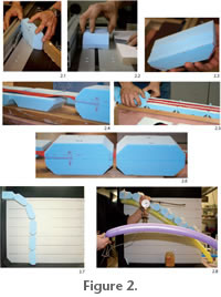
The chain beam was fixed on a vertical plywood pivot board 700 mm above the stand board, on which the vertical pivot board was mounted (Figure 2.7). The polyester belt was inserted into a horizontal slit cut into the pivot board and fixed with screw clamps in a way that the base segment was absolutely immobile. Pivot and stand board were linked to each other by a scale panel with a 100 mm grid, in order to record the load effects during the experiment. Tube-shaped party balloons were tied along the chain beam with gauze bandages according to the reconstructed positions of pneumatic diverticula in sauropod necks. The dorsal balloons were fixed proximally on the shelf by a clothespin. These balloons had a length of about 1.5 m and a diameter of about 50 mm when fully inflated (Figure 2.8). The muzzle of the balloons was connected to a T-switch, which was connected to a compressor and a WIKA manometer (gauge from 0.0-2.5 bar at a scale of 1/10 bar). In one experiment, the balloons were fixed to the segments with double-sided tape, in another experiment they were segmented by twisting parts of the balloons into pneumatic segments. Sometimes intersegmental wedges of Styrodur™ were needed for additional stabilization, especially at its base. At the distalmost segment, a cup was fixed and filled with iron powder to quantify the loads. In order to quantify the sagging effect, the quantity of iron powder was kept constant. For the quantification of the bracing effect, the cup was slowly filled with iron powder until sagging reached a defined level. Implementation of the Cantilever Model and Material ChoicesFor the experiment we did not attempt to simulate any material properties of any organic tissue. Instead we tried to approximate the configuration of a generalized pneumatized chain beam system and its principal functionality as a model for the function of a pneumatized sauropod neck. In the cervical column of a tetrapod, the vertebrae can be regarded as a chain of stable inflexible elements, able to take compression loads and guaranteeing the constancy of length. In the experiment, Styrodur™ blocks formed these incrompressible structures. The complex synovial intervertebral ball and socket articulation of sauropods (Schwarz et al. 2007) would allow movement of the vertebral segments against each other in all directions. However, bending of the vertebrae against each other is limited or nearly blocked in the case of torque movements by the zygapophyseal articulations (see above) (Stevens and Parrish 1999; Stevens 2002; Stevens and Parrish 2005). This kind of movement limitation was simulated with the horizontally orientated polyester belt, which allowed the bending of the segments in the vertical plane until the contact of the Styrodur™ block. Horizontal (lateral) movement and especially torque was limited. An investigation of lateral movements under pneumatic bracing was possible by arranging the chain beam with the polyester belt standing vertically. The most important mechanical property of the chain beam in any arrangement was the strongly limited torque, a feature essential for the position control of sauropod necks. To monitor the support effect of pneumatic tubings alone, all other bracing elements were first omitted from the model. From its fixing point, the experimental chain beam with the polyester belt oriented horizontally would sag completely under its own weight into Styrodor body lock at its base. Later, additional Styrodur™ wedges were placed ventrally between the basal, representing additional ventral compressive bracing elements. The experiment was designed to (1) prove that a chain beam can be braced pneumatically, (2) quantify the effect of different arrangements and pressures of the pneumatic systems around the chain beam and (3) test the option of a pneumatically triggered movement of a chain beam. The results of the experiment are discussed in the frame of an optional utilization of extensive pneumatic systems as found among sauropods in the context of bracing and mobility. Because the simulation of biomaterials is methodologically problematic if not impossible, the choice of the materials for the experiments was restricted to the material properties of the system in general. Bone is a biological composite material with a high compressive and poor tensile strength, therefore being mainly resistant against compression (Koch 1917; Currey 1987, 1999). For producing the simplified vertebral segments in the model, extruded high-resistance polystyrene foam Styrodur™ was chosen. Similar to bone, Styrodur™ has a higher compressive than tensile strength and is resistant against compression. Additionally, the material was cheap and easy to shape. Pneumatic diverticula are air-filled pockets lined by pneumatic epithelium, thus they represent air-filled membrane constructions. The reconstruction of the pneumatic system of sauropods by comparison with extant birds implicates similarities in the properties of the pneumatic diverticula in both groups. In birds, the wall of the diverticula is formed by a smooth, cuboidal to columnar pneumatic epithelium (Duncker 1971; Maina 2005; O'Connor 2006). The pneumatic epithelium is covered by a thin layer of nearly avascular connective tissue, consisting of a loose network of elastic and collagenous fibers and a few blood vessels (Duncker 1971; Carlson and Beggs 1973; Maina 2005). In the air sacs of the body cavity, the external wall of the pneumatic epithelium in many birds touches the peritoneal epithelium, which is a layer of mesothelial cells (Duncker 1971; Carlson and Beggs 1973; Fletcher 1980). The diverticula walls can additionally contain cells of smooth muscle tissue and clusters of adipose cells (Duncker 1971; Maina 2005). Pneumatic diverticula in the neck of extant birds are enwrapped in connective tissue and connected with each other via tiny ducts. The diverticula around the vertebrae can anastomoze with one another (O'Connor 2006), forming larger hose-like diverticula units along the vertebral column. The units of pneumatic diverticula mechanically resemble pneumatic cushions, which can take over loads by using the compressibility of gasses: only part of the load is transmitted to the membrane, the other part increases gas pressure. In the experiment, the complex diverticular system was reduced to its mechanical properties. Therefore, tube-shaped inflated party balloons were used for the simulation. Similar to the pneumatic diverticula, these balloons can take loads by the compressibility of their air filling. The balloons were fixed along the chain beam using a gauze bandage. Such a fixation is necessary to maintain the coherence between the pneumatic system and the chain beam. As was demonstrated above, pneumatic diverticula in tetrapods show a variety of fixing systems, amongst them sheaths of connective tissue. |
|