|
|
|
INTRODUCTION
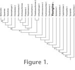
The Late Permian (250 MYA) diapsid reptile Youngina capensis is often regarded as the 'archetypal' basal diapsid (Smith and Evans 1996) and recognized as an "ancestral morphotype" (Carroll 1988b) between more primitive taxa such as parareptiles and captorhinids and modern diapsids (Müller 2003; Figure 1). Its relationships among other diapsids have been disputed. Currie (1981, 1982) posited that Youngina shared a closer relationship with Acerosodontosaurus (Currie 1980), Galesphyrus (Carroll 1976), Hovasaurus (Currie 1981), Kenyasaurus (Harris and Carroll 1977), Tangasaurus (Currie 1982), and Thadeosaurus (Carroll 1981). These taxa collectively were referred to as the Younginiformes, which were variously allied to lepidosauromorphs (Benton 1985; Evans 1988) or as stem-diapsids (Gaffney 1980; Laurin 1991). Their monophyly was never explicitly tested and recently Bickelmann et al. (2009) published the results of their phylogenetic analysis that suggested that these taxa do not form a monophyletic relationship with each other to the exclusion of other diapsid reptiles, though the relationships between all stem-diapsids were highly unresolved in their topology. Youngina is the most derived known stem-diapsid to retain two complete fenestrae in the temporal region, rather than having evolved this condition secondarily (as in derived archosauromorphs and sphenodontids and possibly as in sauropterygians and ichthyopterygians). Thus it appears that the loss of the lower temporal bar occurred in higher stem-diapsids more derived than Youngina (Müller 2003). Youngina is well known from numerous specimens that were previously described as distinct "younginid" or "younginiform" taxa (Gow 1975). Despite its obviously critical position as a stem-diapsid and the existence of multiple specimens, Youngina is in need of a thorough re-description. Formal description of its anatomy is a crucial precursor to understanding: 1) its phylogenetic relationships, 2) its relevance to the interrelationships between other diapsid reptiles and 3) the evolution of the skull in these reptiles (Bickelmann et al. 2009; Modesto and Sues 2002). The first discussion of the braincase of Youngina was carried out by Olsen (1936), who described UC 1528, the holotype of Youngoides romeri. He was limited to observations of the superficial anatomy of the skull, and described it largely in palatal and occipital views. Gow (1975) provided a preliminary discussion of the braincase of TM 3603, which was given more attention later by Evans (1987). Evans sawed this specimen in half to gain access to the internal aspects of the neurocranial bones. Her description is currently the only published, detailed treatment of the braincase of Youngina. While all reptiles (and in fact, all amniotes) ossify the caudoventral portion of the braincase cartilages, ossification patterns differ for the rostral cartilages. Archosauriforms (birds, crocodiles and their ancestors) have uniquely ossified the pila antotica as the laterosphenoid, which encloses the rostral region of the cavum cranii (Clark et al. 1993). Turtles ossify the pila antotica adjoining to the clinoid process of the basisphenoid, similar to the condition found in captorhinids, sauropterygians and many other primitive reptiles, though the clinoid process is much taller in turtles leaving the rostral braincase largely open (Rieppel 1993) and the pila antotica may have also ossified into an archosauriform-like paired "laterosphenoids" primitively in turtles (Proganochelys: Bhullar and Bever 2009; Kayentachelys: Gaffney and Jenkins 2010), though some parareptiles (pareiasaurs) have a similar ossification (Lee 1997). Some lepidosaurs have a prominently ossified rostral expansion of the prootic (i.e., the alar process) which partially closes the braincase wall rostrally (Rieppel 1993). For taxa in which the cavum cranii is extensively enclosed by bone, it is possible to reconstruct the gross shape of the endocranium and understand how it relates to the sensory organs and their associated neurology; for poorly ossified taxa, however, such reconstruction is much more difficult (Hopson 1979; Hopson and Radinsky 1980). Because the braincase of Youngina remains buried in matrix, high resolution X-ray computed tomography (HRXCT) provides an excellent tool for imaging this otherwise inaccessible deep cranial region. Several studies have recently shed new light on braincase anatomy using imaging techniques (Clack et al. 2003; Witmer et al. 2008; Holliday and Witmer 2008; Witmer and Ridgely 2009). Youngina presents an ideal target for HRXCT scanning and a new description of its skull and braincase anatomy. To that end, the holotype of Youngina capensis was successfully scanned, allowing a complete reconstruction of its braincase for the first time. Institutional Abbreviations. AMNH, American Museum of Natural History, New York; UC, University of Chicago, Illinois; TM, Transvaal Museum, South Africa. |
|