EXTRINSIC MUSCLES OF THE SKULL AND NECK
The axial muscles of the neck can be divided into epaxial muscles that run above the ribs and hypaxial muscles that run below the ribs (Gasc 1981;
Tsuihiji 2005,
2007). The epaxial muscles are further divided into three groups, all of which broadly run parallel to the vertebral column (Nishi 1916;
Gasc 1981;
Tsuihiji 2005):
1. m. Transversospinalis (medial column)
2. m. Longissimus (central column)
3. m. Iliocostalis (lateral column)
In general, the m. Transversospinalis sits against the neural spines, the m. Longissimus sits alongside the zygapophyses of the vertebrae, and the m. Iliocostalis is positioned on top of the ribs. The m. Transversospinalis, and more lateral m. Longissimus are separated by the fascial "septum intermusculare dorsi"
(Nishi 1916;
Tsuihiji 2005), but m. Longissimus and m. Iliocostalis are not clearly divided by fascia in Sphenodon
(Nishi 1916;
Tsuihiji 2005). These three groups can also be defined based on their innervation because they receive different twigs or branches from the dorsal rami of the segmental spinal nerves (Gasc 1981;
Tsuihiji 2005).
The axial muscles of Sphenodon have been described several times (Maurer, 1896;
Osawa 1898;
Nishi 1916;
Byerly 1925;
von Wettstein 1931;
Gasc 1981;
Tsuihiji 2005,
2007;
Al-Hassawi 2004,
2007). In his study of non-avian reptile axial musculature,
Gasc (1981) dissected at least one specimen of Sphenodon in addition to other taxa.
Byerly (1925) described some of the neck and shoulder muscles as part of his more general description of the muscles in Sphenodon.
The von Wettstein
(1931) article is a review of previous work, whereas
Al-Hassawi (2004,
2007) specifically concentrated on the cervical muscles in lepidosaurs and partially dissected one Sphenodon specimen. In two detailed works regarding axial muscles in reptiles,
Tsuihiji (2005,
2007) dissected part of one Sphenodon specimen (listed as specimen CAS 20888, SVL 250 mm).
For information regarding the muscles of the neck that do not attach to the skull, such as the intercentral muscle slips, readers are directed towards
Günther (1867),
Maurer (1896),
Osawa (1898),
Nishi (1916),
Byerly (1925),
von Wettstein (1931),
Gasc (1981),
Al-Hassawi (2004,
2007), and
Tsuihiji (2005,
2007).
m. Spinalis (mSp)
The m. Spinalis is a bundle of muscle and tendons within the m. Transversospinalis group (the most medial column of the epaxial
muscles) that runs broadly parallel to the vertebral column alongside the neural
spines (Nishi 1916;
Gasc 1981;
Tsuihiji 2005). It is innervated by medial branches of the dorsal rami
of the spinal nerves (Nishi 1916;
Gasc 1981;
Tsuihiji 2005). Anterior portions of this muscle group connect the axial column to the back of the skull and are presumably used to support and lift the head.
These muscles include the m. Spinalis Capitis and the more laterally positioned m. Semispinalis Capitis. However, these muscles are interconnected and difficult to separate from the rest of the m. Spinalis (Tsuihiji
2005).
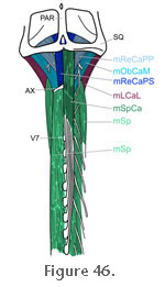 m. Spinalis Capitis (mSpCa). The m. Spinalis Capitis
connects the vertebral column to the skull via tendons that arise from the
neural spines (Figure 21,
Figure 22,
Figure 23, and
Figure 43) (Nishi 1916;
Tsuihiji 2005). This muscle is probably equivalent to the "semi-spinalis capitis" of
Byerly (1925, p. 18). The tendons insert on the posterior surface of the parietal near the midline deep to the m. Depressor Mandibulae and extrinsic muscles of the pectoral girdle (Figure 42) (Byerly
1925; Al-Hassawi 2004,
2007;
Tsuihiji 2005). According to Byerly (1925) the muscle originates on the anterolateral edges of the neural spines between the 4th and 8th vertebrae before inserting on the posterior surface of the parietal. In specimen BMNH 1969.2204 the anteriormost origination, via a tendon, is from the 7th
vertebra (Figure 23 and
Figure 45). This corresponds to the work by
Nishi (1916, p. 261) (Figure 46). m. Spinalis Capitis (mSpCa). The m. Spinalis Capitis
connects the vertebral column to the skull via tendons that arise from the
neural spines (Figure 21,
Figure 22,
Figure 23, and
Figure 43) (Nishi 1916;
Tsuihiji 2005). This muscle is probably equivalent to the "semi-spinalis capitis" of
Byerly (1925, p. 18). The tendons insert on the posterior surface of the parietal near the midline deep to the m. Depressor Mandibulae and extrinsic muscles of the pectoral girdle (Figure 42) (Byerly
1925; Al-Hassawi 2004,
2007;
Tsuihiji 2005). According to Byerly (1925) the muscle originates on the anterolateral edges of the neural spines between the 4th and 8th vertebrae before inserting on the posterior surface of the parietal. In specimen BMNH 1969.2204 the anteriormost origination, via a tendon, is from the 7th
vertebra (Figure 23 and
Figure 45). This corresponds to the work by
Nishi (1916, p. 261) (Figure 46).
m. Semispinalis Capitis (mSspCa). As
Tsuihiji (2005) points out, the m. Semispinalis Capitis muscle was originally described as the m. Semispinalis Dorsi by
Nishi (1916) and corresponds to the "articulo-parietalis" of
Olson (1936), and probably to the "Cervico-capitis" of
Byerly (1925, p. 19), and Longissimus capitis
1 of Al-Hassawi (2004,
2007).
Tsuihiji (2005) argues that it should not be considered part of the longissimus musles and instead has a closer relationship to the spinalis muscles.
 This muscle connects the vertebral column to the skull and originates via tendons from the prezygapophyses (Tsuihiji
2005). According to Al-Hassawi
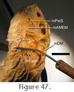
(2004, 2007) this muscle branches from the m. Longissimus Dorsi near the 7th vertebra and inserts on the posterior ventral edge of the parietal and squamosal (Figure 42 and
Figure 43). In specimen BMNH 1972.1223 this muscle can be seen deep to the m. Depressor Mandibulae and m. Episternocleidomastoid (Figure 47). It fans anterolaterally from the axial column and inserts on the posteroventral edge of the parietal-squamosal bar. This muscle connects the vertebral column to the skull and originates via tendons from the prezygapophyses (Tsuihiji
2005). According to Al-Hassawi
(2004, 2007) this muscle branches from the m. Longissimus Dorsi near the 7th vertebra and inserts on the posterior ventral edge of the parietal and squamosal (Figure 42 and
Figure 43). In specimen BMNH 1972.1223 this muscle can be seen deep to the m. Depressor Mandibulae and m. Episternocleidomastoid (Figure 47). It fans anterolaterally from the axial column and inserts on the posteroventral edge of the parietal-squamosal bar.
m. Splenius Capitis (mSplCa). The "splenius capitis"
of Al-Hassawi (2004,
2007) follows a very similar path to the m. Spinalis Capitis along the dorsal part of the vertebral column. It inserts on the posterior surface of the parietal just dorsal to the m. Spinalis Capitis (Al-Hassawi (2004, p. 66,
2007, p. 66) and origination is via tendinous bundles from the tips of the neural spines of the 6th to 12th vertebrae (Al-Hassawi 2004, p. 107,
2007, p. 107).
Tsuihiji (2005) probably includes this muscle within the m. Spinalis Capitis.
m. Axis-Supraoccipital (mAxSu)
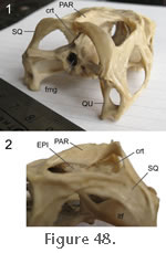 The m. Axis-Supraoccipital originates from the atlantal neural arch and also the anterodorsal margin of the axis and travels anteriorly to insert on the posterodorsal crest of the supraoccipital
(Figure 42,
Figure 43, and
Figure 48) (Al-Hassawi 2004,
2007). It
probably serves to support and lift the head. This muscle was identified by
Al-Hassawi (2004,
2007) and is apparently not known in any other taxon. However, she does not discuss whether this muscle may simply be part of the Rectus Capitis (mReCa).
Tsuihiji (2005) does not describe any muscle fibres that might represent the m. Axis-Supraoccipital so its presence may be subject to individual variation. Examination of BMNH
1969.2204 suggests that what
Al-Hassawi (2004,
2007) observed is probably a tendinous sheet and not a muscle. The m. Axis-Supraoccipital originates from the atlantal neural arch and also the anterodorsal margin of the axis and travels anteriorly to insert on the posterodorsal crest of the supraoccipital
(Figure 42,
Figure 43, and
Figure 48) (Al-Hassawi 2004,
2007). It
probably serves to support and lift the head. This muscle was identified by
Al-Hassawi (2004,
2007) and is apparently not known in any other taxon. However, she does not discuss whether this muscle may simply be part of the Rectus Capitis (mReCa).
Tsuihiji (2005) does not describe any muscle fibres that might represent the m. Axis-Supraoccipital so its presence may be subject to individual variation. Examination of BMNH
1969.2204 suggests that what
Al-Hassawi (2004,
2007) observed is probably a tendinous sheet and not a muscle.
m. Rectus Capitis (mReCa)
The m. Rectus Capitis contributes to lateral movements of the head (Byerly 1925). It originates from the atlantal arch and anterodorsal part of the neural spine of the axis (Figure 43 and
Figure 46) and inserts on the posterior surface of the braincase (Figure 34 and
Figure 42) ("rectus capitis posticus" in
Byerly 1925;
Al-Hassawi 2004,
2007;
Tsuihiji 2005). Based on dissection of specimen CAS20888
Tsuihiji (2005) divided the m. Rectus Capitis into a superficial and deep portion with the superficial portions inserting more medially. This confirmed observations made by
Nishi (1916). The cranial portion of this muscle is still present on the right side of specimen BMNH 1969.2204.
m. Rectus Capitis Posterior Superficialis (mReCaPS).
The m. Rectus Capitis Posterior Superficialis originates from the anterodorsal
portion of the lateral surface of the axis neural spine (Figure 43 and
Figure 46) (Nishi 1916;
Tsuihiji 2005).
Tsuihiji (2005) described the insertion as being lateral to the posterodorsal midline crest of the supraoccipital (Figure 42).
Neither Al-Hassawi (2004,
2007) or
Tsuihiji (2005) report insertions on the squamosal in contrast to
Byerly (1925). In specimen BMNH 1969.2204 the muscle is about 5 mm high and 1 mm wide at mid section.
m. Rectus Capitis Posterior Profundis (mReCaPP).
This muscle originates on the anterodorsal edge of the lateral surface of the axis neural spine ventral to the origin of the m. Rectus Capitis Posterior Superficialis and also from the dorsal surface of the atlas and proatlas (Figure 43 and
Figure 46) (Nishi 1916;
Tsuihiji 2005). Insertion takes place on the posterolateral surface of the supraoccipital lateral to the m. Rectus Capitis Posterior Superficialis (Figure 34,
Figure 42, and
Figure 46)
Tsuihiji (2005). The lateralmost
part of this insertion is associated with a shallow ridge (Al-Hassawi 2004,
2007). This is confirmed by examination of specimen BMNH 1969.2204. In mid section the muscle is about 4 mm wide and less than 1 mm high.
m. Obliquus Capitis Magnus (mObCaM)
The m. Obliquus Capitis Magnus is a flat muscle that spans the gap between the lateral surface of the axis and the posterodorsal surface of the paroccipital process of the opisthotic
(Figure 42 and
Figure 46) (Nishi 1916;
Al-Hassawi 2004,
2007;
Tsuihiji 2005). Its origin is ventral to that of the m. Rectus Capitis whereas its insertion is more lateral to that of the m. Rectus Capitis. A dorsal branch of the dorsal cervical plexus lies between the m. Obliquus Capitis and m. Rectus Capitis
(Nishi 1916;
Tsuihiji 2005).
Al-Hassawi (2004,
2007) reported that the origin took place on the axis neural arch whereas
Tsuihiji (2005) reported the origin as being the the lateral surface of the axis neural spine. The "Obliquus Capitis" of
Byerly (1925) probably corresponds to parts of the m. Longus Capitis and m. Semispinalis Capitis. This muscle is probably partly responsible for lateral head movements and also lifting of the head.
In specimen BMNH 1969.2204 a strap-shaped muscle can be seen to insert on the dorsal surface of the paroccipital process of the opisthotic (Figure 34). In section it is approximately
7 mm wide and 2 mm high. This muscle may be the anterior portion of the m. Obliquus Capitis Magnus.
m. Longissimus (mL)
The m. Longissimus is the central column of the epaxial muscles that runs parallel to the vertebral column, alongside the zygapophyses
of the vertebrae, medial to the iliac blade (Nishi 1916;
Gasc 1981;
Tsuihiji 2005). The muscle group is innervated by medial twigs of the lateral branches of the dorsal rami of the spinal nerves (Tsuihiji
2005). The main part of this muscle (often referred to as the m. Longissimus Dorsi) originates from the ilium and sacrum, and runs anteriorly towards the skull along the lateral surface of the zygapophyses
(Al-Hassawi 2004,
2007). The m. Longissimus cervicus branches away from this muscle and bifurcates into two heads. The anterior head inserts on the proatlas whereas the posterior head inserts on the posterior process of the atlas. Two other muscles branch off from the m. Longissimus and attach to the back of the head: the m. Longissimus Capitis Lateralis (mLCaL) and m. Longissimus Capitis Medialis (mLCaM).
m. Longissimus Capitis Lateralis (mLCaL).
This muscle is equivalent to the Longissimus capitis 2 (mLongiC2) of
Al-Hassawi (2004,
2007, p. 100) and the lateral most part of the "m Transversalis Cervicus" of
Nishi (1916, p. 261). It branches from the m. Longissimus Dorsi (mLD), near the 3rd vertebra and inserts on the lateral tip (or most distal end) of the paroccipital process of the opisthotic
(Al-Hassawi 2004,
2007). The area of insertion illustrated by
Tsuihiji (2005, figure 4) for the "m. Longissimus Capitis pars transversalis" is in agreement with this (Figure 42 and
Figure 43). Evidence of this attachment appears to be present in BMNH 1969.2204.
m. Longissimus Capitis Medialis (mLCaM). This muscle is equivalent to the Longissimus capitis
3 (mLongiC3) of Al-Hassawi (2004,
2007, p. 100). It branches from the m. Longissimus Dorsi
at a point near the axis (Al-Hassawi 2004,
2007), and inserts along the posterior surface on the paroccipital process of the opisthotic below the insertion of the m. Obliqus Capitis
Magnus (Al-Hassawi 2004,
2007). The area of insertion illustrated by
Tsuihiji (2005, figure 4) for the "m. Longissimus Capitis pars transversalis" is in agreement with this (Figure 42 and
Figure 43).
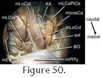
 m. Longissimus Capitis Pars Transversalis Cervicus (mLCaPTCe).
This muscle probably corresponds to the "Longissimus capitis 4" (mLongiC4) of
Al-Hassawi (2004,
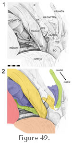
2007, p. 100), which she describes as branching from the m. Longissimus Dorsi near the 2rd vertebra (axis) and inserting on the posterolateral epiphyses of the basal tubera of the basioccipital (Figure 14,
Figure 42,
Figure 43,
Figure 49, and
Figure 50). This corresponds with the descripitions of
Tsuihiji (2007) for the "m Longissimus Capitis Pars Transversalis Cervicus." The cranial portion of this muscle is present on the right side of BMNH 1969.2204 and is triangular in mid-section (Figure 34): the dorsal edge is 4 mm wide whereas the ventrolateral edge and ventromedial edges are both about 6 mm long. m. Longissimus Capitis Pars Transversalis Cervicus (mLCaPTCe).
This muscle probably corresponds to the "Longissimus capitis 4" (mLongiC4) of
Al-Hassawi (2004,
2007, p. 100), which she describes as branching from the m. Longissimus Dorsi near the 2rd vertebra (axis) and inserting on the posterolateral epiphyses of the basal tubera of the basioccipital (Figure 14,
Figure 42,
Figure 43,
Figure 49, and
Figure 50). This corresponds with the descripitions of
Tsuihiji (2007) for the "m Longissimus Capitis Pars Transversalis Cervicus." The cranial portion of this muscle is present on the right side of BMNH 1969.2204 and is triangular in mid-section (Figure 34): the dorsal edge is 4 mm wide whereas the ventrolateral edge and ventromedial edges are both about 6 mm long.
m. Iliocostalis (mIcos).
 The m. Iliocostalis is the lateral column of the epaxial muscles which runs broadly parallel to the vertebral column, on top of the ribs and lateral to the iliac blade (Nishi 1916;
Gasc 1981;
Tsuihiji 2005). The muscle group is innervated by lateral twigs of the lateral branches of the dorsal rami of the spinal nerve (Tsuihiji
2005). The m. Iliocostalis capitis is the part of this muscle that connects the axial skeleton to the head. The m. Iliocostalis is the lateral column of the epaxial muscles which runs broadly parallel to the vertebral column, on top of the ribs and lateral to the iliac blade (Nishi 1916;
Gasc 1981;
Tsuihiji 2005). The muscle group is innervated by lateral twigs of the lateral branches of the dorsal rami of the spinal nerve (Tsuihiji
2005). The m. Iliocostalis capitis is the part of this muscle that connects the axial skeleton to the head.
m. Iliocostalis capitis (mIcosCa). The m. Iliocostalis capitis supposedly originates from the rest of the iliocostalis at about the level of the second and third vertebrae from the fascia separating the m. Iliocostalis and m. Longissimus (Figure
43) (Tsuihiji 2007). It inserts on the basal tubera of the basioccipital anterolateral to the insertions of the m. Longus Capitis Pars Transversalis Cervicus and m. Longus Colli
(Figure 14,
Figure 42,
Figure 43,
Figure 49, and
Figure 50) (Tsuihiji 2007). In BMNH 1969.2204 a muscle that probably represents the cranial half of the m. Iliocostalis capitis is about 2 mm tall by 1 mm wide in section.
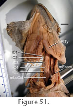
m. Longus Colli (mLoCol)
The m. Longus Colli is the subvertebral layer of the hypaxial musculature. It
arises from a series of slips along the base of the vertebral column that
converge into a single bundle that inserts on the ventral surface of the
braincase (Figure 14,
Figure 22.2,
Figure 42,
Figure 43,
Figure 49,
Figure 50, and
Figure 51) (Osawa
1898; Byerly 1925;
Gasc 1981;
Al-Hassawi 2004,
2007;
Tsuihiji 2007).
Tsuihiji (2007) considers the m. Longus Colli of Sphenodon to include the m. Rectus Capitis of other diapsids. As a result he occasionally refers to the m. Longus Colli as the m. Rectus Capitis (e.g.,
Tsuihiji 2007, figure 4).
 Byerly (1925, p. 18) described the insertion as being on the ventral surface of the axis and occipital condyle. Other authors describe its insertion as taking place on the sphenooccipital tubercle (basal tubercle) of the basisphenoid via a tendon medial to the insertions of the m. Iliocostalis Capitis and the m. Longus Capitis Pars Transversalis (Osawa
1898; Byerly 1925;
Al-Hassawi 2004,
2007;
Tsuihiji 2007). Byerly (1925, p. 18) described the insertion as being on the ventral surface of the axis and occipital condyle. Other authors describe its insertion as taking place on the sphenooccipital tubercle (basal tubercle) of the basisphenoid via a tendon medial to the insertions of the m. Iliocostalis Capitis and the m. Longus Capitis Pars Transversalis (Osawa
1898; Byerly 1925;
Al-Hassawi 2004,
2007;
Tsuihiji 2007).
There is some disagreement with regards to the precise origins of this muscle.
Byerly (1925, p. 17) described it arising from the ventral surfaces of 2nd to 12th vertebrae with an additional slip coming from the rib of the seventh vertebra.
Osawa (1898) also considered some fibres
to originate from the atlas.
Al-Hassawi (2004,
2007) described the posteriormost part of this muscle as originating from the 11th intercentrum (between the 10th and 11th vertebrae) and comprising three subdivisions:
1. A superficial section from intercentrum 8 (equivalent to the 6th keeled intercentrum) between the 7th and 8th vertebrae. It eventually joins the 2nd section.
2. An intermediate section from the flat 10th intercentrum (between the 9th and 10th vertebrae).
3. A third section from the 11th intercentrum (between the 10th and 11th vertebrae) extends laterally to join the other two sections.
Al-Hassawi (2004,
2007, p. 100) also describes two further slips that are probably part of the m. Longus Colli: "first flat intercentral muscle slip to the basioccipital" and "third intercentral muscle slip to the basioccipital."
Examination of BMNH 1969.2204 confirms the presence of a tendon into which the slips converge (Figure 49 and
Figure 50), that the intercentra are an
important site of origin (Al-Hassawi 2004,
2007), and that the most anterior point of origin includes the first intercentrum
(Al-Hassawi 2004,
2007).
Byerly (1925) considered this muscle important for moving the head sideways and keeping it held up, but its placement below the occipital condyle suggests it is better suited for moving the head down.
|