| |
INTRODUCTION
 Sphenodon (tuatara) represents the only living member of the Rhynchocephalia (sensu
Gauthier et al. 1988), a group that was diverse and globally distributed for much of the Mesozoic (Evans et al. 2001;
Jones et al. 2009). Because of its uniqueness, Sphenodon has an iconic status in New Zealand (e.g.,
Acres 1990;
Daugherty and Cree 1990;
Mlot 1997;
Stephens and Lambert 1998;
Baynton 2001;
Parkinson 2002;
Darroch 2005;
Ramstad et al. 2007), and serious efforts are being made for its conservation on the 35 offshore islands it inhabits (e.g.,
Schmidt 1953;
Daugherty et al. 1990;
Mlot 1997;
Gaze 2001;
Nelson et al. 2002;
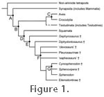
MacAvoy et al. 2007). Phylogenetically, Rhynchocephalia is the sister taxon of Squamata (snakes, lizards, and amphisbaenians), and together both groups make up the larger group Lepidosauria (Figure 1), a monophyletic clade supported by a wealth of morphological and molecular data (Evans 1984,
1988;
Benton 1985;
Schwenk 1986,
1988;
Gauthier et al. 1988;
Rieppel and deBraga 1996;
deBraga and Rieppel 1997;
Zardoya and Meyer 1998,
2001;
Müller 2003;
Rest et al. 2003;
Townsend et al. 2004;
Hill 2005). All lepidosaurs arose from a single common ancestor (independent of all other reptiles sensu
Modesto and Anderson 2004; birds, crocodiles, turtles, and their fossil relatives), approximately 240-250 million years ago (Evans 2003;
Vidal and Hedges 2005; unpublished data). Sphenodon (tuatara) represents the only living member of the Rhynchocephalia (sensu
Gauthier et al. 1988), a group that was diverse and globally distributed for much of the Mesozoic (Evans et al. 2001;
Jones et al. 2009). Because of its uniqueness, Sphenodon has an iconic status in New Zealand (e.g.,
Acres 1990;
Daugherty and Cree 1990;
Mlot 1997;
Stephens and Lambert 1998;
Baynton 2001;
Parkinson 2002;
Darroch 2005;
Ramstad et al. 2007), and serious efforts are being made for its conservation on the 35 offshore islands it inhabits (e.g.,
Schmidt 1953;
Daugherty et al. 1990;
Mlot 1997;
Gaze 2001;
Nelson et al. 2002;
MacAvoy et al. 2007). Phylogenetically, Rhynchocephalia is the sister taxon of Squamata (snakes, lizards, and amphisbaenians), and together both groups make up the larger group Lepidosauria (Figure 1), a monophyletic clade supported by a wealth of morphological and molecular data (Evans 1984,
1988;
Benton 1985;
Schwenk 1986,
1988;
Gauthier et al. 1988;
Rieppel and deBraga 1996;
deBraga and Rieppel 1997;
Zardoya and Meyer 1998,
2001;
Müller 2003;
Rest et al. 2003;
Townsend et al. 2004;
Hill 2005). All lepidosaurs arose from a single common ancestor (independent of all other reptiles sensu
Modesto and Anderson 2004; birds, crocodiles, turtles, and their fossil relatives), approximately 240-250 million years ago (Evans 2003;
Vidal and Hedges 2005; unpublished data).
Sphenodon has long been of interest to anatomists because many aspects of its anatomy, including its muscles, were thought to demonstrate the ancestral condition for amniotes and/or diapsid reptiles (e.g.,
Byerly 1925;
von Wettstein 1931,
1932,
1937;
Anderson 1936;
Sharell 1966;
Barghusen 1973). Accordingly, it has been used in attempts to reconstruct the muscle arrangements of several fossil amniotes including the phytosaur Machaeroprosopus (Anderson
1936) and the early synapsid Dimetrodon (Barghusen 1973). However, as
Schwenk (1986, p. 148) has argued, the primitive nature of Sphenodon has previously been exaggerated. The absence of a tympanic membrane and distinctive physiology are probably both secondary features (Gans 1983;
Whiteside 1986;
Thompson and Daugherty 1992), and osteologically Sphenodon is certainly different from its well-known Mesozoic fossil relatives (e.g.,
Reynoso 2000;
Apesteguía and Novas 2003;
Jones 2008). Nevertheless, the phylogenetic position of Sphenodon as the only extant member of Rhynchocephalia means it is a potentially useful reference taxon for inferring soft tissue arrangement and structure in extinct animals, particularly when used in a phylogenetic bracket (e.g.,
Bryant and Russell 1992;
Witmer 1995,
1997). It is also useful for evaluating the homology of muscles in extant taxa (e.g.,
Schwenk 1986;
Holliday and Witmer 2007). Furthermore, understanding the muscle architecture in Sphenodon may provide clues as to why its close fossil relatives (other rhynchocephlians) demonstrate a variety of skull shapes that coincide with variation in tooth shape and tooth arrangement (Jones 2008).
The complete lower temporal bar means that Sphenodon is structurally analogous to the supposed ancestral condition (Petralacosaurus,
Reisz 1977,
1981) of all diapsid reptiles (lepidosaurs, crocodiles, birds, etc.). Crocodiles and birds also demonstrate the diapsid condition but both are problematic as model organisms as crocodiles possess elongate rostra whereas birds have relatively large braincases and relatively small adductor chambers. It should be stressed, however, the lower temporal bar was absent in basal rhynchocephalians, such as Gephyrosaurus (Evans 1980), and has therefore been secondarily acquired in Sphenodon as repeatedly shown elsewhere (e.g.,
Whiteside 1983,
1986;
Fraser 1988;
Reynoso 2000;
Evans 2003;
Apesteguía and Novas 2003;
Müller 2003;
Wu 2003;
Jones 2008). It is probably a structural feature for supporting the quadrate from joint reaction forces during biting and shearing (Whiteside 1983,
1986;
Fraser 1988;
Rieppel 1992;
Wu 2003;
Jones 2006a,
2008).
The arrangement of jaw and neck muscles dictates how an animal feeds and also how the skull is stressed during feeding. These, in turn, are likely to have a direct effect in forming the shape of the skull during growth (e.g.,
Gregory and Adams 1915;
Adams 1919;
Case 1924;
Olson 1961;
Frazzetta 1968;
Schumacher 1973a;
Oxnard et al. 1995;
Hunt 1998;
Preuschoft and Witzel 2002;
Witzel and Preuschoft 2005;
Jones 2008). The muscle arrangement in Sphenodon is also of particular interest because it reflects a unique feeding mechanism among living organisms (Reilly et al. 2001). Following jaw closure the lower jaw moves forward (prorally) and shears food gripped by the teeth (Farlow 1975;
Robinson 1976;
Gorniak et al. 1982), allowing Sphenodon to deal with prey larger than its gape (Robinson 1973;
Gorniak et al. 1982). Food items are also subjected to three-point bending because there is a row of teeth located on the lateral edge of the palatine bone parallel to the maxillary dentition (Evans 1980;
Jones 2006a,
2007).
Many of the jaw muscles in Sphenodon have been described repeatedly (Byerly 1925;
Lakjer 1926;
Edgeworth 1935;
Anderson 1936;
Poglayen-Neuwall 1953;
Ostrom 1962;
Barghusen 1973;
Haas 1973;
Gorniak et al. 1982;
Wu 2003;
Holliday and Witmer 2007) but the accounts differ, and images are largely limited to lateral views of the skull. Descriptions of the tongue and associated throat muscles have also been made by several authors but are less common and in general are less detailed (e.g.,
Günther 1867;
Lightroller 1939;
Rieppel 1978;
Schwenk 1986). Similarly the neck and pectoral muscles have arguably received less attention than the jaw muscles (Maurer 1896;
Nishi 1916;
Byerly 1925;
von Wettstein 1931;
Gasc 1981;
Al-Hassawi 2004,
2007;
Tsuihiji 2005,
2007). The osteology of Sphenodon has been described by many authors (e.g.,
Günther 1867;
Siebenrock 1893,
1894;
Werner 1962;
Hoffstetter and Gasc 1969;
Rieppel 1992;
Evans 2008;
Jones 2008).
The neck is widely recognised as an important part of an animal's feeding apparatus (e.g.,
Van Damme and Aerts 1997;
Summers et al. 1998;
Stevens and Parrish 1999;
Upchurch and Barrett 2000;
Rayfield et al. 2001;
Anton et al. 2003;
McHenry et al. 2007;
Snively
and Russell 2007a;
2007b). During feeding the positional relationship between the head and neck can change, as can be seen in tiger salamanders (Ambystoma tigrinum,
Larsen and Guthrie 1975) and the Eastern box turtle (Terrapene carolina,
Summers et al. 1998). This will probably affect the magnitude and distribution of strain and stress on the posterior regions of the skull. Most functional studies of skulls do not take into account the neck musculature, and when they do descriptions of its anatomy can be vague (e.g.,
McHenry et al. 2007). The tongue is also important in feeding. It is used to manipulate food items in the mouth during processing (Walls 1981;
Gorniak et al. 1982;
Schwenk 2000; MEHJ pers. obs.), and several studies have observed that the tongue is also employed in pulling small prey into the mouth (e.g.,
Buller 1879;
Gorniak et al. 1982;
Schwenk 2000; pers. obs.), although
Walls (1981, p. 91) did not observe this in the wild population he studied.
Sphenodon is carnivorous and opportunistic, feeding on a wide variety of arthropods, molluscs, and vertebrate material such as lizards, sea birds, and eggs (e.g.,
Günther 1867;
Buller 1877,
1878;
Reischek 1885;
Dawbin 1949,
1962,
1982;
Schmidt 1953;
Farlow 1975;
Walls 1981,
1982;
Ussher 1999;
Schwenk 2000). Examination of faecal pellets from Stephens Island (Walls 1981,
1982;
Newman 1987) demonstrated that relatively slow terrestrial arthropods, such as the large darkling beetle (Mimopeus opaculus), were the most frequent prey items. Rare food items include hatchling Sphenodon, frogs, passerine birds and, on Green Island, the remains of crabs (Walls 1981;
Newman 1987;
Daugherty and Cree 1990;
Blair et al. 2000;
Moore and Godfrey 2006). Sea bird material is important (but not essential) to the larger stronger males on Stephens Island (Walls 1978,
1981;
Cree et al. 1995a,
1999;
Markwell 1998;
Blair et al. 2000;
Gaze 2001),
particularly during spring and summer (Walls 1981). Females and juveniles may also consume more limited amounts of sea bird material but probably as carrion (Cree et al. 1995a).
Plant material is frequently present in Sphenodon faecal pellets (Walls 1981), and certain types of seeds have been found in almost 10% of scats examined as part of one study (I. C. Southey pers. comm. in
Whittaker 1987). Although consumption of plant material may be accidental, it can represent 14% of the total number of items present in faeces (Walls 1981). This places Sphenodon within the generous omnivorous category of
Cooper and Vitt (2002) (diet = >10% plant material). Many of the invertebrates and vertebrates that Sphenodon preys upon are also consumed by the Pacific rats (kiore, Rattus exulans). As a result, for populations of Sphenodon located on islands inhabited with Pacific rats, there is competition for food (Cree et al. 1995b;
Blair et al. 2000).
Adult Sphenodon mainly hunt during the evenings (Walls 1981;
Gans 1983;
Daugherty and Cree 1990) because of their ability to be active in cool temperatures (Thompson and Daugherty 1998). They also possess large eyes that are sensitive to low light levels (Meyer-Rochow et al. 2005). Predation is mainly visual, and in most cases is triggered by movement (e.g.,
Buller 1879;
Farlow 1975;
Walls 1981;
Meyer-Rochow 1988;
Meyer-Rochow and Teh 1991;
Gorniak et al. 1982;
Schwenk 2000) but taste buds are present on the tongue (Schwenk 1986) and consumption of eggs and carrion suggests that smell can also be involved (Walls 1981; see also
Cooper et al. 2001). In terms of feeding strategy (sensu
Pianka 1966;
Huey and Pianka 1981), where "sit and wait" involves ambushing prey from a sedentary position and "widely foraging" involves actively hunting prey, Sphenodon is widely considered to follow a "sit and wait" strategy (e.g.,
McBrayer and Reilly 2002;
Vitt et al. 2003). Nevertheless,
Gans (1983) reported that Sphenodon may also inspect crevices for prey. Young tuatara are more active during the day, possibly to avoid predation from adults (Daugherty and Cree 1990), and this may influence the prey available to them. There is some evidence that they feed on smaller prey items (Ussher 1999, p.123).
The relationship between phenotype and diet remains poorly understood in lepidosaurs, leading some authors to question whether any predicatable relationship exists at all (e.g.,
Schwenk 2000). Broadly comparative work (e.g.,
Metzger and Herrel 2005) is required in order to examine this relationship. It should be remembered, as in other lepidosaurs (e.g.,
Lappin and Husak 2005), Sphenodon also uses its jaws, teeth, and associated muscles in conspecific fighting for burrows, mates, and nesting sites (e.g.,
Newman 1987;
Daugherty and Cree 1990;
Gans et al. 1984;
Gillingham et al. 1995;
Nelson et al. 2004). Computer models as used by
Moazen et al. (2008,
2009) can also provide biomechanical assessment of the hard and soft tissue structures involved and allow specific questions to be tested regarding the function of different anatomical components.
Here we provide a thorough review of Sphenodon osteology and muscle anatomy as relevant to feeding. We evaluate previous discrepancies and, for the first time, provide colour
photographs and three-dimensional imaging to explain complex muscle
arrangements. Our review will provide a basis for future work on muscular biomechanics during feeding.
|