

 Methodology of the micro-computer tomography on foraminifera
Article Number: 15.1.3T
https://doi.org/10.26879/261
Copyright Palaeontological Association, February 2012
Author biographies
Plain-language and multilingual abstracts
PDF version
Submission: 14 December 2010. Acceptance: 14 November 2011
{flike id=121}
ABSTRACT
Research focused on the methodology of the micro-CT device in the foraminifera studies reviewing its advantages and limits. First, the procedure of stable, oriented and removable fixing of foraminifera, washing residue and cuttings in the device was developed, which is fast, cheap, non-destructive and the sample remains re-usable for further investigation. Moreover, a process was developed for foraminifera investigation (e.g., content, amount, identification, description, illustration) in the shortest and simplest way. In the identification of foraminifera as well as in other microfossils group (e.g., radiolaria) the external and internal characteristics together identified the genera, even the species. Additionally, there are several foraminifera species, which are known only from the rock thin sections. The micro-CT was found to be a unique tool for real 3D microscopy and to be able to present the characteristic morphological features when the free movement, rotation or sectioning of the models of the specimens can be performed by interpretation software. However, the biggest advantage of this method is its non-destructive property, which makes it also capable of measuring valuable materials such as holotypes. The relatively small volume analysed and the lack of specific software developed for different micropalaeontological groups are the limits of this method. This visualization technique gives new perspectives in taxonomical studies and in applied micropalaeontology.
Ágnes Görög. Department of Palaeontology, Eötvös University, Pázmány Péter sétány 1/C, Budapest, H-1117, Hungary. gorog@ludens.elte.hu
Balázs Szinger. Department of Palaeontology, Eötvös University, Pázmány Péter sétány 1/C, Budapest, H-1117, Hungary. szinger.balazs@gmail.com
Emőke Tóth. Research Group for Palaeontology, Hungarian Academy of Sciences-Hungarian Natural History Museum - Department of Palaeontology, Eötvös University, Pázmány Péter sétány 1/C, Budapest, H-1117, Hungary. cypridina1981@yahoo.com
János Viszkok. Central Geo Ltd, Mária út 10, Szolnok, H-5000, Hungary. jviszkok@centralgeo.hu
KEYWORDS: micro-CT; micropalaeontology; 2D and 3D visualization; foraminifera; cuttings
FULL CITATION: Görög, Ágnes, Szinger, Balázs, Tóth, Emőke, and Viszkok, János. 2012. Methodology of the micro-computer tomography on foraminifera. Palaeontologia Electronica Vol. 15, Issue 1; 3T, 15p;
palaeo-electronica.org/content/issue-1-2012-technical-articles/121-methodology-of-ct-on-forams
INTRODUCTION
Identification of foraminifera is based on the identification of external as well as internal morphological characteristics. Since Schmidt (1952) and Hay and Sandberg (1967) used an X-ray device and scanning electron microscope (SEM) in micropalaeontological study for the first time, no new methods or technological innovations for observation and illustration of microfossils have been introduced. The scanning electron microscope images provide information only about the external morphology. With an SEM device, further study of the specimens is limited due to the sample preparation, normally due to the gluing on and coating with Au/C. It is also very difficult to get an acceptable quality of the SEM. For the observation of internal morphological features, the conventional method used is the transmitted light microscopic investigation of thin sections of rocks or isolated specimens. There are several disadvantages of this method: it destroys the sample, is time consuming, and demands extensive technical and professional skills.
Study of the internal structure is essential in several micropalaeontological studies:
1. when the extraction of the microfossils from the rock is impossible,
2. for the systematic classification of some groups, e.g., larger foraminifera, radiolaria (entactinarians), Dasycladacea and Rhodophycea, and
3. for ontogenetical studies e.g., the size of the proloculus and the shape of the arrangement of the juvenile chambers of the planktonic and larger foraminifera.
To circumvent the limits of existing techniques, we tested a new method, which is non-destructive and simultaneously allows the study of internal and external morphologies of foraminifera. Theoretically, an appropriate method for these purposes is the Computer Tomograph, which is used worldwide in medical applications. As a result of the technological advances of this device, the micro-Computer Tomograph (micro-CT) was developed at the end of the 1980s. During a micro-CT measurement – similarly to the conventional CT – the sample is irradiated with X-rays, which can provide section images in any spatial direction. From these images it is possible to create 3D models having 1–10 µm spatial resolution, which is the most suitable for the study of microfossils.
The micro-CT has been successfully used in petrography and in some cases in vertebrate palaeontology (e.g., Mees et al., 2003; Rossi et al., 2004; Burrow et al., 2005; Claessens et al., 2009). In fact, the micro-CT can prove its applicability in invertebrate palaeontology, as well as ichnology. It was tested, for example, on brachiopods, echinoderms (Stock et al., 2003a, 2003b), ammonites, insects (e.g., Dierick et al., 2007; Heethoff et al., 2009; Penney et al., 2007; Pakhnevich and Bryantseva, 2007), mollusc shells (Monnet et al., 2009) and microborings (e.g., Tapanila, 2008; Schönberg and Shields, 2008). There are only a few short papers on surface and volumetric quantifications of several foraminifera specimens (e.g., Speijer et al., 2008; Briguglio, 2010). Following our research since 2007 (Szinger et al., 2007), the aim of this work is to develop a general methodology for the use of the micro-CT in foraminifera taxonomy as well as in applied micropalaeontology (e.g., core and cuttings) reflecting both the advantages and the limits.
MATERIAL AND METHODS
In our study isolated specimens of foraminifera, rock fragments, washing residues of the rock, cuttings with mud, washed cuttings and resin-fixed cuttings were measured. Foraminifera are a suitable group for testing the micro-computed tomography. Their average size (50–300 µm), diversity of their external morphology (chamber shape and arrangement), of the internal structure (e.g., septa, beams, rafters, canal and cavity systems), of the material (calcite, aragonite or arenaceous) and of the texture (e.g., perforated, non-perforated, agglutinated, one or more layered) of their skeleton made them promising subjects for these preliminary studies. The specimens originated from different ages and have different states of preservation (empty or filled with matrix of the rock; with original wall texture or with a recrystallised one).
 The studies were performed on a SkyScan-1172 system, which is a high-resolution desktop X-ray microfocus computerized tomography system, and consists of an X-ray microfocus tube (20–100kV, 10W, <5µm spot size, target material of the tube: tungsten), a rotating specimen stage and a two-dimensional X-ray detector. The detector comprises a scintillator screen (gadolinium oxysulphide phosphor, dimension: 25 x 25 mm surface and 25 µm thickness), fibre optics and a CCD sensor (1.3 Mp cooled CCD, 12 bit gray levels). The maximum object size is 20 mm in diameter for a standard scan, and the low-contrast resolution is 5 µm.
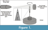
The micro-focus X-ray source illuminates the sample, and the X-rays interact with the matter and attenuate. This attenuation is dependent on the density and atomic number of the object radiated and on the X-ray energies used (Figure 1). The planer X-ray detector collects the magnified projection images. Hundreds of angular shadow images are acquired while the object rotates, thereafter, a computer program synthesizes these images into virtual slices through the X-ray absorbent object. The reconstruction program uses a modified Feldkamp algorithm allowing the simultaneous collection of data throughout the full 3D volume of small specimens. 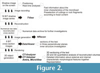 The reconstruction includes alignment optimization, beam-hardening and ring artifact corrections. A detailed description of the basic principle of micro-CT, data acquisition and image reconstruction can be found in the literature (e.g., SkyScan 2005).
The numerical and visual rendering of the acquired datasets were performed and tested in steps by using different commercially available software (TView, NRecon, DataViewer, DICOM-CT, CTAn-CTVol, Osiris4, MVE, MicroView, Amira) (Figure 2).
During the rendering, the setting of the filtering (e.g., density thresholds, ROI, gray scale) was adjusted to obtain the best images.
As a result of software rendering, 2D and 3D images, moreover 3D animations of foraminifera can be generated, or cross-sections can be cut in any directions.
RESULTS OF WORKFLOW OPTIMIZATION
Preparation and Measurement Techniques
During the measuring process, the rock fragments were fixed in a conventional way using X-ray transparent plasticine.
 However, the majority of the micropalaeontological samples are small-sized (up to 300 µm) or consist of small pieces like washings residue and cuttings. We did not find any published method as to how to place such samples in a suitable position in the rotation-axis of the micro-CT holder, to fix them stably for hours of measurements and to remove them without destruction. Another practical question was how to measure the largest volume at the best (or the most suitable) resolution during the shortest time with the simplest preparation for the isolated specimens, for the rock sample or for the cuttings.
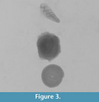
Based on our experiments, a strip (about 0.5 x 2.5 cm) of X-ray celluloid film was found as the most suitable for holding the isolated microfossils and an organic water-soluble glue (gomme adragante applied by wet ultrathin paintbrush) for fixing them. Depending on the required resolution and the size of the samples, three to four specimens can simultaneously be measured. Since the micro-CT device is suitable for serial measurements, by applying the same settings of the measuring parameters, up to 10 to 12 specimens can be fixed on the celluloid film and scanned without any human intervention (Figure 3). Using this preparation technique one isolated specimen requires only about half an hour of measuring time.
For the study of larger amounts of material, like rock fragments and cuttings, a volume of 1–2 cm3 with a cylindrical shape is ideal. To hold the cuttings and to minimize the X-ray attenuation, a thin-walled plastic tube (approximately about d=5 mm and h=30 mm) closed with plasticine is found to be the best solution. At the measuring of cutting, the drilling mud has no disturbing influence on the quality of the shadow image, although it decreases the substantial volume. To avoid the movement of smaller fragments during the rotation of the holder, the cuttings were strongly compacted. A two-component resin was used for fixing, too, but the glue required about 12 hours to bond. Depending on the volume, type of sample and the mode of investigation, the time-span of a measurement is 1–15 hours, the average being 4 hours.
Visualization
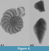 The high-resolution X-ray shadow image can be seen on the monitor of the micro-CT device, which turns around with the adjusted angles in real time, and this series of images can be captured and saved in TIFF format. The screen images are suitable for fast analyses of isolated foraminifera as well as rock fragments or cuttings. The resolution depends on the volume, but for the isolated microfossils, we could get high-quality images. The chamber arrangement, which is essential for the identification of foraminifers in several cases, (e.g., arenaceous forms), is invisible when using a traditional light or a scanning electron microscope. This feature, however, can easily be studied with micro-CT, especially when a convenient orientation is used (Figure 4). Due to the easily removable gluing, the observation of the same specimen in different views is also possible.
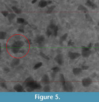 Another advantage of the method is the ability for the preliminary investigation of small quantities (~1–2 cm3) of rock fragments or of cuttings without any preparation. Due to the ability to measure very quickly, these preliminary results can help to decide the further treatment or investigation of the material. Information can easily be acquired on quantity and quality of the microfossil, along with the dimensions, volume and shape of the grains, referring also the sedimentological features. However, in these cases the spatial resolution is not good enough for the exact identification. With the help of washing the valuable sample volume can be increased. This way, micro-CT is an ideal tool for preselection (Figure 5).
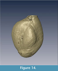
Opportunities for the volumetric rendering using micro-CT are illustrated with case studies on isolated microfossils and cuttings.
First, we chose a group of foraminifera, the miliolids (studied genera are Triloculina and Spiroloculina) where both the external and internal characteristics are required for the identification of species, and even the genera. Traditionally, these characteristics could be observed with two different processes: using a light microscope on isolated specimens on the one hand and on oriented thin sections on the other. The latter preparation technique is delicate, time consuming and destructive for the sample.
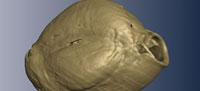
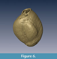
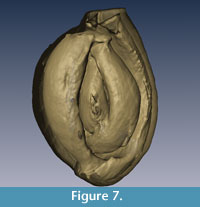
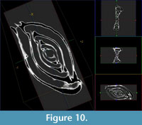
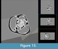
Miliolids have an imperforate calcite wall and their main diagnostic characteristics on the generic level are the chamber arrangement and the shape and restricted (or auxiliary) elements (e.g., tooth) of the aperture (Łuczkowska, 1972; 1974; Loeblich and Tappan, 1988). There are two diagnostic sections of the miliolids, one which penetrates the embryonic chamber (it is in the innermost part of the test) in the coiling axis, across the aperture and the other is perpendicular to it (Figure 6, Figure 7, Figure 8, Figure 9, Figure 10, Figure 11, Figure 12, Figure 13, Figure 14, Figure 15, Figure 16, Figure 17, and Figure 18).
 |
 |
 |
 |
 |
 |
 |
 |
 |
 |
 |
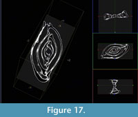 |
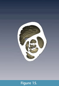
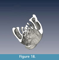 The micro-CT provides an easy option to fix the coiling axis and adds the position of the virtual section, thus the characteristic sections can easily be produced. For example, in Figure 8 applying angle metering or any graphic program it is easy to measure that by each chamber on the last whorl the coiling plane rotates by 120°, which is the triloculine chamber arrangement. It is possible to make the last, outermost chambers translucent. In this way, the inner chambers, and hence the early quinqueloculine stage, can be observed (Figure 7).
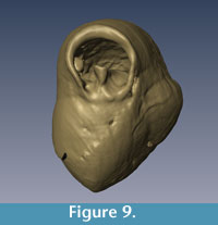
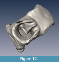
 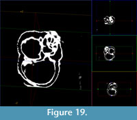 Another diagnostic characteristic, the tooth, can also be studied by using the 3D rendering programs (Figure 9 and Figure 12). Measuring the size or the volume of the proloculus and the first chambers can give information about the ontogeny or distinguish the microsphaeric and macrosphaeric generations.
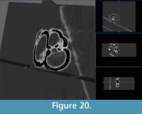 In the case of planktonic foraminifers (studied specimens are Globigerina sp.), the small size and the fragility of the skeleton make it very difficult to produce thin sections from isolated specimens. This problem could also be solved by using micro-CT (Figure 19 and Figure 20).
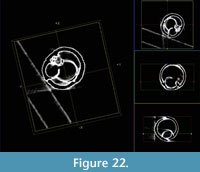
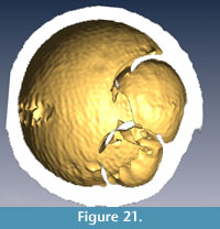 The sectioning is especially important in some unique planktonic forms like orbulinids (Figure 21 and Figure 22) having the last large, spherical chamber, which covers all the former diagnostic ones. It is a time-consuming and very delicate task to remove mechanically the wall of the last chamber under microscope. This work can be avoided applying micro-CT.
 For the taxonomical study of trochospiral foraminifera, three views (spiral, umbilical and side) are necessary. Due to the small-sized planktonic foraminifers, the diagnostic characteristics (e.g., the orientation of the coiling axis, the position and the shape of the aperture, and the surface ornamentation) are usually difficult to observe under the light microscope. It is also problematic to fix the small-sized (50-150µ) benthic specimens in the suitable position for the SEM pictures.
 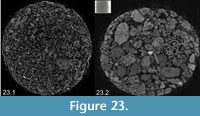 Even if successful, it is also questionable whether the three specimens, from which the three views are obtained, belong to the same species. With the help of volumetric rendering, these characteristic views of the same specimen can be better studied.
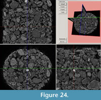 In the investigation of cuttings, washing residues and rock fragments, the visualisation technique allows the making of virtual sections in any direction, similarly to the isolated forms as discussed above. These sections provide useful information about the bio- and lithoclasts composition of the studied material, as demonstrated in the washing residue of selected Carpathian (Lower Miocene) and Sarmatian (Middle Miocene) beds (Figure 23 and Figure 24).
DISCUSSION
Based on our research, we could not find any technical drawbacks for the application of the micro-CT in micropalaeontology. Its advantages and limitations are summarized in the following paragraphs.
Advantages
The sample preparation proposed in this study is fast, cheap, non-destructive and re-usable for further investigation.
One of the main advantages of this visualization technique in the classical micropalaeontological studies is to save the valuable and irreplaceable specimens from destruction during preparation or measurement. Thus, it is possible to study the structural details of precious type specimens/holotypes.
According to the aim of the foraminifera investigation (e.g., content, amount, identification, description, illustration) the process can be stopped at the required point of the workflow (Figure 2). Dimensions and volume calculations are the basic function of all programs which we used.
In real time the investigation of the samples (position, identification of Region of Interest (ROI), along with the quality of the final results) can be checked, the first analyses of the microfossils can be done on the monitor of the device.
In a short time, high-resolution 2D X-ray shadow images could give important information about the inner structure of the microfossils and the sedimentological characteristics of the rocks and cuttings due to the short and easy preparation procedures and the high speed of measurement.
Even the less advanced software packages make it possible to cut the investigated specimen in three perpendicular planes at the beginning of the workflow. Since both the cutting points and planes can be optionally positioned, it is easy to get any desired section of the foraminifera.
An advanced way to present the characteristic morphological features is the 3D reconstruction. Free movement, rotation, zooming, cutting-off or sectioning of the models of the specimens can be performed on the rendered object. This method obviously makes it possible to receive more detailed information on both the external and internal characteristics of the test. It can facilitate the taxonomical studies (identification and classification of the taxa) of some microfossil groups because it is possible to make 3D rendering and sectional images of the skeleton in any direction with the help of different software. By obtaining these spatial slices, the external and internal morphological features could be observed together. In addition, as a proof for the high quality of these rendered 3D models, it is possible to measure any quantitative parameters of the skeleton with high precision. Moreover, this method allows the 3D reconstruction of the species described only from thin sections and unknown as isolated form. Vice versa, to help the identification from thin sections, a rendered microfossil section atlas would be suitable for both scientific and educational purposes. The technique facilitates the ontogenetic studies and thus it helps in the investigation of phylogenetic relationships.
In applied micropalaeontology, the X-ray images of the cuttings permit the fast pre-selection of the faunal bearing samples for further investigation. A portable micro-CT device would be suitable for the continuous observation of the cuttings during well drilling. The results of the measurements can be stored in digital format, which are easy to handle with computers and can be sent over the Internet to a specialist anywhere in the world.
The measured numerical data can be processed using different software packages to get the best results or they can be re-processed as new software appears on the market. It is possible to transform the data into different formats (images, movies) for publication or presentation.
Limitations
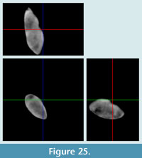 Since the image construction is based on the scanning of areas with different average density and atomic number (Z), if the carbonate skeleton is recrystallised and filled with sparrycalcite, it is not suited to micro-CT investigation (Figure 25).
Depending on the resolution, it is difficult to measure very small-sized (<50 µm) foraminifers with some micro-CT devices. In this case, to achieve the required resolution, it is necessary to reduce the distance between the detector and the sample. This modification can cause contact between the detector and the sample holder, such that the rotation of the sample is hindered.
Because of the attenuation of the different frequency X-ray waves, smaller samples yield better resolution. Smaller samples yield better resolution, which is why the measurement of rocks, cuttings or core samples with larger volume can provide only indicative information about the amount of microfossils.
The resolution of the 3D reconstruction is not enough for the study of the surface ultra-structure.
Historically, the CT devices and visualization software were developed for medical uses and their related specific requirements. Similar development of software or modules (e.g., settings of the filters for the material of different skeletons) would be helpful to assist its widespread application in micropalaeontology.
CONCLUSIONS
Our study demonstrates the usability of micro-CT in micropalaeontology on isolated foraminifera tests as well as on the microfossil content of small-sized rock fragments and cuttings. First, a method of stable, oriented and removable fixing of the microfossils and cuttings in the device were developed.
A workflow is proposed for foraminiferologists as well as other micropalaeontologists, which can be finished at any step in the procedure when the aim of the study is reached.
The high-resolution X-ray image series is well suited to the study of cuttings, and for the pre-selection of samples for further micropalaeontological investigations. Additionally, these types of images show more deterministic characteristics about foraminifera than those from the light microscope and even those from SEM pictures (e.g., at the specimens with invisible suture because of the smooth or roughly agglutinated surface).
Compared to conventional methods, the volumetric rendering offers opportunities to make any number of arbitrary oriented sections from isolated, as well as, rock-embedded microfossils without destruction. Furthermore, only this method is capable of creating 3D models of the latter kind of sample, also showing the ornamentation of the skeleton surface, which is essential for species identification.
Our results proved that the application of micro-CT device in palaeontological research can replace some of those investigations which require difficult professional aptitudes (skill, routine and time).
This visualization technique gives new perspectives in taxonomical studies of unique specimens, species bearing characteristic internal structure or which are known only from thin sections. In addition, it increases the efficiency of the cuttings studies in drilling geology.
ACKNOWLEDGMENTS
The authors would like to sincerely thank M. Kovács and Cs. Dobó Nagy (Semmelweis University, Budapest) for their help in using the micro-CT device, T. Földes (Tomo Geo Kft., Szolnok) for the consultations on CT technology and to J. Pálfy (MTA-ELTE, Budapest) and R. Wernli (University of Geneva) for their helpful advices. Thanks to R. Pitt and J.-M. Jaquet, who greatly improved the English. J. Fábián-Lukács (Praxis pro Aqua, INNO-5-2006-0022, OMFB-0147/2006) OTKA (68791) and Hantken Foundation are acknowledged for the financial support.
E. Tóth is thankful for the contribution of HAS-HNHM Research Group for Paleontology (Paleo-contribution No. 147.).
REFERENCES
Briguglio, A. 2010. Hydrodynamic behaviour of nummulitids. Unpublished PhD Thesis, Universität Wien, Austria. othes.univie.ac.at/12131/1/2010-11-08_0749105.pdf
Burrow, C.J., Jones, A.S., and Young, G.C. 2005. X-ray microtomography of 410 million-year-old optic capsules from placoderm fishes. Micron, 36:551-557.
Claessens, L.P.A.M., O'Connor, P.M., and Unwin, D.M. 2009. Respiratory evolution facilitated the origin of pterosaur flight and aerial gigantism. PLoS ONE, 4(2):e4497. doi:10.1371/journal.pone.0004497.
Dierick, M., Cnudde, V., Masschaele, B., Vlassenbroeck, J., Van Hoorebeke, L., and Jacobs, P. 2007. Micro-CT of fossils preserved in amber. Nuclear Instruments and Methods in Physics Research Section A: Accelerators, Spectrometers, Detectors and Associated Equipment, Proceedings of the 10 th International Symposium on Radiation Physics - ISRP 10, 580(1):641-643.
d'Orbigny, A. 1839. Foraminifères, p. 1-224. In de la Sagra, R. (ed.), Histoire physique, politique et naturelle de l'île de Cuba. Arthus Bertrand, Paris, 8.
Hay, W.W. and Sandberg, P.A. 1967. The scanning electron microscope, a major break-through for micropaleontology. Micropaleontology, 13:407-418.
Heethoff, M., Helfen, L., and Norton, R.A. 2009. Description of Neoliodes dominicus n. sp. (Acari, Oribatida) from Dominican Amber, aided by synchrotron x-ray microtomography. Journal of Paleontology, 83:153-159.
Loeblich, A.R. and Tappan, H. 1988. Foraminiferal Genera and Their Classification. Van Nostrand Reinhold, New York, 2 vols.
Łuczkowska, E. 1972. Miliolidae (Foraminiferida) from Miocene of Poland, part I. Revision of the classification. Acta Palaeontologica Polonica, 17:341-377.
Łuczkowska, E. 1974. Miliolidae (Foraminiferida) from Miocene of Poland, part II. Biostratigraphy, Palaeoecolgy and Systematics. Acta Palaeontologica Polonica, 19:1-176.
Mees, F., Swennen, R., Van Geet, M., and Jacobs, P. 2003. Applications of x-ray computed tomography in the geosciences. Geological Society, London, Special Publication 215.
Monnet, C., Zollikofer, C., Bucher, H., and Goudemand, N. 2009. Three dimensional morphometric ontogeny of mollusc shells by micro-computed tomography and geometric analysis. Palaeontologia Electronica, 12(3), 12A:1-13.
Paalzow, R. 1917. Beiträge zur Kenntnis der Foraminiferen fauna der Schwammergel des Unteren Weissen Jura in Süddeutschland. Abhandlungen der Naturhistorischen Gesellschaft zu Nürnberg, 17:201-249.
Pakhnevich, A.V. and Bryantseva, A. 2007. Studying of fossil invertebrates for example brachiopods by using of the X-ray micro-CT SkyScan 1172. SkyScan User Meeting 2007, Abstract, www.skyscan.be/company/usersmeeting2007.htm (2010-03-21)
Penney, D., Dierick, M., Cnudde, V., Masschaele, B., Vlassenbroeck, J., Van Hoorebeke, L., and Jacobs, P. 2007. First fossil Micropholcommatidae (Araneae), imaged in Eocene Paris amber using x-ray computed tomography. Zootaxa, 1623:47-53.
Reuss, A.E. 1844. Geognostische Skizzen aus Böhmen. II. Theil- Die Kreidegebilde des westlichen Böhmens. C.W. Medau & Comp, Prag.
Reuss, A.E. 1850. Neue Foraminiferen aus den Schichten des Österreichischen Tertiärbeckens. Denkschriften der Akademie der Wissenschaften, Wien, Mathematisch-Naturwissenschaftliche Klasse, 1:365-390.
Rossi, M., Casali, F., Romani, D., Bondioli, L., Macchiarelli, R., and Rook, L. 2004. MicroCT Scan in paleobiology: application to the study of dental tissues. Nuclear Instruments and Methods in Physics Research B, 213:747-750.
Schönberg, C.H.L. and Shields, G. 2008. Micro-computed tomography for studies on Entobia: transparent substrate versus modern technology, p.147-164. In Wisshak, M. and Tapanila, L. (eds.), Current Developments in Bioerosion. Erlangen Earth Conference Series, Springer-Verlag, Berlin, Heidelberg.
Schmidt, R.A.M. 1952. Microradiography of microfossils with X-ray diffraction equipment, Science, 115 (1952):94-95.
SkyScan 2005. SkyScan 1172. Desktop X-ray Microtomograph. Instruction Manual. www.itg.uiuc.edu/help/microct/1172_Manual.pdf (2010-03-21)
Speijer, R.P., Van Loo, D., Masschaele, B., Vlassenbroeck, J., Cnudde, V., and Jacobs P. 2008. Quantifying foraminiferal growth with high-resolution x-ray computed tomography: New opportunities in foraminiferal ontogeny, phylogeny, and paleoceanographic applications. Geosphere; 4:760-763.
Stock, S.R., Ignatiev, K.I., Dahl, T., Veis, A., and De Carlo, F. 2003a. Three-dimensional microarchitecture of the plates (primary, secondary, and carinar process) in the developing tooth of Lytechinus variegatus revealed by synchrotron x-ray absorption microtomography (microCT). Journal of Structural Biology, 144:282-300.
Stock, S.R., Nagaraja, S., Barss, J., Dahl, T., and Veis, A. 2003b. X-ray microCT study of pyramids of the sea urchin Lytechinus variegatus. Journal of Structural Biology, 141:9-21.
www.sciencedirect.com/science/journal/10478477
Strickland, H. E. 1846. On two species of microscopic shells found in the Lias. Quarterly Journal of the Geological Society, 2:30-31.
Szinger B., Görög Á., Tóth, E., and Viszkok, J. 2007. Mikro-CT alkalmazása, mint új lehetőség az őslénytani kutatásban, p. 34-35. In Pálfy, J., Bosnakoff, M., and Pazonyi, P. (eds.), 10. Program, előadáskivonatok, kirándulásvezető. Magyar Õslénytani Vándorgyũlés, 2007. május 24-26. Budapest (In Hungarian)
Tapanila, L. 2008. The medium is the message: imaging a complex microboring (Pyrodendrina cupra igen. n., isp. n.) from the early Paleozoic of Anticosti Island, Canada, p. 123-145. In Wisshak, M. and Tapanila, L. (eds.), Current Developments in Bioerosion. Erlangen Earth Conference Series, Springer-Verlag, Berlin, Heidelberg.
von Fichtel, L. and von Moll, J.P.C. 1798. Testacea microscopica, aliaque minuta ex generibus Argonauta et Nautilus, ad naturam picta et descripta (Mikroskopische und andere kleine Schalthiere aus den Geschlechtern Argonaute und Schiffer nach der Natur gemahlet und beschrieben). Gedruckt bei Anton Pichler, Wien.
