Issue
Table of Contents

Cranial joints in Sphenodon:
JONES ET AL.

Plain-Language &
Multilingual  Abstracts

Abstract

Introduction

Material and Method

Results

Summary of Results

Skull Mechanics in Sphenodon

Conclusions

Acknowledgements

References

Appendix 1

Appendix 2


Print article


MATERIAL AND METHOD

Skeletal material of Sphenodon from a number of collections was examined:

Angela Milner Personal Collection, NHM, UK (AMPC); Auckland Museum, New Zealand (AIM); University of Auckland, New Zealand (AUP); Booth Museum of Natural History, Brighton, UK (BMB); Grant Museum of Zoology, UCL, London, UK (LDUCZ); Kings College London, Life Sciences, London, UK (KCL); The Manchester Museum, University of Manchester, Manchester, UK (MANCH); Natural History Museum, London, UK (BMNH); Oxford Museum of Natural History, Oxford, UK (OUMNH); David Gower Personal Collection, NHM, UK (DGPC); University Museum of Zoology, Cambridge, UK (UMZC); The Field Museum, Chicago, USA (FMNH); Museum of New Zealand Te Papa Tongarewa, Wellington, New Zealand (NMNZ); Yale Peabody Museum of Natural History, New Haven, USA (YPM). The main specimens used are listed in Table 2. Unfortunately, much of this material lacks locality or sex data. Most specimens probably represent Sphenodon punctatus Gray, 1831, rather than the rarer second species S. guntheri Buller, 1877, but this is not always certain. Moreover, the status of S. guntheri as a valid species has once again been recently questioned by Hay et al. (2010).

A Sphenodon skull, DGPC1, was kindly provided by David Gower (NHM) from his personal collection, with permission for it to be disarticulated. Prior to disarticulation this skull was drawn in several views to illustrate the external appearance of the cranial joints (e.g., Figure 5, Figure 6). The skull was cleaned in a solution of 5% tergazyme heated to 50 ºC, and individual joints were additionally cleaned using acetone. Disarticulation was achieved using 48 h submersion in pectinase (pig gut enzyme) with the assistance of Wendy Birch (UCL). The individual bones were rinsed thoroughly in running water for a week and left to dry in air. The DGPC1 skull had already been sagittally sectioned and therefore information on the midline articulations was obtained from other specimens.

All drawings were made using a Wild stereomicroscope with camera lucida. Sand was used to control and maintain orientation. In general, lighting was directed on to the specimens from the top left hand corner, although for some facets low angled lighting was used to examine and draw particularly subtle texture. As stated in some figure captions, drawings were occasionally made in a view perpendicular to the surface of a facet rather than of the bone as a whole.

The sutures of adult Sphenodon were also manually segmented in two specimens using data from microCT. The first specimen, YPM 9194, was segmented using VG Studio MAX (Volume Graphics GmbH, Heidelberg, Germany) under the supervision of Dr Jessie Maisano at the High-Resolution X-ray Computed Tomography Facility, University of Texas, Austin, USA. The second specimen, LDUCZ x036, was segmented at UCL using AMIRA (Visualization Sciences Group, Burlington MA, USA) after micro-CT scanning at the University of Hull, UK.

The descriptions are organised joint by joint rather than bone by bone (following Herring [1972] and Weishampel [1984]). Each joint is also categorised depending on its general location within the skull (roughly following Weishampel [1984]). Descriptions of each joint generally progress from anterior to posterior and include location; basic joint type e.g., butt, overlap, interdigitated; length and shape of the seam; exact shape and orientation of facets; type and degree of overlap at the joint; texture of the facets; tightness of fit (may relate to the extent of soft tissue); and movements that are prevented or permitted (without soft tissue).

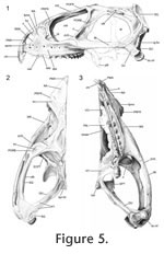
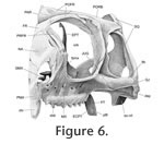
Digital imaging software, 'Adobe Photoshop 2.0' (Adobe Systems, San Jose, California), was used to combine drawings of bones with the "ghosts" of overlying or underlying bones. Once all the sutures had been assessed in detail, summary diagrams were produced. These include outlines of the skulls in lateral, ventral, dorsal, and occipital view with the inferred areas of underlying bone superimposed (e.g., Bolt and Wassersug 1975; Busbey 1995; Clack 2002; Jenkins et al. 2002). In addition, diagrammatic cross-sections were produced. A consistent colour code is used throughout for each bone (Table 3).

A small problem requiring consideration is the slight distortion of bone shape caused by dehydration (Tyler 1976, p. 6), particularly in long and thin bones or processes. Together with loss of soft tissue this is probably why bones in articulated dry skulls may separate slightly (e.g., maxillae in LDUCZ x343). This distortion can make rearticulation difficult. The anterior superior process of the squamosal in DGPC1, for example, has split, probably due to the loss of the organic component and subsequent dehydration. Another factor inhibiting accurate rearticulation is that in life soft tissue may push individual bones against each other or pull bones away from each other. In addition, small thin processes are delicate and liable to breakage, e.g., the triangular tips of the anterodorsal processes of the squamosal in DGPC1. Cleaning and disarticulation of the skull may also produce artificial surface texture. The full history of DGPC1 is unknown, including how the bulk of the soft tissue was removed and whether solvents were used to clean the surface.


Next Section

Cranial joints in Sphenodon
Plain-Language & Multilingual  Abstracts | Abstract | Introduction | Material and Method
Results| Summary of Results | Skull Mechanics in Sphenodon
Conclusions | Acknowledgements | References | Appendix 1 | Appendix 2
Print article