

Volume 28.2
May–August 2025
Full table of contents
ISSN: 1094-8074, web version;
1935-3952, print version
Recent Research Articles
See all articles in 28.2 May-August 2025
See all articles in 28.1 January-April 2025
See all articles in 27.3 September-December 2024
See all articles in 27.2 May-August 2024
Interested in submitting a paper to Palaeontologia Electronica?
Click here to register and submit.
 |
||||
 |
||||
Article Search
APPENDIX
Catalog of specimen numbers and morphotypes. DIC = dicotyledonous angiosperm, MON = monocotyledonous angiosperm, DIC FRAG = distinct dicotyledonous angiosperm. (*indicates morphotype exemplar).
|
Affinity |
Specimen Number |
Morphotype Number |
|
DIC |
RU-2010-849* |
KP-01 (12) |
|
DIC |
RU-2010-832 |
KP-01 |
|
DIC |
RU-2010-833 |
KP-01 |
|
DIC |
RU-2010-834 |
KP-01 |
|
DIC |
RU-2010-835 |
KP-01 |
|
DIC |
RU-2010-836 |
KP-01 |
|
DIC |
RU-2010-864 |
KP-01 |
|
DIC |
RU-2010-865 |
KP-01 |
|
DIC |
RU-2010-857 |
KP-01 |
|
DIC |
RU-2010-858 |
KP-01 |
|
DIC |
RU-2010-852 |
KP-01 |
|
DIC |
RU-2010-853 |
KP-01 |
|
DIC |
RU-2010-838* |
KP-02 (1) |
|
DIC |
RU-2010-267* |
KP-03 (3) |
|
DIC |
RU-2010-839 |
KP-03 |
|
DIC |
RU-2010-841 |
KP-03 |
|
DIC |
RU-2010-840* |
KP-04 (5) |
|
DIC |
RU-2010-842 |
KP-04 |
|
DIC |
RU-2010-843 |
KP-04 |
|
DIC |
RU-2010-985 |
KP-04 |
|
DIC |
RU-2010-986 |
KP-04 |
|
DIC |
RU-2010-844* |
KP-05 |
|
DIC |
RU-2010-845* |
KP-06 (4) |
|
DIC |
RU-2010-846 |
KP-06 |
|
DIC |
RU-2010-987 |
KP-06 |
|
DIC |
RU-2010-862 |
KP-06 |
|
DIC |
RU-2010-848* |
KP-07 (1) |
|
DIC |
RU-2010-860* |
KP-08 (1) |
|
DIC |
RU-2010-850* |
KP-09 (2) |
|
DIC |
RU-2010-851 |
KP-09 |
|
DIC |
RU-2010-859* |
KP-10 (1) |
|
DIC |
RU-2010-861* |
KP-11 (1) |
|
DIC |
RU-2010-863* |
KP-12 (1) |
|
MON |
RU-2010-866* |
KP-13 (3) |
|
MON |
RU-2010-867* |
KP-14 (8) |
|
DIC FRAG |
RU-2010-837* |
KP-15 (1) |
|
DIC FRAG |
RU-2010-847* |
KP-16 (1) |
|
DIC |
RU-2010-988 |
Unidentifiable fragements (41) |
TABLE 1. Descriptions of stratigraphic layers of the exposed section of the Grit Member (GM) at the study site.
|
Layer |
Thickness |
Description |
|
GBM-05 |
70 |
Bluish, greenish light grey. Fine to very fine sandstone. Ripple marks present. |
|
GBM-04 |
25 |
Greenish light grey. Finely laminated, medium fine sandstone, mudstone. |
|
GBM-03 |
1 |
Dark grey. Silty sandstone that in areas shows signs of mud cracks. |
|
GBM-02 |
40 |
Greenish light grey. Fine grained sandstone. Massively bedded. Organic material, including leaves, was found within this layer. |
|
GBM-01 |
30 |
Dark grey. Fine grained silty sandstone. |
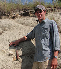 Daniel P. Maxbauer
Department of Biology
Saint John's University
Collegeville, Minnesota, USA
and Department of Earth and Environmental Sciences,
Wesleyan University
Middletown, Connecticut, USA
and Department of Earth Sciences
University of Minnesota
Minneapolis, Minnesota, USA
maxba001@umn.edu
Daniel Maxbauer is a PhD student in earth sciences at University of Minnesota. He studied earth and environmental sciences at Wesleyan Universtiy (CT) for his Master’s degree and natural sciences as an undergraduate at Saint John’s University (MN). His research has focused on using fossil plants to reconstruct past climates and environments.

 Daniel J. Peppe
Department of Geology
Baylor University
Waco, Texas, USA
daniel_peppe@baylor.edu
Daniel Peppe is an assistant professor of Geology at Baylor University. He received his degrees from St. Lawrence University (BS) and Yale University (MPhil, PhD), and was a postdoctoral researcher at Wesleyan University. His research program uses a collaborative, interdisciplinary approach to address how environmental change drives evolutionary processes in plants and animals. In his research, Peppe integrates methods in paleobotany, ecology, sedimentology, stratigraphy, and paleomagnetism to address questions about the underlying dynamics of environmental, biotic, and climatic change through time. Peppe's research is focused on reconstructing ancient climates and ecosystems in North America and East Africa, and on developing better and more accurate paleoclimate and paleoecological proxies.
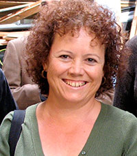 Marion Bamford
Bernard Price Institute for Palaeontology
University of the Witwatersrand
Johannesburg, South Africa
Marion.Bamford@wits.ac.za
Professor Marion Bamford is a palaeobotanist at the Bernard Price Institute, University of the Witwatersrand, Johannesburg, where she lectures to undergraduate students, supervises post-graduates and carries out research. Her specialty is fossil wood but she also works on charcoal, leaves, seeds, phytoliths and pollen. She works with multidisciplinary teams in South Africa on the Karoo, Cretaceous and Quaternary floras. She also works on projects in East African hominid sites such as Rusinga, Laetoli, Olduvai Gorge, and Koobi Fora.
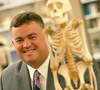 Kieran P. McNulty
Evolutionary Anthropology Lab
Department of Anthropology
University of Minnesota
Minneapolis, Minnesota, USA
kmcnulty@umn.edu
Kieran McNulty is an associate professor working in the Evolutionary Anthropology Laboratory at the University of Minnesota. His study of ape and human evolution is conducted, in part, using modern quantitative techniques for understanding components of cranial variation. He also conducts paleontological research in the early Miocene deposits of Western Kenya with the aim of better documenting the early diversification of hominoid primates.
 William E.H. Harcourt-Smith
Department of Anthropology
Lehman College CUNY
Bronx, New York, USA
and Department of Anthropology
Graduate Center CUNY
New York, New York, USA
and Division of Paleontology
American Museum of Natural History
New York, New York, USA
willhs@amnh.org
William Harcourt-Smith is a paleoanthropologist specializing in the evolution of fossil apes and humans. He earned his Ph.D. in vertebrate paleontology from University College London and now has joint appointments at the American Museum of Natural History in New York and Lehman College. He helped to curate the new permanent Hall of Human Evolution and co-directs paleontological research on Rusinga Island, Kenya.
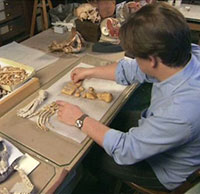
 Larry E. Davis
Department of Biology
Saint John's University
Collegeville, Minnesota, USA
ldavis@csbsju.edu
Larry Davis is a Professor of Geology/Biology at the College of Saint Benedict and Saint John’s University and received his PhD from Washington State University. He teaches courses in invertebrate and vertebrate paleontology, evolution and diversity, carbonate depositional environments, and reef ecology. He has conducted studies on the Chambered Nautilus in Papua New Guinea and Palau.
FIGURE 1. 1. A map showing Africa, star indicates approximate location of Lake Victoria, Rusinga Island and Mfangano Island. 2. Generalized map of Rusinga Island including basic stratigraphic distributions and general site locations. Star indicates the approximate location of this studies location. GPS coordinates for the study site: S 00° 24.350' E 034° 8.834'. 3. Generalized Miocene stratigraphy on Rusinga Island. Star indicates stratigraphic position of fossil leaf locality. Mbr. = member, Fm. = formation.
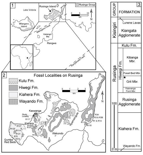
FIGURE 2. Stratigraphic section of the Grit Member exposed at fossil leaf locality. Ripple marks were found in GM-05, salt hoppers in GM-03, and fossil leaves in GM-02. GM = Grit Member.
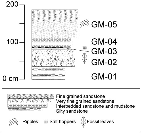
FIGURE 3. 1. Ripple marks a top GM-05. 2. Salt hoppers from GM-03. Scale bar = 1 cm.
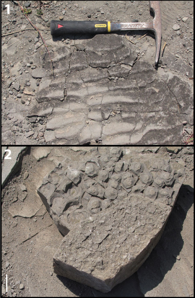
FIGURE 4. 1. Morphotype exemplar for KP-01, RU-2010-849. 2. RU-2010-864. 3. RU-2010-853. All specimens in 4.1-4.3 belong to KP-01 and display brochidodromous secondary venation, elliptic laminar shape, and cuneate base shape. 4. KP-02 morphotype exemplar, RU-2010-838, leaf showing oblong laminar shape, cordate base, pinnate primary venation, and brochidodromous secondary venation. All scales in 4.1-4.4 = 1 cm. 5. Enlarged portion of 4.4 showing cordate base with a naked basal vein, at least four basal veins, and brochidodromous secondary venation. Scale = 2 mm.
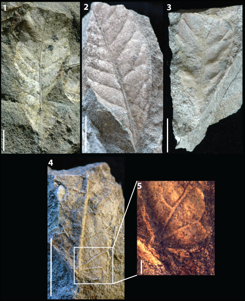
FIGURE 5. 1. KP-03 morphotype exemplar, RU-2010-267, showing eucamptodromous secondaries, highly ascending secondary angle, and abundant intersecondaries. Scale bar = 1 cm. 2. Enlarged portion of 5.1 showing straight opposite percurrent intercostal tertiaries and regular reticulate quaternary vein fabric. Scale = 2 mm. 3. RU-2010-839, leaf shows the diagnostic highly ascending secondaries unique to KP-03 along with the ovate laminar shape and straight apex shape. Scale bar = 1 cm. 4. Enlarged portion of 5.3 showing major secondaries becoming brochidodromous distally. Scale = 2 mm. 5. KP-04 morphotype exemplar, RU-2010-840, showing eucamptodromous secondary venation, low angle of divergence of secondaries that turn up abruptly near margin, and cordate base shape. Scale = 1 cm. 6. RU-2010-842, further showing the diagnostic secondary vein course and cordate base shape characteristic of KP-04. Scale = 1 cm.
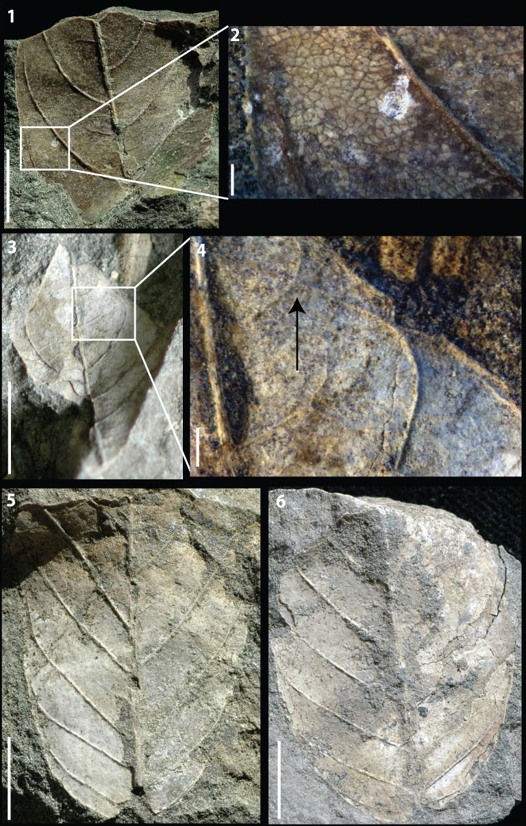
FIGURE 6. 1. KP-05 morphotype exemplar, RU-2010-844, view of whole leaf showing ovate to elliptic shape, pinnate primary venation with a convex to rounded base. Note that the leaf is slightly folded on underlying rock, causing deformation of the overall shape in photo. 2. RU-2010-844, right side of leaf showing brochidodromous secondary venation. 3. Line drawing showing brochidodromous secondary venation, simple agrophic veins, intersecondaries, and opposite sinuous percurrent tertiaries with obtuse angles. All scale bars = 1 cm.
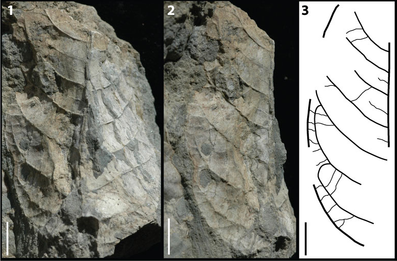
FIGURE 7. 1. KP-06 morphotype exemplar, RU-2010-845, leaf showing brochidodromous secondary venation, oblong laminar shape, and convex base shape. 2. Line drawing showing brochidodromous secondary venation, intersecondaries, and opposite sinuous percurrent tertiaries with angles varying from perpendicular to obtuse. 3. RU-2010-862, basal end of leaf showing convex base shape and brochidodromous secondary venation. 4. RU-2010-862, apical end of leaf showing acute apex angle. All scale bars = 1 cm.
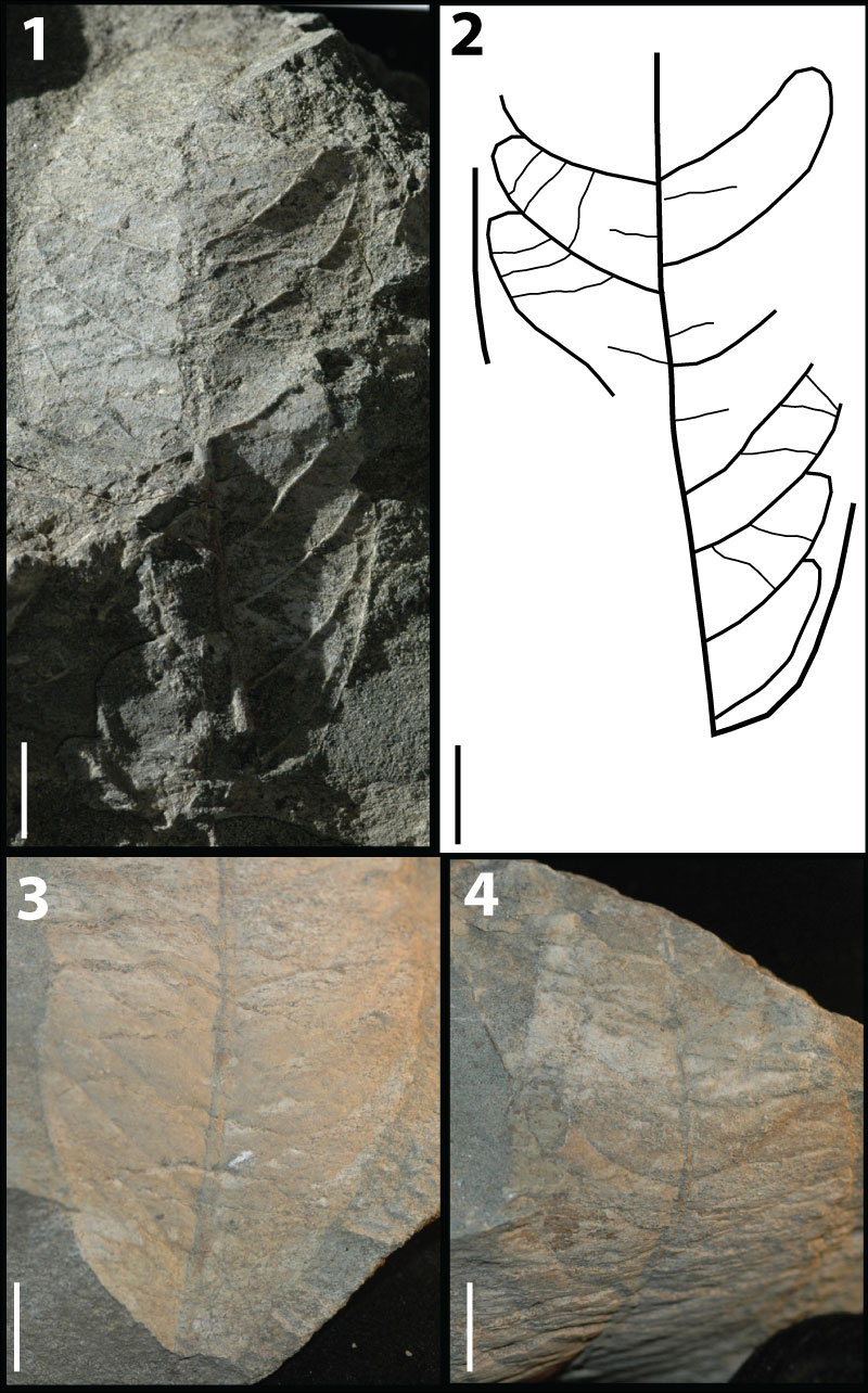
FIGURE 8. 1. KP-07 morphotype exemplar, RU-2010-848, showing ovate laminar shape, concave base shape, pinnate primary venation, and brochidodromous secondary venation. 2. Line drawing showing brochidodromous secondary venation, intersecondaries, and mixed percurrent intercostal tertiaries. 3. KP-08 morphotype exemplar, RU-2010-860, showing elliptic to oblong laminar shape, straight apex shape, and weak brochidodromous secondaries with close, uniform, spacing. All scale bars = 1 cm.
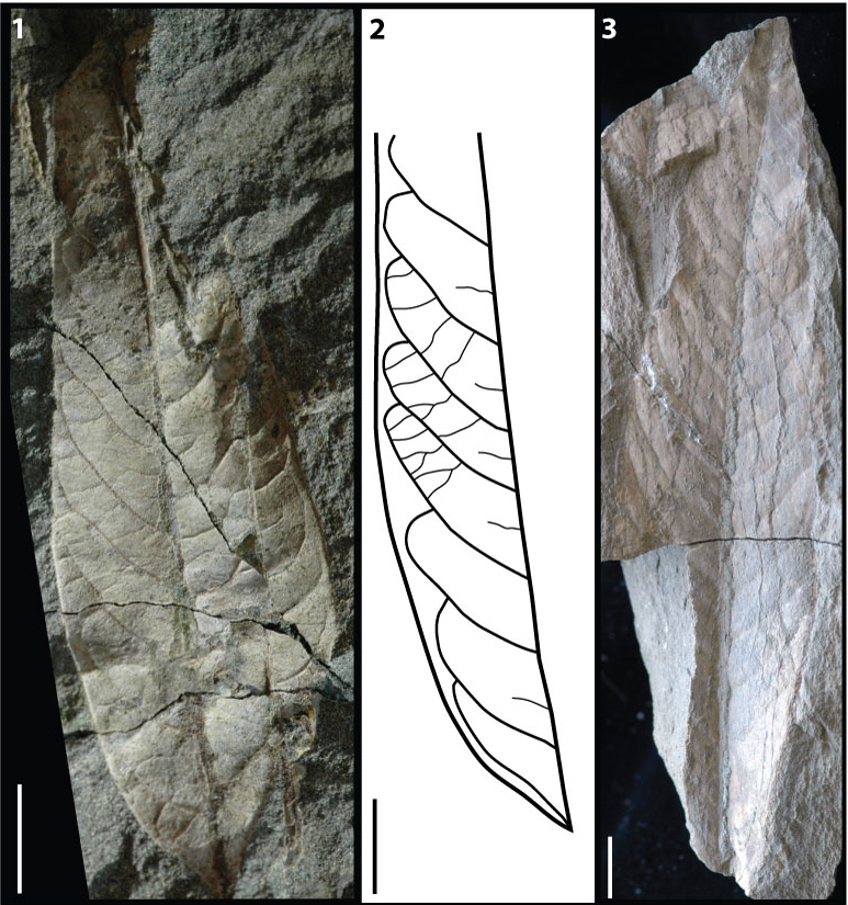
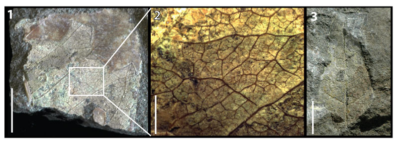
FIGURE 9. 1. KP-09 morphotype exemplar, RU-2010-850, showing ovate laminar shape, straight apex and pinnate primary venation. 2. KP-10 morphotype exemplar, RU-2010-859, showing brochidodromous secondaries with uniform angles and spacing. 3. Counterpart to RU-2010-859 showing acute apex angle and acuminate apex shape. 4. KP-11 morphotype exemplar, RU-2010-861, showing convex base shape and pinnate primary venation. 5. Enlarged portion of 9.4 showing irregular reticulate intercostal tertiaries. Scale = 2 mm. 6. KP-12 morphotype exemplar, RU-2010-863, showing regularly spaced secondaries diverging at a low angle, and a stout midvein. 7. Enlarged portion of 9.6 showing mixed percurrent intercostal tertiaries. 8. Line drawing highlighting the uniform obtuse angles of the intercostal tertiaries. All scales in 9.1-9.4 and 9.6-9.8 = 1 cm.
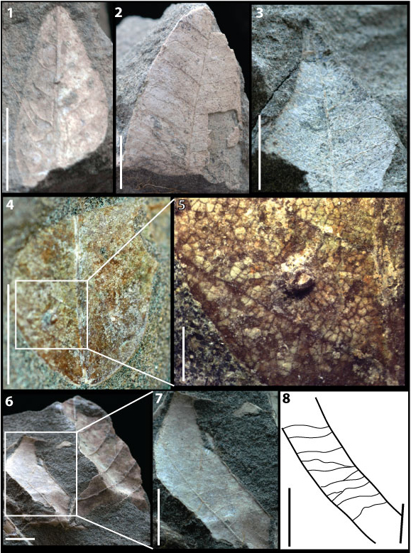
FIGURE 10. 1. KP-13 morphotype exemplar, RU-2010-866, aff. Typha sp. showing major linear veins parallel and evenly spaced. Scale = 1 cm. 2. aff. Typha sp. close up showing minor linear veins running parallel and evenly spaced between major linear veins. Scale = 2 mm. 3. KP-14 morphotype exemplar, RU-2010-867, aff. Phragmites sp. showing parallel major linear veins with midrib. Scale =1 cm. 4. Additional specimen of KP-14, aff. Phragmites sp., to further demonstrate presence of a midrib, distinguishing KP-14 from KP-13.
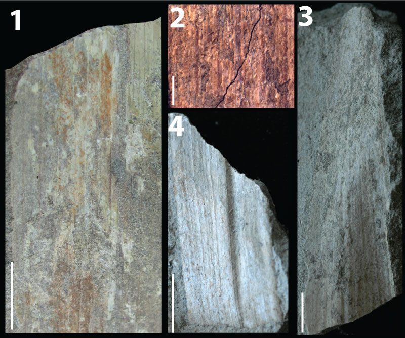
FIGURE 11. 1. KP-15 distinct dicotyledonous fragment, RU-2010-837, showing brochidodromous secondary venation and well preserved higher order venation. Scale bar = 1 cm. 2. Enlarged portion of 11.1 showing intercostal tertiaries irregular reticulate, epimedial tertiaries reticulate, exterior tertiaries variable, and quaternary vein fabric irregular reticulate. 3. KP-16, distinct dicotyledonous fragment, RU-2010-847, showing cladodromous secondary vein course and potential ovate laminar shape. This fragment is potentially the apex of a larger leaf.
A morphotype catalog and paleoenvironmental interpretations of early Miocene fossil leaves from the Hiwegi Formation, Rusinga Island, Lake Victoria, Kenya
Plain Language Abstract
There is an abundance of early Miocene (~18-20 million years ago) faunal and floral remains from deposits on Rusinga Island (Lake Victoria, Kenya). Past research has overwhelmingly focused on the early Miocene fauna from Rusing Island, and much less is known about the flora. Furthermore, no studies have thoroughly documented fossil leaves from Rusinga Island. Here, we describe the first collection of early Miocene fossil leaves from Rusinga Island. Characteristics of the flora along with sedimentological evidence within the collection area suggest that the local paleoenvironment was a patchwork of woodland and forested habitats in what was likely a warm climate. This work highlights the need for more research into the Rusinga Island floras, with a particular focus on fossil leaves.
Resumen en Español
Catálogo de morfotipos e interpretaciones ambientales de las hojas fósiles del Mioceno temprano de la Formación Hiwegi (isla de Rusinga, lago Victoria, Kenia)
Los depósitos del Mioceno temprano de la isla de Rusinga (lago Victoria, Kenia) contienen abundantes restos de fauna y flora. A pesar del interés que históricamente ha suscitado la fauna del Mioceno inferior de Rusinga, se ha prestado poca atención a la flora fósil y, hasta ahora, no se habían descrito los morfotipos de hojas fósiles de la isla. En este trabajo presentamos un catálogo de los morftoipos de hojas fósiles colectadas en el Miembro Grit de la Formación Hiwegi de Rusinga. Describimos catorce morfotipos, que comprenden doce angiospermas dicotiledóneas y dos monocotiledóneas, así como dos fragmentos de hojas de dicotiledóneas. Las características de la flora y las evidencias sedimentológicas, junto con las investigaciones previas, sugieren que el paleoambiente local era una hábitat de ribera dentro de un mosaico de áreas boscosas de densidad variable en un clima probablemente cálido. Este trabajo representa un importante primer paso en el conocimiento de la vegetación del Mioceno temprano de la isla de Rusinga y pone de manifiesto tanto la necesidad como el potencial de futuras investigaciones sobre estas floras.
Palabras clave: Mioceno temprano; isla de Rusinga; paleobotánica; megaflora; paleoambiente
Traducción: Miguel Company
Résumé en Français
Un catalogue de morphotypes et interprétations environnementales des feuilles fossiles miocènes inférieures de la Formation Hiwegi, île de Rusinga, Lac Victoria, Kenya
Les dépôts miocènes inférieurs sur l'île de Rusinga (Lac Victoria, Kenya) contiennent d'abondants restes de faune et de flore. Malgré l'attention particulière qui a été historiquement portée sur la faune miocène inférieure de l'île de Rusinga, la flore miocène inférieure a en revanche fait l'objet de beaucoup moins d'attention et aucune étude n'a à ce jour décrit les morphotypes de feuilles fossiles de cette île. Nous présentons ici un catalogue de morphotypes de feuilles fossiles collectées dans le Membre Grit de la Formation Hiwegi sur l'île de Rusinga. Nous décrivons 14 morphotypes, comprenant 12 angiospermes dicotylédones et deux angiospermes monocotylédones, ainsi que deux fragments de feuilles angiospermes dicotylédones distinctes. Les caractéristiques de la flore et les preuves sédimentaires, couplées aux précédentes études, suggèrent que le paléoenvironnement local était un habitat riparien au sein d'une mosaïque de bois et de biomes forestiers, le tout probablement dominé pas un climat chaud. Ce travail constitue une première étape importante dans la connaissance de la végétation miocène inferieure de l'île de Rusinga, et met en évidence le besoin et l'intérêt de nouvelles recherches sur ces flores du Miocène inférieur.
Mots clés : Miocène inférieur ; île de Rusinga ; mégaflore paléobotanique ; paléoenvironnement
Translator: Olivier Maridet
Deutsche Zusammenfassung
In progress
Translator: Eva Gebauer
Arabic

Translator: Ashraf M.T. Elewa
-
-
PE: An influential journal
 Palaeontologia Electronica among the most influential palaeontological journals
Article number: 27.2.2E
July 2024

 A Review of Handbook of Paleoichthyology Volume 8a: Actinopterygii I, Palaeoniscimorpha, Stem Neopterygii, Chondrostei