

Volume 28.2
May–August 2025
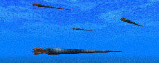
Full table of contents
ISSN: 1094-8074, web version;
1935-3952, print version
Recent Research Articles
See all articles in 28.2 May-August 2025
See all articles in 28.1 January-April 2025
See all articles in 27.3 September-December 2024
See all articles in 27.2 May-August 2024
Interested in submitting a paper to Palaeontologia Electronica?
Click here to register and submit.
 |
||||
 |
||||
Article Search
TABLE 1. Linear regression of the number of pectoral fin spines related to length of the pectoral fin segment of Bothriolepis canadensis. *p < 0.05
|
Fin segment |
Position |
n |
Equation |
p |
R2 |
|
proximal |
lateral |
39 |
number of spines = 0.54 * (proximal segment length) + 19.6682 ± 0.04 |
1.65e-14* |
0.79 |
|
proximal |
medial |
8 |
number of spines = 0.09 * (proximal segment length) + 32.08 ± 0.07 |
0.23 |
0.10 |
|
distal |
lateral |
11 |
log number of spines = 30.42 * (log distal segment length) – 24.79 ± 8.26 |
0.005* |
0.56 |
FIGURE 1. Reconstructions of Bothriolepis canadensis: 1.1, redrawn from Patten (1904, figure 1); 1.2, redrawn from Stensiö (1948, text-figure 38); 1.3, redrawn from Vézina (1996, figure 1); 1.4, redrawn from Arsenault et al. (2004, figure 8C-8D); 1.5, new reconstruction based on 3D model. Alignment and scaling based on the dorsal thoracic armor length.
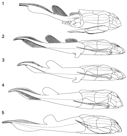
FIGURE 2. 3D reconstruction of Bothriolepis canadensis. Odd numbers without pectoral fins showing the brachial process, even numbers with pectoral fins; 1-2, dorsal view; 3-4 lateral view; 5-6, ventral view; 7-8, front view; 9-10, posterior view; 11, lateral view with posterior part of the body. Scale bar equals 1 cm. Use this figure as a target for the PaleoAR mobile application to get the 3D model.
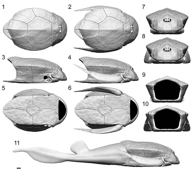
FIGURE 3. New reconstruction of Bothriolepis canadensis. Odd numbers without pectoral fins showing the brachial process, even numbers with pectoral fins; 1-2, dorsal view; 3-4 lateral view; 5-6, ventral view; 7-8, front view; 9-10, posterior view. Scale bar equals 1 cm.
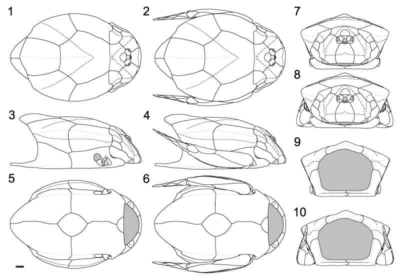
FIGURE 4. Gill opening of Bothriolepis canadensis. Head plates and pectoral fin in transparency to illustrate the relationships between the anterior ventrolateral plate (AVL) and the submarginal plate (SM).
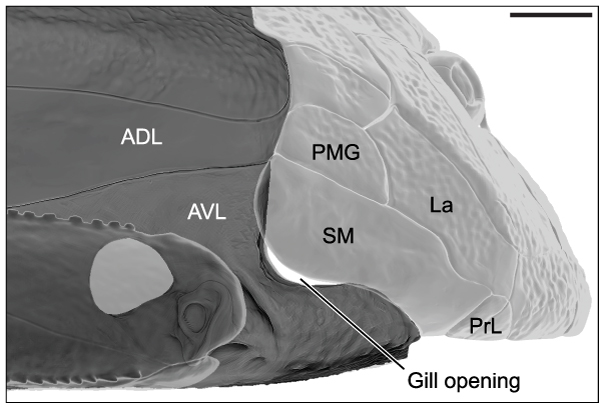
FIGURE 5. 3D reconstruction of the right pectoral fin of Bothriolepis canadensis: 1, lateral view; 2, medial view; 3, ventral view; 4, dorsal view. Scale bar equals 1 cm.
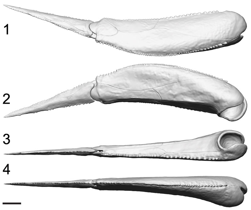
FIGURE 6. 3D reconstruction of the right pectoral fin segments of Bothriolepis canadensis: 1, lateral view of proximal segment; 2, medial view of proximal segment; 3, ventral view of proximal segment; 4, lateral view of distal segment; 5, medial view of distal segment. Scale bar equals 1 cm.
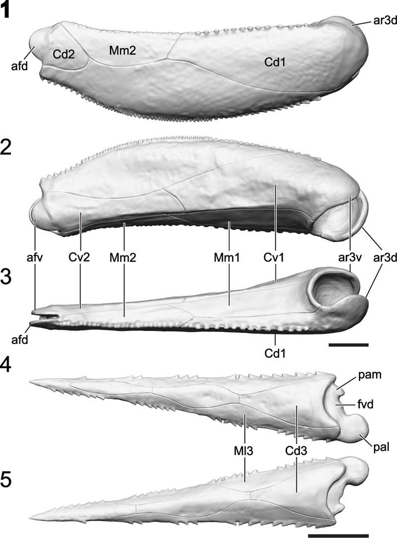
FIGURE 7. Range of movements for the pectoral fin of Bothriolepis canadensis: 1, rotation around the brachial process in fully retracted position; 2, fully protracted position; 3, fully protracted position front view; 4, latero-medially movement of the distal segment; 5, minimum angle of protraction for maximum mobility of the pectoral fin; 6, rotation around the brachial process in a protracted angle of 16°; 7, up-and-down movement in a protracted angle of 16°. Use this figure as a target for the PaleoAR mobile application to get a video of pectoral fins possible movement range.
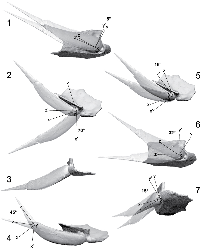
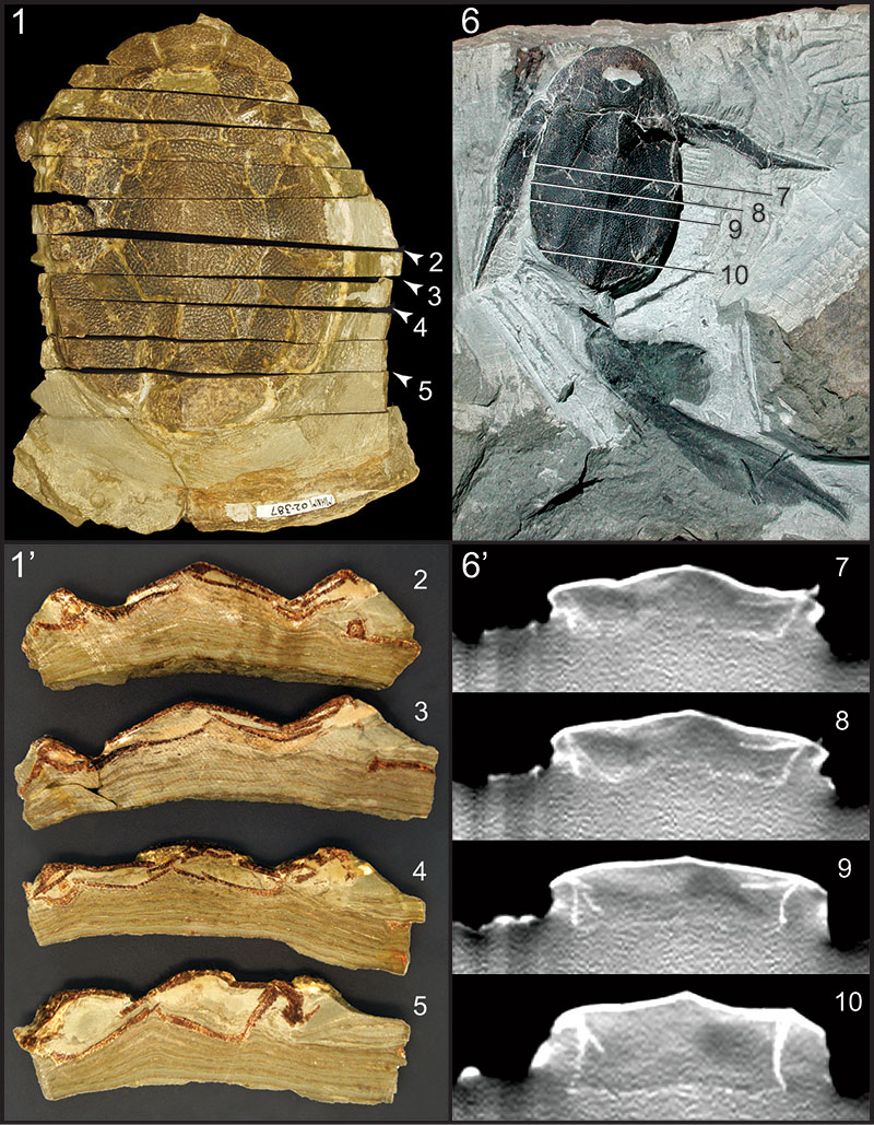
FIGURE 8. Taphonomic differences with reference to two types of sediment compaction for Bothriolepis canadensis: 1 and 1’, specimen MHNM 02-387 in dorsal view preserved in laminites with coronal sectioned with positioning of the sections illustrated in 2-5; 2-5, coronal sections showing the flattening and distortion of the specimen; 6 and 6’, specimen MHNM 02-2676 in dorsal view preserved in siltstone with positioning of the CT-scan coronal sections 7-10; 7-10, coronal sections showing the 3D condition of preservation and a weak lateral compaction.
 Isabelle Béchard
Centre de développement et de recherche en imagerie numérique
608 avenue Saint-Rédempteur
Matane
Québec, G4W 0E1
Canada
bechard.isabelle@cdrin.com
Isabelle Béchard graduated from the Université du Québec à Rimouski in Marine biology (BSc) in 2009 and Wildlife and habitat management (MSc) in 2011. Her M.Sc. thesis dealt with the ontogeny of the Late Devonian dipnoan Scaumenacia curta (Miguasha, Canada) with one of the most complete size series for a Paleozoic fish. Working now as a research assistant for Dr. Richard Cloutier at the Université du Québec à Rimouski, her work focus on Evo-Devo in paleontology and on the anatomy of the vertebrate species from the Miguasha Laggersttäte. In October 2013, she joined the Centre de développement et de recherche en imagerie numérique (CDRIN) in Matane (Québec) as the scientific director.

 Félix Arsenault
Centre de développement et de recherche en imagerie numérique
608 avenue Saint-Rédempteur
Matane
Québec, G4W 0E1
Canada
arsenault.felix@cdrin.com
Félix Arsenault is a 3D artist and technician who received is Collegial degree (DEC) in 3D animation and computer graphics at CEGEP de Matane in 2012. Félix is specialized in character art, which include having a strong knowledge of 3D softwares used in cinema, video game and research as well as knowledge of human and animal anatomy. In November 2012, he joined the Centre de développement et de recherche en imagerie numérique (CDRIN) in Matane where he is working on several projects
 Richard Cloutier
Département de Biologie
Chimie et Géographie
Université du Québec à Rimouski
300 allée des Ursulines
Rimouski
Québec, G5L 3A1
Canada
richard_cloutier@uqar.ca
Since 2000, Richard Cloutier is a professor-researcher at the University du Québec à Rimouski, where he is teaching Evolutionary Biology, Comparative Anatomy and Ichthyology. Richard graduated from the Université du Québec à Montréal (BSc, Geology, Biology), the Université de Montréal (MSc, biostatistics), and the University of Kansas (MPh, PhD, Systematics and Ecology). He received numerous grants in evolutionary biology, evo-devo, paleontology and virtual paleontology. Most of his research deals with various aspects of the evolutionary biology of lower vertebrates including systematics, evo-devo, and paleobiology.
 Johanne Kerr
Parc national de Miguasha
231 route Miguasha Ouest
Nouvelle
Québec, G0C 2E0
Canada
kerr.johanne@sepaq.com
In 1988, Johanne Kerr started working for the Parc national de Miguasha. Since 1996, she is the collection manager. Johanne participated with Dr. Richard Cloutier to establish a new classification system for Miguasha’s collections. She managed the computerization of the entire collection. Johanne also contributes to research in paleontology, geology and related topics.
The Devonian placoderm fish Bothriolepis canadensis revisited with three-dimensional digital imagery
Plain Language Abstract
The external morphology of the Late Devonian placoderm fish Bothriolepis canadensis from the Escuminac Formation (Miguasha, Canada) is reanalyzed using cutting-edge technology in three-dimensional (3D) digital imagery such as 3D surface scanner and 3D modeling software to create a 3D digital model. Digital manipulation of the model allows us to investigate some biomechanical aspects and constraints of the morphology. In contrast to previous interpretations, there is no indication of mobility between the cephalic and thoracic armors. A gill opening is clearly visible and the cephalic plates surrounding it are immobile. The dorsal plates of the thoracic armor form a hydrodynamic dorsal crest, which most likely has an important role in locomotion. Investigation of pectoral fin movement excluded the possibility of stroking and using them as anchoring devises. The 3D model of B. canadensis brings out unexpected novelties on one of the supposedly best-known Devonian fish.
Resumen en Español
Revisión, con imágenes digitales tridimensionales, del placodermo devónico Bothriolepis
En este artículo se analiza de nuevo la morfología externa del pez placodermo Bothriolepis del Devónico tardío de la Formación Escuminac (Miguasha, Canadá) empleando tecnología de imágenes digitales tridimensionales de última generación como escáner de superficie 3D y software de modelización tridimensional. Se han usado diecinueve ejemplares bien preservados de B. canadensis para reconstruir un modelo digital en tres dimensiones de la armadura dérmica y cuatro para reconstruir la parte carnosa posterior del cuerpo. La manipulación digital del modelo nos ha permitido analizar algunos aspectos biomecánicos y limitaciones de la morfología. En trabajos anteriores se había conjeturado, a partir de reconstrucciones inexactas, sobre la movilidad de la armadura cefálica, las placas submarginales y las aletas pectorales. En contra de las interpretaciones previas, no hemos hallado indicios de movilidad entre las armaduras cefálica y torácica. La placa submarginal queda inmovilizada en la armadura cefálica, y la abertura branquial se localiza entre las placas submarginal y ventrolateral anterior de la armadura torácica. La elevación medio-dorsal de la armadura torácica forma una cresta dorsal hidrodinámica con su máxima altura situada a lo largo de la placa media dorsal, y que, muy probablemente, tenía un papel importante en la locomoción. Las posiciones completamente retraída y extendida (70º) de las aletas pectorales permiten solo un movimiento restringido, excluyendo la posibilidad de utilizarlas para la propulsión o como elementos de anclado. El máximo de movilidad se alcanza en un ángulo extendido de 16º, que permite una rotación de 32º alrededor del proceso braquial y de 15º en un movimiento de arriba abajo. En resumen, el modelo tridimensional de B. canadensis pone de manifiesto novedades inesperadas con respecto al pez devónico supuestamente mejor conocido.
Palabras clave: Bothriolepis canadensis; Placodermi; reconstrucción; modelo tridimensional: escáner 3D; locomoción; realidad aumentada
Traducción: Miguel Company
Résumé en Français
Le poisson placoderme du Dévonien Bothriolepis canadensis revisité à partir d'imagerie numérique en trois dimensions
La morphologie externe du placoderme Bothriolepis canadensis du Dévonien supérieur de la Formation d'Escuminac (Miguasha, Canada) a été révisée à l'aide d'instruments à la fine pointe de la technologie dans le domaine de l'imagerie 3D. Dix-neuf spécimens bien conservés de B. canadensis ont été utilisés afin de construire un modèle numérique 3D de l'armure dermique et quatre spécimens ont été utilisés afin de reconstruire la partie postérieure du corps. La manipulation virtuelle du modèle 3D a permis de réévaluer certaines fonctions et contraintes biomécaniques de la morphologie du B. canadensis. Contrairement aux interprétations précédentes, formulées à partir de reconstructions erronées, aucun indice ne laisse croire à la possibilité de mouvement entre les armures céphalique et thoracique. Au niveau de l'armure céphalique, la plaque submarginale est immobile et une ouverture branchiale distincte est située entre la plaque submarginal et la plaque antérieure ventrolatérale de l'armure thoracique. La crête médiane dorsale de l'armure thoracique atteint son maximum de hauteur au niveau de la plaque médiane dorsale et forme une structure dorsale hydrodynamique qui joue un rôle stabilisateur lors de la locomotion. En position fermée et ouverte au maximum (70°), les nageoires pectorales ne peuvent presque pas bouger, ce qui exclue la possibilité de les utiliser d'une quelconque façon pour la propulsion ou comme système d'encrage. Le maximum de mobilité est permit par un angle d'ouverture d'au moins 16° qui permet un mouvement de rotation de 32° autour du processus brachial et de 15° dans un mouvement de bas-en-haut. Bothriolepis canadensis est souvent qualifié comme étant le poisson du Dévonien le plus connu, malgré cela, le modèle 3D a tout de même permis de découvrir des nouveautés.
Mots-clés: Bothriolepis canadensis; Placodermi; reconstruction; trois dimensions (3D); modèle 3D; scanner 3D; locomotion; réalité augmentée
Translator: Isabelle Béchard and Kenny J. Travouillon
Deutsche Zusammenfassung
Der devonische Placoderme Bothriolepis canadensis neu-untersucht mit dreidimensionaler digitaler Bildgebung
Die extreme Morphologie des spätdevonischen Placodermen Bothriolepis canadensis aus der Escuminac Formation (Miguasha, Kanada) wird mit Cutting-Edge Technologie wie 3D Oberflächenscanner und 3D Modellierungssoftware neu-analysiert. Für die Rekonstruktion eines 3D Modells der Panzerplatten wurden neunzehn gut erhaltene Stücke von B. canadensis verwendet, wobei anhand von vier Stücken der fleischige hintere Teil des Körpers rekonstruiert wurde. Digitale Manipulation des Modells erlaubt es uns einige biomechanische Aspekte und Constraints der Morphologie zu untersuchen. Über die Mobilität des Kopfpanzers, der submarginalen Platten und der Brustflossen wurden vorher Hypothesen aufgestellt, die auf inakkuraten Rekonstruktionen basierten. Im Gegensatz zu vorhergehenden Interpretationen gibt es keine Hinweise auf eine Beweglichkeit zwischen Kopf-und Brustpanzer. Die submarginale Platte sitzt bewegungsunfähig auf dem Kopfpanzer; eine Kiemenöffnung befindet sich zwischen den submarginalen und anterioren ventrolateralen Platten des Brustpanzers. Der mediane dorsale Kamm auf dem Brustpanzer formt einen hydrodynamischen dorsalen Kamm. Dessen maximale Höhe verläuft entlang der posterioren medianen Dorsalplatte, die höchstwahrscheinlich eine wichtige Rolle bei der Lokomotion spielt. Bei kompletter Retraktion und Protraktion (70°) der Brustflosse ist nur eine beschränkte Bewegung möglich und ein Auf-und Abschlagen oder die Nutzung als Ankerhilfe ist ausgeschlossen. Das Maximum an Mobilität wird bei einer Protraktion von 16° erreicht, was eine Rotation von 32° um den Brachialprozess erlaubt und 15° an Auf-und Abbewegung. Das 3D Modell von B. canadensis verdeutlicht unerwartete Neuerungen zu einem der angeblich bestbekanntesten devonischen Fische.
Keywords: Bothriolepis canadensis; Placodermi; Rekonstruktion; dreidimensional (3-D); 3D Modell; 3D Scanner; Lokomotion; erweiterte Realität
Translator: Eva Gebauer
Arabic
Translator: Ashraf M.T. Elewa
-
-
PE: An influential journal
 Palaeontologia Electronica among the most influential palaeontological journals
Article number: 27.2.2E
July 2024

 A Review of Handbook of Paleoichthyology Volume 8a: Actinopterygii I, Palaeoniscimorpha, Stem Neopterygii, Chondrostei