

Volume 28.2
May–August 2025
Full table of contents
ISSN: 1094-8074, web version;
1935-3952, print version
Recent Research Articles
See all articles in 28.2 May-August 2025
See all articles in 28.1 January-April 2025
See all articles in 27.3 September-December 2024
See all articles in 27.2 May-August 2024
Interested in submitting a paper to Palaeontologia Electronica?
Click here to register and submit.
 |
||||
 |
||||
Article Search
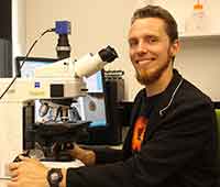 Chris Mays. Department of Palaeobiology, Naturhistoriska riksmuseet, Box 50007, S-104 05 Stockholm, Sweden; School of Earth, Atmosphere and Environment, Monash University, 9 Rainforest Walk, Clayton, Victoria 3800, Australia. chris.mays@nrm.se
Chris Mays’ research interests have focused on the palaeoenvironments and floral ecosystems of the south polar and sub-polar regions during past greenhouse intervals. From 2012–2017, Chris was undergraduate lecturer and postdoctoral researcher at Monash University’s School of Earth, Atmosphere & Environment. During this time, his research involved the sedimentary successions and both palynological and macrofloral fossils of New Zealand (the Chatham Islands, South Island), Australia (Victoria, Tasmania), and the Antarctic Peninsula. In order to document the fossils from a series of previously unknown localities, he employed a range of emerging imaging and ‘virtual palaeontology’ techniques, such as neutron tomography and synchrotron X-ray tomography. In 2016, he was awarded the inaugural Mary Wade Award by the Australian Association of Palaeontologists (AAP) for the publication of his monograph on the mid-Cretaceous palynology of the Chatham Islands. Most recently, he has undertaken a postdoctoral fellowship at the Naturhistoriska riksmuseet (the Swedish Museum of Natural History), Stockholm, Sweden. This project focuses on the extinction and recovery trends of the terrestrial flora during the end-Permian mass extinction. He is also Associate Editor for Alcheringa: An Australasian Journal of Palaeontology. His work has been supported by the Australian Research Council, the National Science Foundation, the Australian Nuclear Science and Technology Organisation, AAP, Monash University, National Geographic and the Paleontological Society.
Chris received his B.Sc. (Hons) in 2008 from the University of Melbourne, Australia, and his Ph.D. in 2012 from Monash University, Australia.

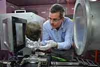 Joseph J. Bevitt. Australian Nuclear Science and Technology Organisation, Research Office, B3, New Illawarra Road, Lucas Heights, NSW 2234, Australia. jbv@ansto.gov.au
Joseph is a neutron-scattering scientist at the Australian Centre for Neutron Scattering (ACNS) and manager of the Research Office of the Australian Nuclear Science and Technology Organisation. This incorporates the User Office, Grants Office and Knowledge Centre (Australia's nuclear library) functions. These functions support the user programs of the landmark facilities for Australian science; the OPAL research reactor, Centre for Accelerator Science, ACNS, cyclotron facility and the Australian Synchrotron.
Joseph is an instrument scientist on the DINGO radiography/tomography/imaging station at the OPAL Research Reactor, Deputy President of the International Society for Neutron Radiology and a guest research fellow of the Dinosaur Evolution Research Centre at Jilin University, China. He collaborates with museums and universities to pioneer the use of neutron microCT (3-D imaging of objects using neutrons with micrometre resolution) in the areas of palaeontology, archaeology and cultural heritage. Specific applications are to: 1) digitally excavate and reconstruct fossilised remains; 2) investigate disease and medical practice in ancient time; and 3) determine methods of manufacturing ancient cultural artefacts.
Joseph received his B.Sc. and Ph.D. from the University of Sydney, Australia.
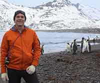 Jeffrey D. Stilwell. School of Earth, Atmosphere and Environment, Monash University, 9 Rainforest Walk, Clayton, Victoria 3800, Australia and Australian Museum, 1 William Street, Sydney NSW 2010, Australia. jeffrey.stilwell@monash.edu
Jeffrey Stilwell’s current research embraces the integration of palaeontological data in 'big picture science', especially the potential to employ applied palaeontology methods in deciphering ancient greenhouse earth systems. He is renowned for his long-term research on ancient polar greenhouse Earth environments and ecosystems, especially in southern latitudes in Antarctica, New Zealand-Chatham Islands and Australia. Professor Stilwell has published >70 peer reviewed articles and five books/monographs in the fields of geology and palaeontology. His research portfolio includes many Australian government grants since 1996 (Australian Research Council [ARC] grants, including APD, ARF, DP, LP, and LIEF schemes; National Geographic Society Committee for Research and Exploration grants (x3); ANSTO awards; Australian Synchrotron award; and many industry grants). Stilwell discovered the first in situ Mesozoic Australian amber in May 2011, as part of a study on Cretaceous-aged sediments in the Otway Basin. This major discovery led to a 2014–17 ARC-DP project on the abundant amber, which in turn, has resulted in comparable deposits with diverse inclusions of animals, plants and microorganisms—many groups of which have no prior fossil record in Australia, and in some instances, the entire Southern Hemisphere. He is currently working with many colleagues from Australia and Europe to learn more about southern Australia’s evolutionary heritage during critical intervals in Earth history from ~230 to 40 million years ago.
Jeff received his B.Sc. and M.Sc. from Purdue University, Indiana, and Ph.D. from the University of Otago, New Zealand.
APPENDIX
Austrosequoia novae-zeelandiae (Ettingshausen, 1887) Mays et al., 2017, animated volume rendering of neutron tomographic reconstruction, Relative Neutron Attenuation (RNA) spectrum and scale as per Figure 2, PL1227. Click on image to download or run animation.
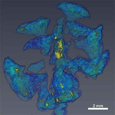
FIGURE 1. 1) Map of eastern Zealandia including New Zealand and the Chatham Islands, grey areas = emergent, grey outline = 2000 m isobath, boxed area is displayed in Figure 1.2. 2) Map of the Chatham Islands, grey areas = emergent, boxed area is displayed in Figure 1.3. 3) Geological map of the Waihere Bay area, northwest Pitt Island, fossil locality recorded in this study is indicated, age estimates from the following sources: Tupuangi Formation (Mildenhall, 1994; Mays and Stilwell, 2013), Kahuitara Tuff (Mildenhall, 1994; Stilwell, 1998), other estimates (Campbell et al., 1993; Panter et al., 2006). Modified from figures 1 and 3 of Mays et al. (2015b) with permission.
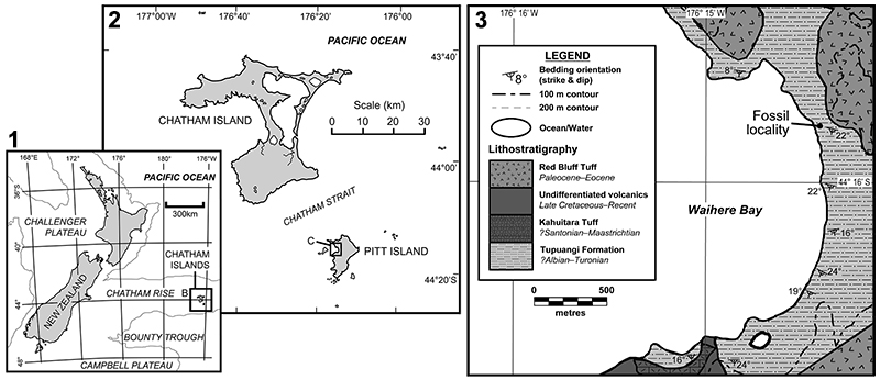
FIGURE 2. Austrosequoia novae-zeelandiae (Ettingshausen, 1887) Mays et al., 2017, PL1227. 1) Transverse section of a partially exposed, desiccated ovulate cone. 2) Neutron tomographic reconstruction largely encapsulated in sedimentary matrix, white indicates high neutron attenuation, oblique-transverse view. 3) Volume rendering of neutron tomographic reconstruction, RNA = Relative Neutron Attenuation, grid texture on RNA spectrum indicates relative transparency, regions of highest neutron attenuation represent in situ resin within cone axis and minor enclaves of resin near the distal ends of the bract-scale complexes, desiccation exhibited by large gaps in coalified organic remains (blue/green), oblique-transverse view. 4) Greyscale histogram from neutron tomographic reconstruction of PL1227 (16-bit) these values represent the neutron attenuation of the reconstructed volume, colours and transparency textures as per Figure 2.3, threshold values presented in Table 2, the spectrum has been cropped at the extremes for this graphical representation.
See Appendix for an animation of the virtually extracted specimen illustrated in Figure 2.3.
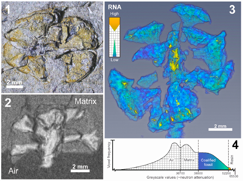
FIGURE 3. Artist’s reconstruction of ovuliferous cone and fertile shoot of Austrosequoia novae-zeelandiae (Ettingshausen, 1887) Mays et al., 2017, artist: Mali Moir.
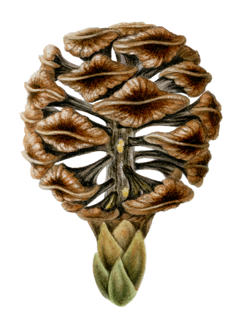
TABLE 1. Neutron radiograph data collection parameters for illustrated specimen (PL1227).
| Scan parameter | Value |
| Total sample dimensions (mm) | 62×58×14 |
| Scintillator composition | ZnS/6LiF |
| Scintillator dimensions (mm) | 100x100x0.05 |
| Rotation angle (°) | 0.25 |
| Total rotation (°) | 180 |
| Projections | 720 |
| No. of frames/projection | 3 |
| Frame exposure (secs) | 60 |
| No. of dark and beam profile images | 6 each |
| Radiograph pixel width (mm) | 0.0161 |
TABLE 2. Resin volume estimates and segmentation thresholds, all values are from visualisations performed with Avizo 9.0.1, volumes based on voxel diameter = 16.1 μm, “max.” and “min.” refer to the software maximum and minimum absolute spectrum values (0 and 65535, respectively), total = coalified plant remains + fossil resin, relative resin volume (%) = (fossil resin / total) × 100.
| Neutron attenuation segmentation thresholds | Volume (no. of voxels) |
Volume (μm3) |
||
| Medium | Lower | Upper | ||
| Air | Min. | 30700 | - | - |
| Matrix | 30700 | 38500 | 1.60×10 | 6.67×10¹² |
| Bract-scale complex 1 | ||||
| Coalified plant remains | 38500 | 52250 | 1.59×10 | 6.65×10 |
| Fossil resin | 52250 | Max. | 2.61×10 | 1.09×10 |
| Total | 38500 | Max. | 1.62×10 | 6.76×10 |
| Relative resin volume (%) | 1.61 | |||
| Bract-scale complex 2 | ||||
| Coalified plant remains | 38500 | 52250 | 7.05×10 | 2.94×10 |
| Fossil resin | 52250 | Max. | 6.65×10 | 2.78×10 |
| Total | 38500 | Max. | 7.12×10 | 2.97×10 |
| Relative resin volume (%) | 0.94 | |||
| Bract-scale complex 3 | ||||
| Coalified plant remains | 38500 | 52250 | 1.14×10 | 4.74×10 |
| Fossil resin | 52250 | Max. | 9.98×10 | 4.16×10 |
| Total | 38500 | Max. | 1.15×10 | 4.78×10 |
| Relative resin volume (%) | 0.87 | |||
| Bract-scale complex 4 | ||||
| Coalified cone scale | 38500 | 52250 | 8.49×10 | 3.54×10 |
| Fossil resin | 52250 | Max. | 1.13×10 | 4.73×10 |
| Total | 38500 | Max. | 8.60×10 | 3.59×10 |
| Relative resin volume (%) | 1.32 | |||
Pushing the limits of neutron tomography in palaeontology: Three-dimensional modelling of in situ resin within fossil plants
Plain Language Abstract
A high-resolution, three-dimensional model of a fossil seed cone from the Chatham Islands revealed the presence of internal fossil resin (amber). Its distribution of resin bodies supports the affinity with the modern conifer genus Sequoia (Family Cupressaceae). The model was constructed employing neutron tomography (a technique analogous to X-ray computed tomography [CT], but with neutrons instead of X-rays), and the high neutron attenuation contrasts between the resin, the enclosing organically preserved (coalified) plant remains, and sediment matrix allowed for a clear-cut segmentation of these different components. The results demonstrate the feasibility of detecting different organic compounds within a given fossil specimen using neutrons. With emerging advances in this technology, a wider variety of organic compounds and anatomical features within a given specimen should be discernible; this will have far-reaching implications for the forms and functions of fossils, as well as their post-burial histories and conservation.
Resumen en Español
En los límites de la tomografía de neutrones en paleontología: modelado tridimensional de resina in situ en el interior de plantas fósiles
La tomografía computarizada es una técnica cada vez más utilizada para el estudio no destructivo de fósiles. Mientras que la ciencia de la tomografía computarizada (TC) de rayos X ha avanzado mucho desde sus primeras aplicaciones en fósiles a principios de la década de 1980, las aplicaciones y limitaciones de la tomografía de neutrones (TN) permanecen relativamente inexploradas en paleontología. Las tomografías de neutrones de resolución más alta hasta la fecha en paleontología se llevaron a cabo en un ejemplar de Austrosequoia novae-zeelandiae (Ettingshausen) Mays y Cantrill obtenido de los estratos del Cretácico medio (Cenomaniense; ~ 100-94 Ma) de las Islas Chatham, este de Zelandia. Previamente, la especie ha sido identificada con resina fósil in situ (ámbar); los nuevos análisis de tomografía de neutrones mostraron una señal de atenuación de neutrones anómalamente alta para la resina fósil. Los datos resultantes proporcionaron un fuerte contraste entre los materiales: 1) resina fósil; 2) materia vegetal carbonificada; y 3) matriz sedimentaria. Ello supuso obtener distintas representaciones tridimensionales de los mismos. Estos datos facilitaron un modelo anatómico de masas de resina endógenas dentro del eje del cono y los conjuntos de brácteas. Los tipos de masas de resina y sus distribuciones respaldan una estrecha relación con Sequoia Endlicher (Cupressaceae), un grupo de coníferas cuyos representantes actuales solo se encuentran en el Hemisferio norte. Este estudio demuestra la viabilidad de la TN como un medio para diferenciar compuestos orgánicos químicamente distintos dentro de los fósiles. Aquí, hacemos recomendaciones específicas con respecto a: 1) la idoneidad de los diferentes tipos de preservación fósil para utilizar la TN; 2) la conservación de ejemplares orgánicos con consolidantes y adhesivos hidrogenados; y 3) la aplicación de métodos emergentes (por ejemplo, contraste de fase de neutrones) para mejoras adicionales cuando se obtienen imágenes de estructuras anatómicas con pequeños detalles. Estos hallazgos demuestran que todavía estamos lejos de alcanzar los límites conceptuales de la TN como medio para extraer fósiles virtualmente, o para obtener imágenes de su anatomía interna, incluso cuando están presentes dentro de la matriz de roca.
Palabras clave: tomografía de neutrones; resina; cono de semilla; Cupressaceae; Cretácico; conservación
Traducción: Enrique Peñalver (Sociedad Española de Paleontología) or Diana Elizabeth Fernández
Résumé en Français
Repousser les limites de la tomographie par neutrons en paléontologie : modélisation tridimensionnelle de la résine in situ dans des plantes fossiles
La tomographie assistée par ordinateur est une technique de plus en plus populaire pour l’étude non invasive des fossiles. Alors que la science de la tomodensitométrie par rayons X (CT) a grandement mûri depuis ses premières applications à des fossiles dans les années 1980, les applications et limitations de la tomographie par neutrons (NT) restent relativement peu explorées en paléontologie. Des scans par tomographie par neutrons avec la plus haute résolution jamais atteinte en paléontologie ont été effectués sur un spécimen d’Austrosequoia novae-zeelandiae (Ettingshausen) Mays et Cantrill trouvé dans des couches du Crétacé moyen (Cénomanien ; ~100 Ma–94 Ma) des îles Chatham, à l’est de la Zealandia. Précédemment, cette espèce a été identifiée avec de la résine fossile (ambre) in situ ; les nouvelles analyses par tomographie par neutrons démontrent une atténuation anormalement forte du signal de neutrons pour la résine fossile. Les données obtenues ont fourni un fort contraste entre les représentations tridimensionnelles de : 1) la résine fossile ; 2) la matière végétale carbonifiée ; et 3) la matrice sédimentaire. Ces données ont facilité la construction d’un modèle anatomique des corps de résine endogènes au sein de l’axe du cône et des complexes d’écailles de bractée. Les types et les distributions des corps de résine soutiennent une forte affinité avec Sequoia Endlicher (Cupressaceae), un groupe de conifères dont les membres actuels sont seulement trouvés dans l’hémisphère nord. Cette étude démontre la faisabilité de la NT comme moyen de différencier des composés organiques distincts au sein des fossiles. Dans cet article, nous formulons des recommandations spécifiques concernant : 1) l’adéquation des styles de préservation des fossiles pour la NT ; 2) la préservation des spécimens organiques avec des consolidants hydrogènes et des colles ; et 3) l’application des méthodes récemment développées (e.g., NT par contraste de phase) pour encore améliorer l’illustration très détaillée des structures anatomiques. Ces découvertes démontrent que nous sommes encore loin d’atteindre les limites conceptuelles de la NT comme moyen d’extraire virtuellement les fossiles, ou de capturer leur anatomie interne même quand ils sont pris dans une matrice rocheuse.
Mots-clés : tomographie par neutrons ; résine ; cône ; Cupressaceae ; Crétacé ; préservation
Translator: Antoine Souron
Deutsche Zusammenfassung
Ausloten der Grenzen von Neutronen-Tomographie in der Paläontologie: dreidimensionales Modellieren von in situ Harzen innerhalb fossiler Pflanzen
Computer-Tomographie ist eine zunehmend beliebte Technik um Fossilien zerstörungsfrei zu untersuchen. Während sich die Röntgen-Computer-Tomographie (CT) seit der ersten Anwendung bei Fossilien in den frühen 1980er Jahren stark weiterentwickelt hat, bleiben Anwendung und Grenzen von Neutronen-Tomopgraphie (NT) relativ unerforscht in der Paläontologie. Die höchstauflösenden neutronentomographischen Scans in der Paläontologie bislang wurden von Austrosequoia novae-zeelandiae (Ettingshausen) Mays and Cantrill gemacht, ein Stück, das aus den mittelkretazischen Schichten (Cenoman; ~100–94 Ma) der Stratham Inseln (östliches Zealandia) stammt. Bislang wurde die Art mit in situ Harz (Bernstein) identifiziert. Die neue neutronentomographische Analyse wies ein ungewöhnlich hohes Neutronendämpfungssignal für fossiles Harz auf. Die resultierenden Daten lieferten einen starken Kontrast und deutliche dreidimensionale Repräsentanzen: 1) des fossilen Harzes; 2) es inkohlten Pflanzenmaterials und 3) der Sedimentmatrix. Diese Daten vereinfachten das anatomische Modell eines endogenen Harzkörpers innerhalb der Zapfen-Achse und des Deckblattkomplexes. Form und Verteilung der Harzkörper unterstützen eine nahe Verbindung mit Sequoia Endlicher (Cupressaceae), einer Gruppe von Koniferen, deren heutige Mitglieder nur in der nördlichen Hemisphäre gefunden werden. Diese Untersuchung demonstriert die Umsetzbarkeit von NT als Möglichkeit chemisch verschiedene organische Verbindungen innerhalb von Fossilien zu unterscheiden. Wir machen hier konkrete Empfehlungen in Bezug auf: 1) die Eignung von fossilen Erhaltungsarten für NT; 2) die Erhaltung von organischen Stücken mit wasserstoffhaltigen Konsolidierungsmitteln und Klebemitteln; und 3) die Anwendung von Zukunftsmethoden (Neutronenphasenkontrast) für weitere Verbesserungen bei der Abbildung von fein-detaillierten anatomischen Strukturen. Diese Funde zeigen, dass wir die konzeptionellen Grenzen von NT als ein Mittel zum virtuellen Extrahieren von Fossilien oder zum Abbilden ihrer internen Anatomie selbst wenn sie in Matrix eingeschossen sind, noch lange nicht erreicht haben.
Schlüsselwörter: Neutronen-Tomographie; Harz; Zapfen; Cupressaceae; Kreide; Erhaltung
Translator: Eva Gebauer
Arabic
Translator: Ashraf M.T. Elewa
-
-
PE: An influential journal
 Palaeontologia Electronica among the most influential palaeontological journals
Article number: 27.2.2E
July 2024

 A Review of Handbook of Paleoichthyology Volume 8a: Actinopterygii I, Palaeoniscimorpha, Stem Neopterygii, Chondrostei