

Article Search
Volume 27.2
May–August 2024
Full table of contents
ISSN: 1094-8074, web version;
1935-3952, print version
Recent Research Articles
See all articles in 27.2 May-August 2024
See all articles in 27.1 January-April 2024
See all articles in 26.3 September-December 2023
FIGURE 1. Sketch of the measurement techniques in micro-CT device.
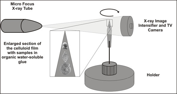
FIGURE 2. Workflow diagram of acquisition and numerical and visual rendering with proposed software packages (modified after SkyScan 2005).
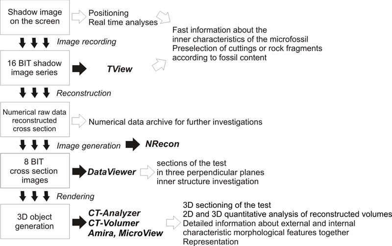
FIGURE 3. Specimens with different wall material fixed with gomme adragante (water-soluble glue) on celluloid films. Bottom to top: Spirillina infima (Strickland, 1846) (Jurassic, Som Hill, Transdanubian Range, Hungary), Paalzowella scalariformis Paalzow, 1917 (Jurassic, Som Hill, Transdanubian Range, Hungary), Bolivina dilatata Reuss, 1850 (Badenian, North Hungarian Range). The largest diameter is 300 μm.
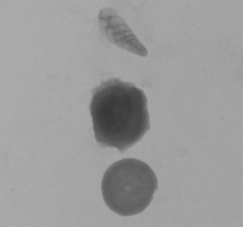
FIGURE 4. X-ray shadow images of (4.1) hyaline Elphidium macellum (von Fichtel and Moll, 1798) (Sarmatian, Zsámbék Basin, North Hungary) (the largest diameter is 400 μm) and (4.2) agglutinated Tritaxia tricarinata (Reuss, 1844) (Cretaceous, Magyarpolány, Transdanubian Range) test (the largest diameter is 250 μm) of foraminifera.
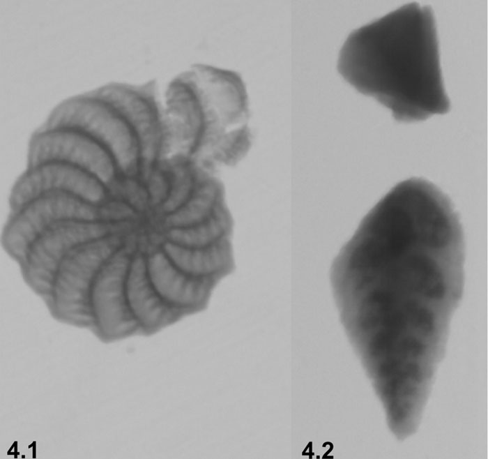
FIGURE 5. X-ray shadow image of the Badenian (Székkutas, SE-Hungary) cuttings in plastic tube holder. Red circles indicate the foraminifera. The largest diameter of the foraminifera is 300 μm.
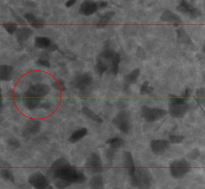
FIGURE 6. 3D model of Triloculina schreiberiana d'Orbigny, 1839 (Recent, Zadar, Adriatic Sea), side view (Amira software). The largest diameter is 350 μm.
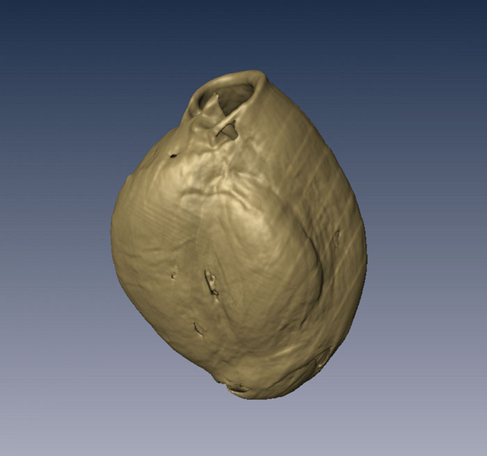
FIGURE 7. 3D model of Triloculina schreiberiana d'Orbigny, 1839 figured on Figure 6, with translucent last chambers, side view. (Amira software). The largest diameter is 350 μm.
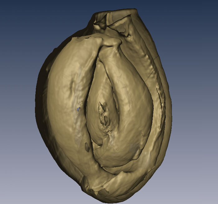
FIGURE 8. Section across the embryonic chamber of the 3D models of Triloculina schreiberiana d'Orbigny, 1839 illustrated on Figure 6 (Amira software). The largest diameter is 350 μm.

FIGURE 9. 3D model of of Triloculina schreiberiana d'Orbigny, 1839 illustrated on Figure 6, apertural view (Amira software). The largest diameter is 350 μm.
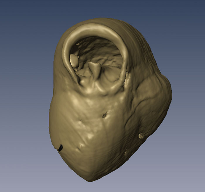
FIGURE 10. Apertural section penetrating the embryonic chamber (large image) of Spiroloculina cymbium d'Orbigny, 1839 (Recent, Zadar, Adriatic Sea) and three perpendicular views (right side) (MicroView software). The largest diameter is 350 μm.
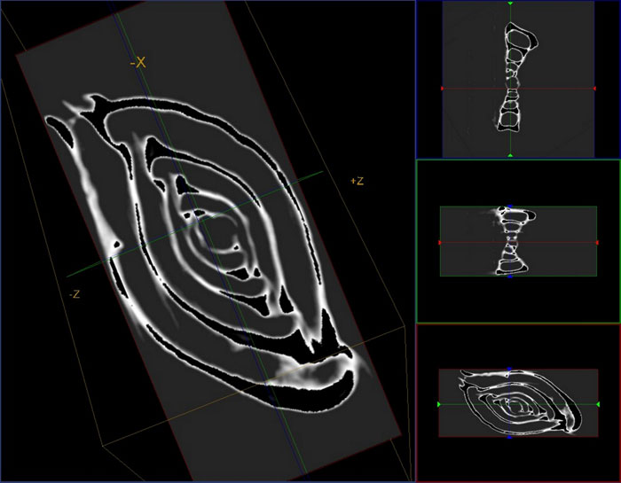
FIGURE 11. Section across the embryonic chamber of the 3D models of Spiroloculina cymbium d'Orbigny, 1839 figured on Figure 10 (Amira software). The largest diameter is 350 μm.
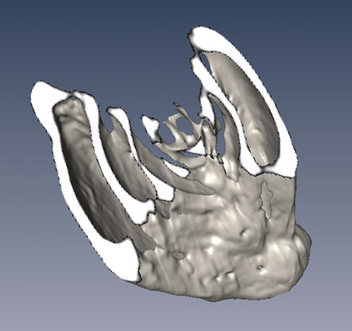
FIGURE 12. 3D model of of Spiroloculina cymbium d'Orbigny, 1839 illustrated on Figure 10, apertural view (Amira software). The largest diameter is 350 μm.
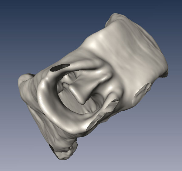
FIGURE 19. Axial section of Globigerinoides trilobus (Reuss, 1850) (Badenian, North Hungarian Range) and three perpendicular views (right side) (MicroView software). The largest diameter is 200 μm.
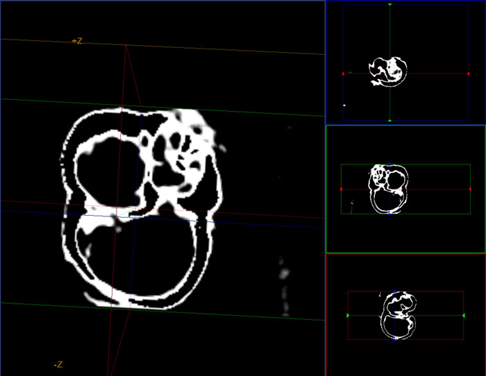
FIGURE 21. Section across the juvenile chambers of the 3D models of Orbulina universa d'Orbigny, 1839 (Badenian, North Hungarian Range) (Amira software). The largest diameter is 150 μm.
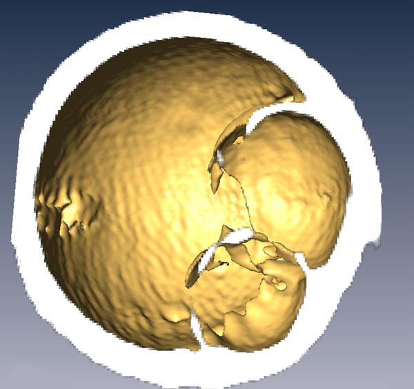
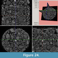
FIGURE 23. Cross section images of Carpathian (North Hungarian Range) (23.1) and Sarmatian (Zsámbék Basin, Hungary) (23.2) washing residue in plastic tube holder (DataViewer software). Diameter of the plastic tube is 3.5 mm.
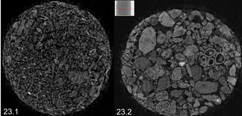
FIGURE 25. Sectioning in three perpendicular planes of recrystallized test and infilled by sparrycalcite of Eoguttulina sp. (Cretaceous, Calvaria Hill, Transdanubian Range) (DataViewer software). The height of the test is 310 μm.
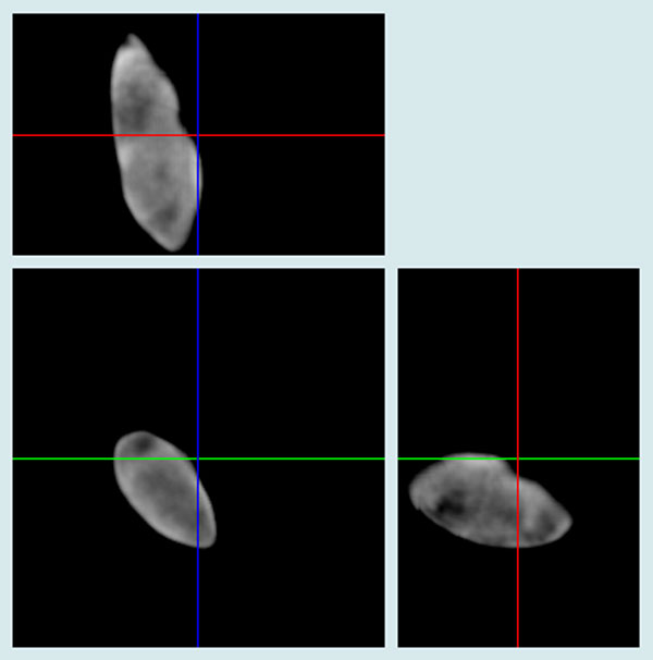
FIGURE 13. Sectioning in three perpendicular planes of Triloculina schreiberiana d'Orbigny, 1839 illustrated on Figure 6 (MicroView software).
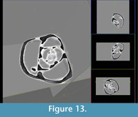
Figure 14. Animation of the 3D model of Triloculina schreiberiana d'Orbigny, 1839 illustrated on Figure 6 (Amira software).
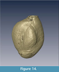
FIGURE 15. Arbitrary sectioning of the 3D model of Triloculina schreiberiana d'Orbigny, 1839 illustrated on Figure 6 (Amira software).
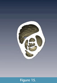
FIGURE 16. 3D animation of Triloculina schreiberiana d'Orbigny, 1839 illustrated on Figure 6, with translucent last chambers (Amira software).

FIGURE 17. Sectioning of Spiroloculina cymbium d'Orbigny, 1839 illustrated on Figure 10 and three perpendicular views (right side) (MicroView software).
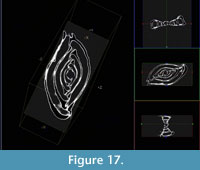
FIGURE 18. 3D animation of Spiroloculina cymbium d'Orbigny, 1839 illustrated on Figure 10, with translucent last chambers (Amira software).
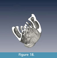
FIGURE 20. Sectioning of Globigerinoides trilobus (Reuss, 1850) illustrated on Figure 19 and three perpendicular views (right side) (MicroView software).
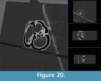
FIGURE 22. Sectioning of Orbulina universa d'Orbigny, 1839 illustrated on Figure 21 and three perpendicular views (right side) (MicroView software).
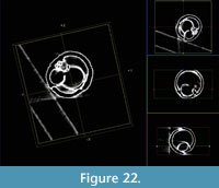
FIGURE 24. Sectioning in three perpendicular views of Sarmatian cuttings (DataViewer software).
Ágnes Görög Department of Palaeontology
Eötvös University
Pázmány Péter sétány 1/C
Budapest, H-1117
Hungary
Micropalaeontologist Ágnes Görög is an assistant professor in the Palaeontological Department at Eötvös Loránd University, Budapest, Hungary. Her diploma work was on the Sarmatian (Late Miocene) benthic foraminifera. After she defended her PhD. Theses on Cretaceous Orbitolinids of Hungary. Her research concerns the taxonomy, stratigraphy and ecology of foraminifera from Late Triassic, Jurassic, Cretaceous and Miocene. Since 1994 together with professor Roland Wernli (Univ. Genève) she is focused on the taxonomy, palaeobiogeography and evolution of early planktonic foraminifera. She is the leader of the Hantken Foundation.
Balázs Szinger.  Department of Palaeontology
Eötvös University,
Pázmány Péter sétány 1/C,
Budapest, H-1117
Hungary
Balázs Szinger is a micropalaeontologist. Currently he is doing a Ph.D. research in Eötvös University of Budapest. He also got his M.S. degree (2004) in Geology at the Eötvös University of Budapest, his thesis was about Lower Cretaceous foraminifera. Since 2007 he is micropalaentologist in the MOL Hungarian Oil and Gas Plc. His major research topic is sedimentology and micropalaeontology of the Hungarian Upper Jurassic and Lower Cretaceous.
Emőke Tóth Research Group for Palaeontology
Hungarian Academy of Sciences-Hungarian Natural History Museum - Department of Palaeontology
Eötvös University
Pázmány Péter sétány 1/C
Budapest, H-1117
Hungary
Emőke Tóth is a micropalaeontologist and first lecturer at Department of Palaeontology, Eötvös University, Budapest. She received her M.S. (2005) in Geology from the Eötvös University of Budapest and her Ph.D. (2009) in Palaeontology from the Eötvös University and from the University Claude Bernard-Lyon1 (France). She worked 2009 to 2011 at HAS-HNHM Research Group for Palaeontology, Budapest. Her current research interests include the systematics, evolution and palaeoecology of Mesozoic ostracods. Other interests relate to the palaeoenvironmental reconstruction of Middle Miocene Paratethys based on palaeontological and geochemical analyses (stable isotopes and trace elements) of ostracods and foraminifers.
János Viszkok Central Geo Ltd
Mária út 10,
Szolnok, H-5000
Hungary
János Viszkok, Dr. es Sci., head of Geological Division, having finished his M.Sc. degree he started his career at MOL Plc. (and its predecessor) in 1987. He participated - in different levels - as well-site, research and reservoir geologist in Hungarian and International oil prospects. He got his Dr. of Sciences degree at University of Geneva in 2000. He fulfilled a post-doc status at Total (former ELF, later Totalfinaelf) at its research center in France. From 2002 he was appointed to the head of Aquaplus Ltd. Hydrogeological Division, in 2006 he moved to Central Geo Ltd. (Hungary) as chief geologist.
Methodology of the micro-computer tomography on foraminifera
Plain Language Abstract
Methodological and technological innovations significantly advance the development of Earth sciences. Micropalaeontology, as well as foraminiferology, is the study of external and internal morphological characteristics of the fossils and the basis of identification and systematic description, which first revolutionized with the use of light microscopes. In the 1960s the scanning electron microscopy (SEM) employed a radically new concept for the observation and illustration of microfossils due to their higher resolution (c. 10 μm). However, the SEM or environmental SEM images give information only about the external morphology and as a result of sample preparation for SEM (fixing and coating with Au/C), the further study of the specimens is limited. Since the use of SEM in micropalaeontology, no new methods or technological innovations for observation and documentation of microfossils have been introduced. For the observation of internal morphological features the conventional method is the transmitted light microscopy of thin sections of rocks or isolated specimens. Unfortunately this method is highly destructive, time consuming and too demanding of extensive professional skills. These characteristics led our research team to test the usability of micro-CT in foraminifera studies because this method is non-destructive and allows the simultaneous study of both the internal and external morphologies.
In our study isolated specimens of foraminifera, rock fragments, washing residues of the rock, cuttings with mud, washed cuttings and resin fixed cuttings were measured. The average size of foraminifera (50-300 µm), diversity of their external morphology (chamber’s shape, arrangement and size) and internal structure (e.g., septa), the material (calcite, aragonite or arenaceous) and textural (e.g., perforated, non-perforated, agglutinated, one or more layered) diversity of their external skeleton made them promising for these preliminary studies. The specimens originated from different ages and have different preservation (empty and filled with matrix of the rock, with original wall texture and with recrystallised one). The studies were performed on a SkyScan-1172 (100kV) system with 1.3 Megapixel camera. The numerical and visual rendering of the acquired datasets were performed and tested by using different commercial software.
First, by solving several technical problems (e.g., fixing the specimen on an appropriate holder) we developed a fast and non-destructive preparation technique for the measurements where the samples remain re-usable for other investigations. A workflow was developed to reach the objective of the foraminifera investigation (e.g., content, amount, identification, description, illustration) in the shortest and simplest way.
The image on the screen has already been used for the fast analysis of isolated microfossils as well as rock fragments or cuttings, along with visualising the characteristic internal structure of the specimens and the presence or absence of the skeletons in the volume of the rocks.
In some foraminifera group (e.g., porcelaneous) the external and internal characteristics together identified the genera, and even the species. Compared to conventional methods, the volumetric rendering permits opportunities to make any number of arbitrarily oriented sections from isolated, as well as rock-embedded microfossils, without destruction. Vice versa, a rendered microfossil atlas (sections’ collection) would help the identification from thin sections, and would be suitable for both scientific and educational purposes. Since the image construction is based on the scanning of areas with different average density and atomic number (Z); if the carbonate skeleton is recrystallised and filled with calcite, it is not suitable for micro-CT investigation. The resolution of the 3D reconstruction is not good enough for the study of the surface ultra-structure.
Historically the Computed Tomography and software were developed for medical uses and their related specific requirements. Similar developments of software or modules (e.g., settings of filters for the material of different skeletons) would also be helpful in taxonomical studies of unique specimens stored in the collections of different museums and the widespread application of micro-CT in classical and applied micropalaeontology.
Resumen en Español
Metodología de la tomografía microcomputarizada en foraminíferos
Este trabajo se ha centrado en la aplicación de los escáneres de tomografía microcomputarizada para el estudio de foraminíferos, analizando sus ventajas y limitaciones. En primer lugar se ha desarrollado un procedimiento para colocar en el aparato de manera estable, orientada y fácilmente desmontable, foraminíferos, residuos de levigados y secciones. El procedimiento es rápido, barato, no destructivo y los restos de las muestras son reutilizables para otros estudios. Se ha desarrollado además un proceso extremadamente rápido y simple para la investigación de los foraminíferos (p. ej., contenido, abundancia, identificación, descripción, ilustración). En la identificación de los foraminíferos, al igual que en otros grupos de microfósiles (p. ej., radiolarios) el conjunto de características externas e internas identifican los géneros, e incluso las especies. Existen además algunas especies de foraminíferos que sólo se conocen en lámina delgada. La tomografía computarizada resulta ser una herramienta única para la microscopía real en 3D y poner de manifiesto los rasgos morfológicos más característicos cuando se puede recrear el movimiento libre, la rotación o el seccionamiento de modelos de los ejemplares mediante el software de interpretación. Sin embargo, la principal ventaja de este método es su carácter no destructivo, que posibilita la medida de material valioso como los holotipos. Las limitaciones de este método están ligadas al volumen relativamente pequeño que puede ser analizado y a la falta de software específico para los diferentes grupos micropaleontológicos. Esta técnica de visualización aporta nuevas perspectivas a los estudios taxonómicos y de micropaleontología aplicada.
PALABRAS CLAVE: tomografía microcomputarizada; micropaleontología; visualización 2D y 3D; foraminíferos; secciones
Traducción: Miguel Company
Résumé en Français
Méthodologie de micro-tomographie calculée par ordinateur appliquée aux foraminifères.
La recherche c’est concentré sur la méthodologie d’utilisation d’un appareil de micro-CT dans l’étude des foraminifera, faisant l’inventaire des ses avantages et de ses limites. D’abord la procédure de fixation stable, orientée et détachable des foraminifères, de coupe, et lavage des résidus dans l’appareil a été développée, de manière a être rapide, peu coûteuse, non destructive et que les restes soient re-utilisables dans d’autres études. De plus une méthode a été développée pour l’étude des foraminifera (e.g, contenu, nombre, identification, description, illustration) de la manière la plus simple et le plus courte. Dans l’identification des foraminifera ainsi que d’autres groupes de microfossiles (e.g., radiolaria), les caractéristiques externes et internes ont permis ensemble l’identification au niveau générique, et même spécifique. De plus, il y a plusieurs espèces de foraminifères qui ne sont connues qu’a partir de fines sections de roches. La micro-CT s’est révélée comme le seul outil permettant une vraie microscopie 3D et capable de montrer les caractéristiques morphologiques quand le mouvement libre, rotation ou section des modèles peut être réalisé par un logiciel d’interprétation. Toutefois, le plus grand avantage de cette méthode reste sa propriété non destructive, nous permettant d’analyser du matériel de valeur tel que les holotypes. Les volumes relativement faibles analysés et le manque de logiciel spécifiquement développé pour les groupes micropaléontologique sont les limites de cette méthode. Ces techniques de visualisation fournissent de nouvelles perceptives dans l’étude taxonomique dans la micropaléontologie appliquée.
MOTS CLES: micro-CT; micropalaeontologie; visualisation 2D et 3D; foraminifera; coupe
Translator: Olivier Maridet
Deutsche Zusammenfassung
Methodik der Mikro-Computertomographie anhand von Foraminiferen
Die vorliegende Studie befasst sich mit der Methodik von Mikro-CT beim Studium von Foraminiferen und arbeitet Vorteile und Grenzen heraus. Zuerst wurde ein Verfahren entwickelt, das eine stabile, räumlich orientierte und entfernbare Fixierung der Foraminifera erlaubt. Dabei ist es möglich, Reste und Bruchstücke innerhalb der Apparatur zu entfernen. Die Methode ist schnell, günstig und nicht-destruktiv, d.h. die Probe bleibt wiederverwendbar für weitere Studien. Desweiteren wurde ein Verfahren entwickelt, das die Untersuchung der Foraminiferen (z.B. Inhalt, Menge, Identifizierung, Beschreibung, Darstellung) auf einfachste und kürzeste Weise erlaubt. Bei der Bestimmung von Foraminiferen und anderen Mikrofossil-Gruppen (z.B. Radiolarien) ergibt sich die Gattung, teilweise auch die Art, anhand der Kombination der externen und internen Merkmale. Zusätzlich gibt es viele Foraminiferenarten, die nur aus Gesteinsdünnschliffen bekannt sind. Das Mikro-CT ermöglicht als einziges Gerät echte 3D-Mikroskopie. Es kann charakteristische morphologische Züge abbilden, indem es die freie Bewegung, Rotation oder schnittweise Darstellung eines Modells durch Interpretations-Software ergänzt und wiedergibt.
Der größte Vorteil dieser Methode liegt jedoch in der nicht-destruktiven Untersuchungsmethode, wodurch sogar wertvolle Stücke, wie z.B. Holotypen, untersucht werden können. Einschränkungen ergeben sich durch die noch kleine Menge analysierter mikropaläontologischer Funde und dem Fehlen entsprechender Interpretations-Software für unterschiedliche Gruppen.
Diese Visualisierungstechnik gibt neue Perspektiven für taxonomische Studien und angewandte Mikropaläontologie.
KEYWORS: Mikro-CT, Mikropaläontologie, 2D und 3D Visualisierung, Foraminiferen, Schnitte
Translator: Anke Konietzka
Arabic
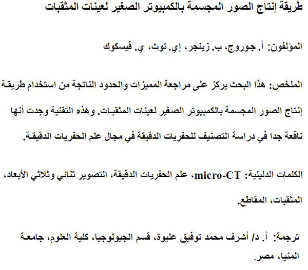
Translator: Ashraf M.T. Elewa
Polski Abstrakt
METODOLOGIA MIKRO-TOMOGRAFII KOMPUTEROWEJ OTWORNIC
Przedstawione badanie skupiało się na metodologii obsługi mikro-tomografu komputerowego podczas pracy z otwornicami, rewidując jego zalety oraz ograniczenia. Po pierwsze, opracowano szybką, tanią, nieniszczącą procedurę stabilnego mocowania w odpowiedniej pozycji i zdejmowania otwornic oraz pozbywania się reziduum i okruchów powstałych podczas cięcia. Dodatkowo metoda ta zapewnia ponowne użycie próbek. Dodatkowo, opracowano krótszą i prostszą metodę do badań otwornic (np. badania składu, ilości, identyfikacji, opisu, ilustracji). Podczas oznaczania otwornic, jak również innych grup mikroskamieniałości (takich jak np. promienice), zarówno cechy zewnętrzne jak i wewnętrzne identyfikują rodzaj ale również gatunek. Na dodatek, jest kilka gatunków otwornic znanych jedynie na podstawie przekrojów obecnych w płytkach cienkich skał. Mikro-tomografia komputerowa okazała się wyjątkowym narzędziem do faktycznej mikroskopii trójwymiarowej i mogącym przedstawić cechy morfologiczne dzięki użyciu oprogramowania umożliwiającego swobodny ruch, obracanie i krojenie stworzonych modeli okazów. Jednakże, najważniejszą zaletą tej metody są jej nieniszczące okazów właściwości, dzięki czemu możliwe staje się dokonywanie pomiarów na holotypach. Stosunkowo mała liczba przeanalizowanych próbek oraz brak odpowiedniego oprogramowania do badań różnych grup mikropaleontologicznych stanowią ograniczenia tej metody. Przedstawiona metoda wizualizacji daje nowe możliwości jeśli chodzi o taksonomię oraz badania w dziedzinie mikropaleontologii stosowanej.
Słowa kluczowe: mikro-tomografia komputerowa, mikropaleontologia, wizualizacja 2D i 3D, otwornice, ścinki
Translation: Dawid Mazurek and Robert Bronowicz
-
-
-
Review: The Princeton Field Guide to Mesozoic Sea Reptiles
 The Princeton Field Guide to Mesozoic Sea Reptiles
Article number: 26.1.1R
April 2023

 Poster Winners 2024