

Article Search
Volume 27.2
May–August 2024
Full table of contents
ISSN: 1094-8074, web version;
1935-3952, print version
Recent Research Articles
See all articles in 27.2 May-August 2024
See all articles in 27.1 January-April 2024
See all articles in 26.3 September-December 2023
Both of these fossils are included in the supplementary material here as downloadable virtual models. Both are in the VAXML interchange suggested by Sutton et al. (2012; see also www.spiers-software.org). The models are zipped; when extracted and SPIERS is installed, models can be viewed by double-clicking on the .vaxml files. Low-performance systems may struggle to render the model.
FIGURE 1. A reconstruction of Camptophyllia eltringhami from Gill (1924).
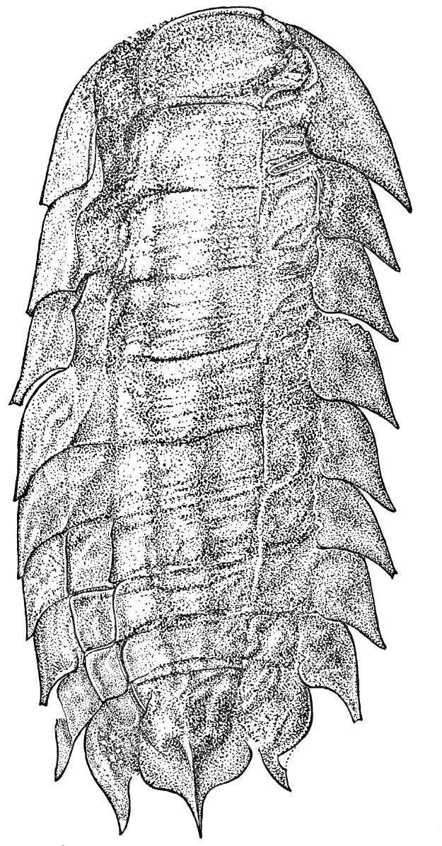
FIGURE 2. Fossil examples of the genus Camptophyllia. 1, NHM In41505, a specimen from the Tyne Coalfield. Visible fossil 44 mm in length. 2, NHM In 41503, the holotype of Camptophyllia fallax Gill 1924, Tyne. Fossil 28 mm in length. 3, Counterpart to 1 showing only posterior segments. Fossil 30 mm in length. 4, NHM In 41504, the holotype of Camptophyllia eltringhami Gill 1924, Tyne. Fossil 39 mm in length. 5, CH3, a specimen from the private collection of Mr. Stephen Livesley, Crock Hey. Fossil 42 mm in length. 6, CH304a, a specimen from the private collection of Mr. Sean Sale, Crock Hey. Fossil 35 mm in length. 7, NHM I. 13951, a specimen from Coseley. Fossil 18 mm in length. 8, NHM I. 13952, a specimen from Coseley. Fossil 13 mm in length. 9, NHM In 22843, a specimen from Coseley. Fossil 20 mm in length. 10, NHM In 22844, a specimen from Coseley. Visible fossil 20 mm in length.
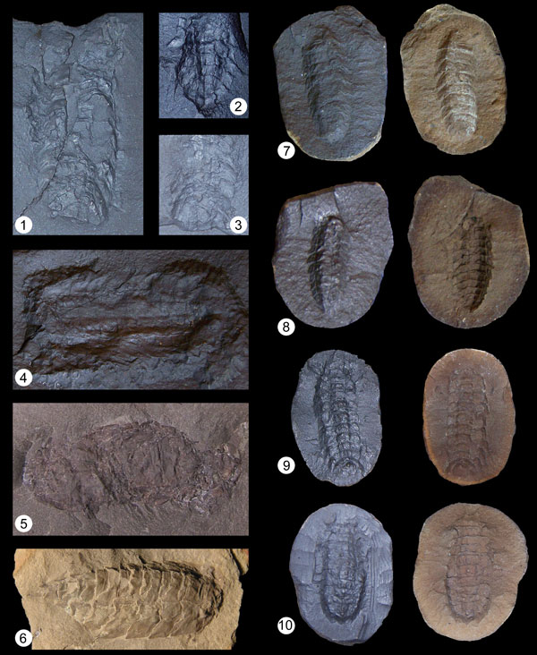
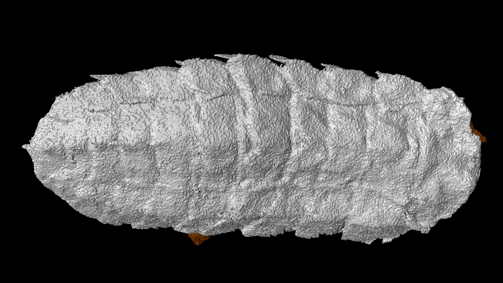
FIGURE 3. Computer reconstruction of Camptophyllia specimen NHM In 22843 from Coseley. 1, Dorsal view. 2, Lateral view. 3, Ventral view. 1-10 = segment numbers; AR = artefact; LS = lateral spine; LT = lateral tubercle; TR = transverse (cephalic) ridge; TT = terminal tubercle. Fossil 20 mm in length.
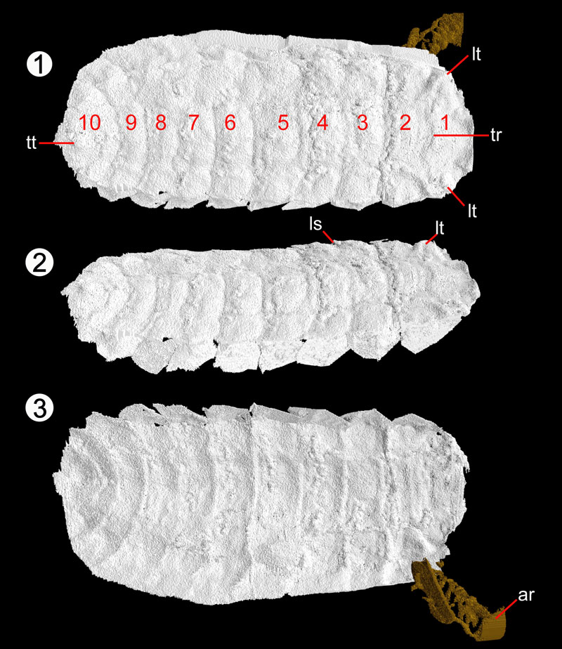
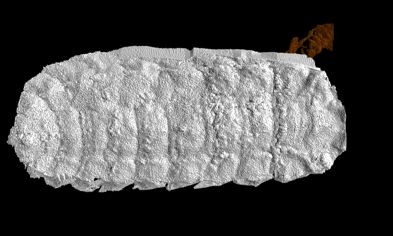
FIGURE 4. Computer reconstruction of Camptophyllia specimen NHM I. 13952 from Coseley. 1, Dorsal view; note lateral curvature of specimen visible from this orientation. 2, Lateral view showing dorsal curvature.Posterior margin of overlapping lateral plates marked by red dashed lines. 3, Zoom of dorsal surface of segments 5-7 showing more complex tergal boundaries accommodating dorsal curvature. 4, Ventral view. 1-10 = segment numbers; ABR = anterior boundary ridge; AR = artefact; DC = line showing zone of dorsal curvature (coiling); LM = lateral margin (ventral); LP = lateral plate; LS = lateral spine; LT = lateral tubercle; M = median depression; MM = median margin (ventral); MS = median spike; MT = median tubercle; PBR = posterior boundary ridge; TR = transverse (cephalic) ridge; TS = terminal spike; TT = terminal tubercle. Fossil is 12.9 mm in length.
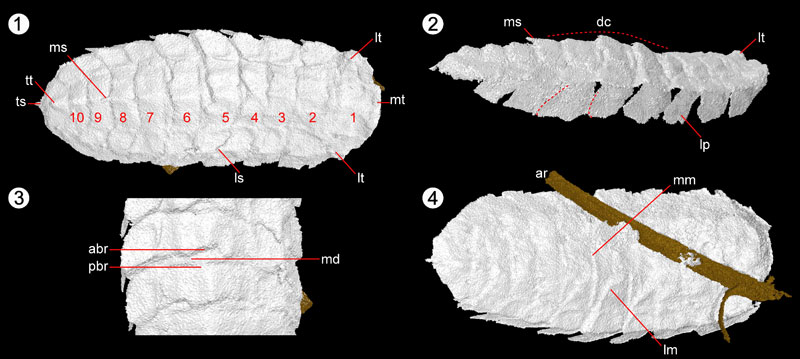
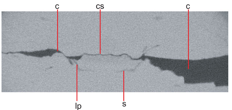
FIGURE 5. XMT slice image through cephalic region of NHM In 22843, showing box-like arrangement of head. C = crack; CS = central sclerite; LP = lateral plate; S = sternite; T = tergite. Fossil is 3.46 mm across.
Animation 1. Tomographic reconstruction of Camptophyllia specimen NHM I.13952 from Coseley.
Animation 2. Tomographic reconstruction of Camptophyllia specimen NHM In 22843 from Coseley.
Russell J. Garwood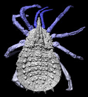 Manchester X-ray Imaging Facility
School of Materials
The University of Manchester
Oxford Rd.,
Manchester M13 9PL
UK
Russell Garwood is currently a research associate in 3D and 4D geological materials imaging, based at the Research Complex at Harwell (Rutherford Appleton Laboratory, Didcot) and part of the Manchester X-Ray Imaging Facility. He studied at Imperial College, London as an undergraduate, and a PhD student, completing a thesis on the 3D reconstruction of Carboniferous fossils in Late 2010. His research to date has focused on the palaeobiology of early terrestrial arthropods as revealed by CT scanning and computer reconstruction. More recently he has acted as the computed tomography lab manager at the Natural History Museum, London, which has allowed him to apply the same techniques to a wide range of geological, mineralogical, palaeontological, archaeological and historical problems. His other research interests include the computer modelling of evolution, abiogenesis, and early evolution.
![]()
Mark Sutton Department of Earth Science and Engineering
Imperial College
London SW7 2AZ
UK
Mark Sutton is a Senior Lecturer in the Department of Earth Science & Engineering, Imperial College, London. He received his PhD on brachiopod taxonomy from the University of Cardiff in 1996, and his subsequent research centres around three-dimensional reconstruction and phylogenetic analysis of Palaeozoic invertebrates, including arthropods, molluscs, brachiopods, echinoderms and more besides. His research interests extend other applications of three-dimensional reconstruction and computer modelling in Palaeontology. He is best known for work on the Silurian-aged Herefordshire Lagerstätte. Mark is an editor for Palaeontologia Electronica, and a member of the Palaeontological Association Council.
The enigmatic arthropod Camptophyllia
Plain Language Abstract
The Carboniferous (359-299 million years ago) provides the earliest truly widespread fossil record of life on land. In numerous UK deposits, palaeontologists have found fossils of an organism known as Camptophyllia, whose evolutionary origin and relationships remain obscure. This poor understanding is partly because only its top surface is known – no fossils show its underside, leaving this part of its anatomy entirely unrecorded. We report the use of a high-resolution form of CT scanning to try and reveal this anatomy, and help deduce the relationships of this unusual creature. Despite scanning almost all known fossils, and revealing some new aspects of the anatomy of Camptophyllia, the underside remains unresolved. While there are a number of reasons this could be the case, we suggest that it is possible the underside did not possess a thick exoskeleton, while the top surface did, allowing the preservation of the latter, not the former, during fossilisation. The technique has revealed new aspects of Camptophyllia’s anatomy, however, and the enhanced detail has allowed us to provide some suggestions as to how the organism lived.
Resumen en Español
El artrópodo enigmático Camptophyllia
El género Camptophyllia es un artrópodo enigmático del Carbonífero superior del que sólo se conocen 11 ejemplares procedentes de cinco yacimientos de tipo Konservat-Lagerstätten de los depósitos conocidos como Coal Measures. Estos fósiles, preservados en concreciones sideríticas, muestran sólo la superficie dorsal del organismo, desconociéndose totalmente la morfología de la región ventral y de los apéndices, lo que dificulta la ubicación taxonómica y filogenética del género. En este estudio se ha aplicado la microtomografía de rayos X de alta resolución (XMT) a seis ejemplares de Camptophyllia procedentes de cuatro yacimientos. Esta técnica ha proporcionado nuevos detalles sobre la morfología, confirmando que el primer segmento es cefálico y facilitando nuevas informaciones sobre el modo de vida del organismo. Sin embargo, a pesar de haber analizado todos los ejemplares conocidos del género, excepto uno, no se ha podido reconstruir su anatomía ventral. Esto es posiblemente debido a un sesgo tafonómico relacionado con una débil esclerotización de la región ventral. Las afinidades de Camptophyllia permanecen, por tanto, oscuras, a la espera del hallazgo de nuevos materiales.
PALABRAS CLAVE: Camptophyllia; Carbonífero; siderita; tomografía computarizada; VAXML
Traducción: Miguel Company
Résumé en Français
L’énigme de l’arthropode Camptophyllia
L’énigmatique genre d’arthropode Camptophylliais du Carbonifère supérieur est connu à partir de 11 fossiles, provenant de cinq Lagerstätten des houilles à charbon. Ces fossiles inclus dans la sidérite révèlent uniquement la surface dorsale de l’organisme – la morphologie ventrale et ses appendices étant totalement inconnus, gênant les efforts pour placer taxonomiquement et phylogénétiquement le genre. Cette étude présente une utilisation de la micro-tomographie haute résolution à rayons X (XMT) de six spécimens de Camptophyllia, provenant de Lagerstätten carbonifères. La re-étude sur la base de la XMT a fourni de nouveaux détails de la morphologie, confirmant que le segment le plus antérieur est céphalique et permettant des hypothèses mieux documentées sur le mode de vie de l’organisme. Toutefois, bien qu’aillant scanné tous les représentants connus du genre sauf un, l’anatomie ventrale reste indéterminée ; il est possible que cet état taphonomique résulte d’une faible sclérotisation de la région ventrale. En attendant la découverte de plus de matériel, les affinités de Camptophyllia restent incertaines.
MOTS CLES : Camptophyllia; Carbonifère; sidérite; tomographie par ordinateur; VAXML
Translator: Olivier Maridet
Deutsche Zusammenfassung
Der rätselhafte Arthropode Camptophyllia
Von der rätselhaften Arthropoden-Gattung Camptophyllia aus dem Oberkarbon sind 11 Fossilien aus fünf Kohlelagerstätten bekannt. Diese in Siderit eingebetteten Fossilien zeigen nur die Dorsalseite des Organismus – die Morphologie der Ventralseite und der Anhänge ist komplett unbekannt, was es schwierig macht die Gattung taxonomisch und phylogenetisch einzuordnen. Diese Untersuchung informiert über die Anwendung von hochauflösender Röntgenmikrotomographie (XMT) an sechs Camptophyllia –Stücken aus vier karbonischen Lagerstätten. Untersuchungen mit XMT lieferten neue morphologische Details. So konnte bestätigt werden, dass das vorderste Segment dem Schädel angehört und es können informativere Überlegungen über die Lebensweise des Organismus angestellt werden. Allerdings konnte die ventrale Anatomie nicht gelöst werden, obwohl alle Stücke außer einem eingescannt wurden. Dies hat möglicherweise einen taphonomischen Hintergrund resultierend in der nur spärlich sklerosierten Ventralregion. Bis zur Entdeckung weiteren Materials bleiben die Verwandtschaftsbeziehungen von Camptophyllia daher auch künftig unklar.
SCHLÜSSELWÖRTER: Camptophyllia; Karbon; Siderit; Computertomographie; VAXML
Translators: Eva Gebauer and Anke Konietzka
Arabic
Translator: Ashraf M.T. Elewa
Polski Abstrakt
Enigmatyczny stawonóg Camptophyllia
Enigmatyczny rodzaj późnokarbońskiego stawonoga Camptophyllia znany jest z 11 skamieniałości znalezionych w pięciu stanowiskach typu Coal Measures Lagerstätten. Te zawarte w syderycie skamieniałości ukazują tylko grzbietową powierzchnię organizmu – morfologia jego spodniej część i odnóży jest całkowicie nieznana, utrudniając próby umieszczenia rodzaju taksonomicznie lub filogenetycznie. Niniejsze badanie donosi o wykorzystaniu wysokorozdzielczej mikrotomografii rentgenowskiej (XMT) do zbadania sześciu okazów Camptophyllia z czterech karbońskich Lagerstätten. Rewizja w oparciu o XMT zapewniła nowe szczegóły morfologiczne, potwierdzając, że położony najbardziej z przodu segment jest głowowym i umożliwiła bardziej rzeczowe spekulacje dotyczące sposobu życia organizmu. Jednak pomimo zeskanowania wszystkich poza jednym reprezentantów rodzaju, anatomia spodniej strony nie została rozwikłana; możliwe że przyczyny są tafonomiczne, wynikające ze słabej sklerytyzacji wewnętrznej strony. Do czasu odkrycia dalszych materiałów, pokrewieństwa Camptophyllia pozostają niejasne.
SŁOWA KLUCZOWE: Camptophyllia; karbon; syderyt; tomografia komputerowa; VAXML
Translators: Dawid Mazurek, Robert Bronowicz, and Daniel Madzia
Riassunto in Italiano
L’enigmatico artropode Camptophyllia
L’enigmatico genere Camptophyllia, un artropode del Carbonifero superiore, è conosciuto sulla base di 11 fossili rinvenuti in cinque Lagerstätten di Coal Measures. Tali fossili, inclusi in siderite, mostrano solo la superficie dorsale degli organismi, quindi la loro morfologia ventrale e appendicolare è completamente sconosciuta. Ciò impedisce di dare al genere una collocazione tassonomica o filogenetica.
In questo studio viene utilizzata la microtomografia a raggi X ad alta risoluzione (XMT) su sei esemplari di Camptophyllia provenienti da quattro Lagerstätten carboniferi. I risultati hanno confermato che il segmento anteriore è quello cefalico e hanno facilitato le speculazioni riguardanti il modo di vita di tale organismo. Purtroppo però, nonostante le scansioni, in tutti i casi tranne uno l’anatomia ventrale non è stata visualizzata. E’ possibile che ciò sia dovuto a ragioni tafonomiche, conseguenza di una ridotta sclerotizzazione della regione ventrale. Per il momento, quindi, a meno di rinvenire nuovo materiale, le affinità di Camptophyllia rimangono sconosciute.
PAROLE CHIAVE: Camptophyllia; Carbonifero; siderite; tomografia computerizzata; VAXML
Translator: Chiara Angelone
-
-
-
Review: The Princeton Field Guide to Mesozoic Sea Reptiles
 The Princeton Field Guide to Mesozoic Sea Reptiles
Article number: 26.1.1R
April 2023

 Poster Winners 2024