

FIGURE 3. Variation in enamel thickness in the teeth of Revueltosaurus callenderi 1-9, premaxillary tooth (P-33799--longitudinal section); 10-14, maxillary tooth (P-33798--transverse section). 1, overview of premaxillary tooth indicating approximate place where measurements and micrographs shown in this figure and Figure 4 were taken; 2-9, close-up views showing enamel thickness variation, with enamel-dentine junction (EDJ) oriented relative to overview in (1); and 10, overview of maxillary tooth section, indicating approximate place where measurements and micrographs shown in this figure and Figure 5 were taken; 11-14, close-up views showing enamel thickness variation, with enamel-dentine junction (EDJ) oriented relative to overview in (10). Scale bars = 100 µm except for 1 (5 mm), 2 (10 µm) and 10 (2 mm). Numbers in image are linked to further enlargements. Author note: Figure 3.10 is part of overview.
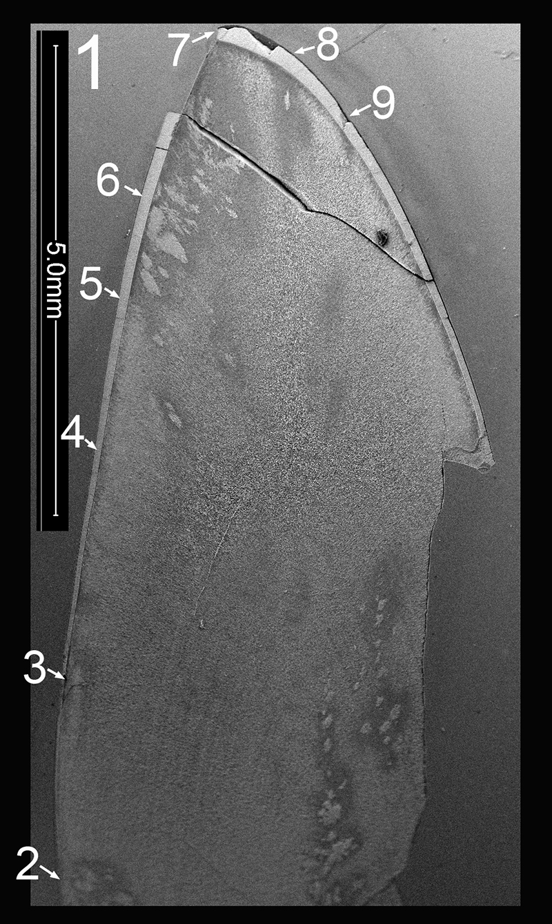
FIGURE 3.2. Original micrograph showing tooth enamel microstructure of premaxillary tooth of Revueltosaurus callenderi (P-33799) in longitudinal section near the base of the tooth; EDJ oriented to bottom of picture.
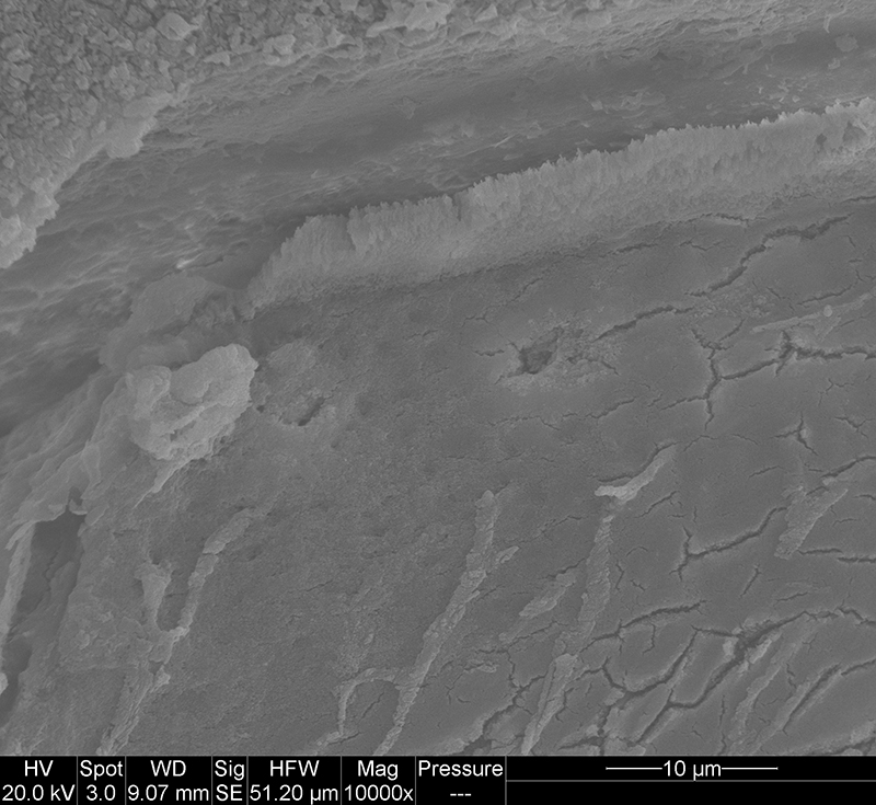
FIGURE 3.3. Original micrograph showing tooth enamel microstructure of premaxillary tooth of Revueltosaurus callenderi (P-33799) in longitudinal section near the base of the tooth; EDJ oriented to bottom of picture.
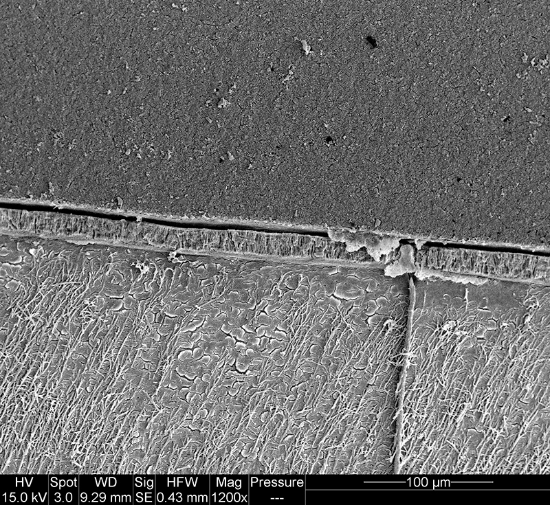
FIGURE 3.4. Original micrograph showing tooth enamel microstructure of premaxillary tooth of Revueltosaurus callenderi (P-33799) in longitudinal section near the middle of the tooth; EDJ oriented to bottom of picture.
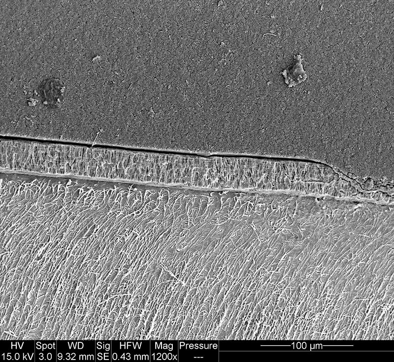
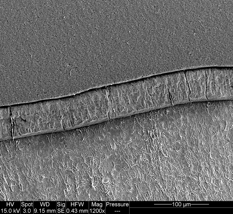
FIGURE 3.5. Original micrograph showing tooth enamel microstructure of premaxillary tooth of Revueltosaurus callenderi (P-33799) in longitudinal section near the middle of the tooth; EDJ oriented to bottom of picture.
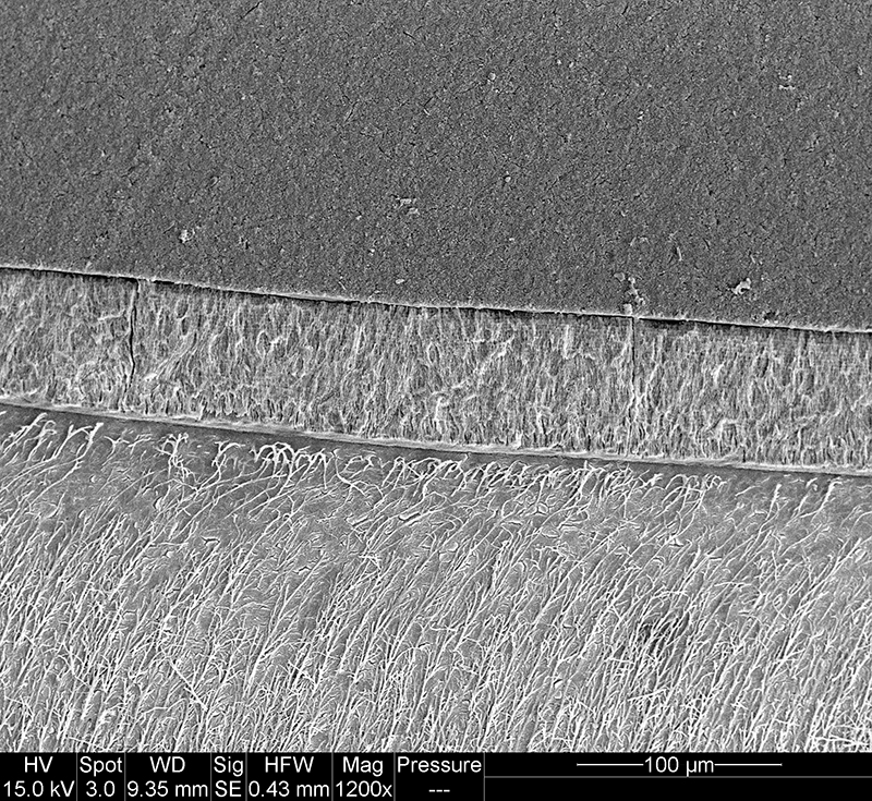
FIGURE 3.6. Original micrograph showing tooth enamel microstructure of premaxillary tooth of Revueltosaurus callenderi (P-33799) in longitudinal section high on the crown; EDJ oriented to bottom of picture.
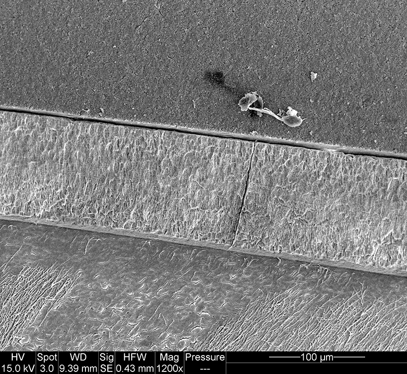
FIGURE 3.7. Original micrograph showing tooth enamel microstructure of premaxillary tooth of Revueltosaurus callenderi (P-33799) in longitudinal section at the tip of the crown; EDJ oriented to left of picture.
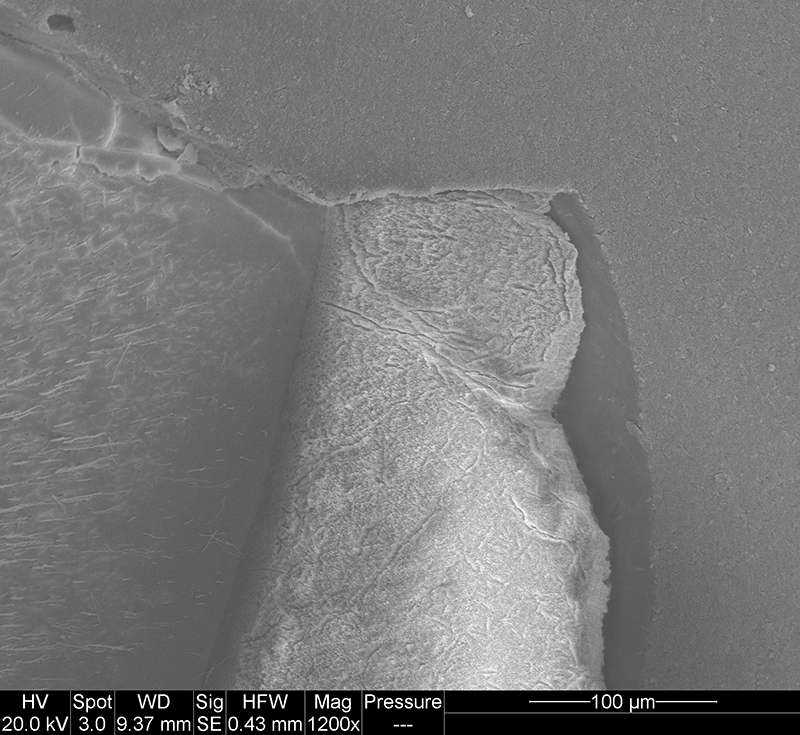
FIGURE 3.8. Original micrograph showing tooth enamel microstructure of premaxillary tooth of Revueltosaurus callenderi (P-33799) in longitudinal section near the tip of the crown; EDJ oriented to bottom of picture.
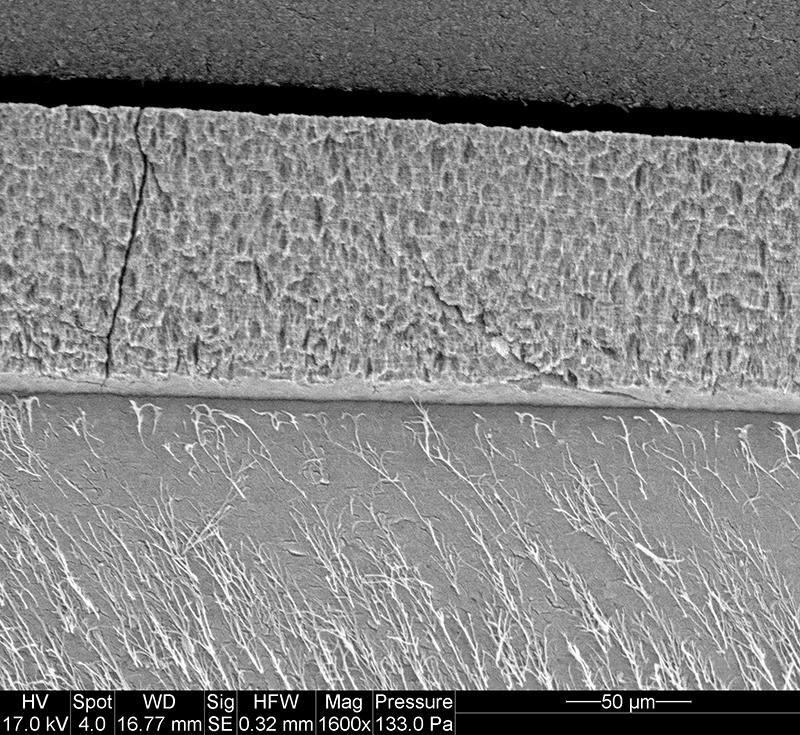
FIGURE 3.9. Original micrograph showing tooth enamel microstructure of premaxillary tooth of Revueltosaurus callenderi (P-33799) in longitudinal section near the base of the tooth; EDJ oriented to upper left of picture.
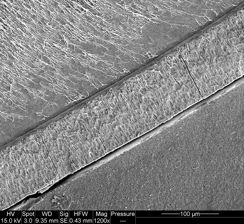
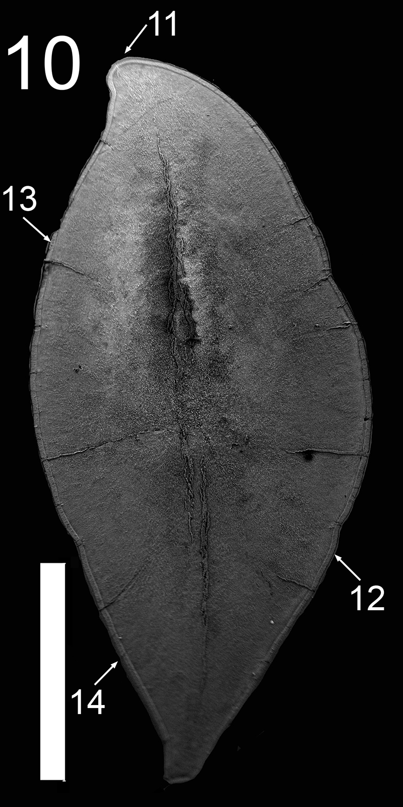
FIGURE 3.11. Original micrograph showing tooth enamel microstructure of maxillary tooth of Revueltosaurus callenderi (P-33798) in transverse section across one denticle.
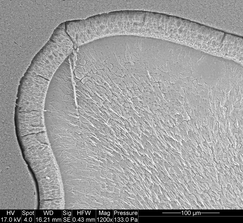
FIGURE 3.12. Original micrograph showing tooth enamel microstructure of maxillary tooth of Revueltosaurus callenderi (P-33798) in transverse section on labial side; EDJ to upper right.
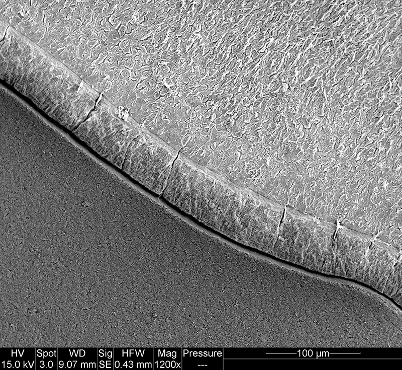
FIGURE 3.13. Original micrograph showing tooth enamel microstructure of maxillary tooth of Revueltosaurus callenderi (P-33798) in transverse section on lingual side; EDJ to bottom.
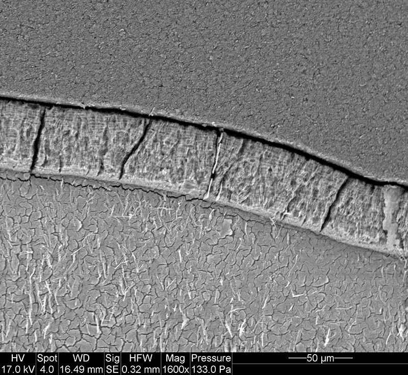
FIGURE 3.14. Original micrograph showing tooth enamel microstructure of maxillary tooth of Revueltosaurus callenderi (P-33798) in transverse section on labial side; EDJ to bottom.

 Poster Winners 2024
 The Princeton Field Guide to Mesozoic Sea Reptiles