

FIGURE 7. Scanning electron microscope images of (UCMP 165211), Krzyzanowskisaurus hunti premaxillary tooth enamel microstructure in transverse section. 1, overview of tooth indicating approximate place where measurements and micrographs shown in this figure were taken; 2-7, close-up views showing enamel thickness variation, with EDJ oriented relative to overview in (1) and OES away from the same overview. Scale bars = 20 µm except 1 (2 mm). Numbers in image are linked to further enlargements.
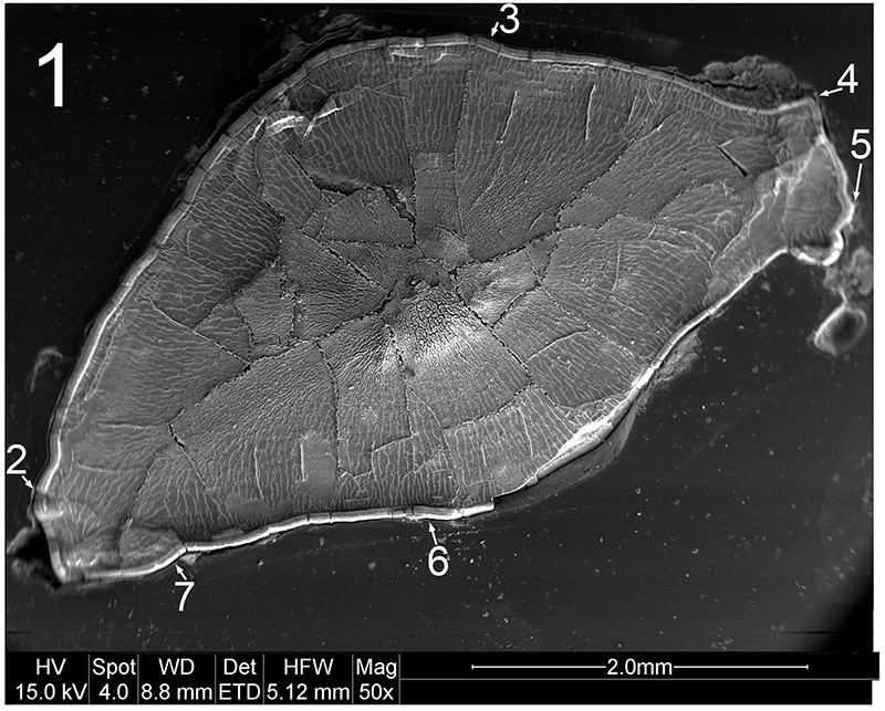
FIGURE 7.2. Original micrograph showing tooth enamel microstructure of (UCMP 165211), Krzyzanowskisaurus hunti in transverse section on the labial side; EDJ oriented to right of picture.
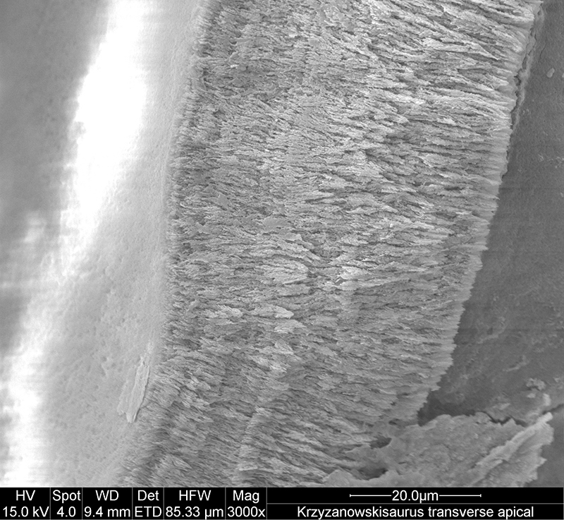
FIGURE 7.3. Original micrograph showing tooth enamel microstructure of (UCMP 165211), Krzyzanowskisaurus hunti in transverse section on the labial side; EDJ oriented to left of picture.
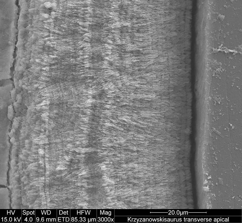
FIGURE 7.4. Original micrograph showing tooth enamel microstructure of (UCMP 165211), Krzyzanowskisaurus hunti in transverse section across a denticle; EDJ oriented to lower left of picture.
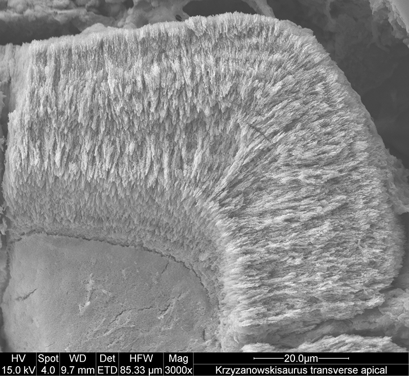
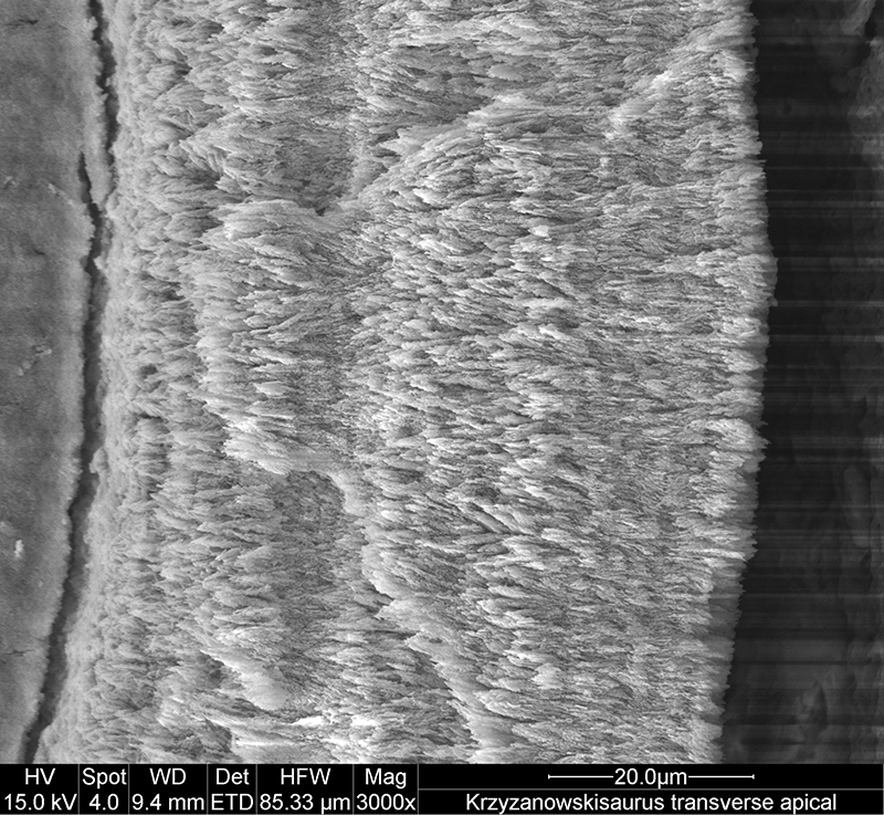
FIGURE 7.5. Original micrograph showing tooth enamel microstructure of (UCMP 165211), Krzyzanowskisaurus hunti in transverse section across a denticle; EDJ oriented to left of picture.
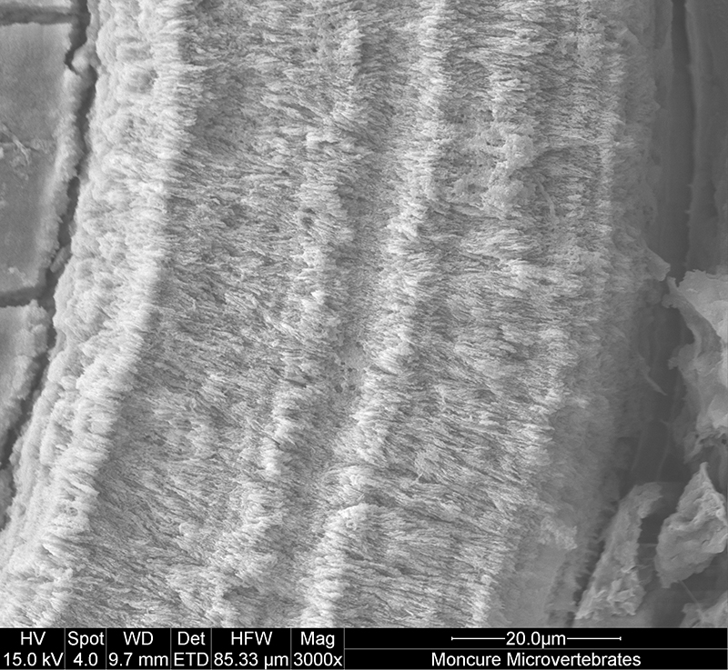
FIGURE 7.6. Original micrograph showing tooth enamel microstructure of (UCMP 165211), Krzyzanowskisaurus hunti in transverse section on the lingual side; EDJ oriented to left of picture.
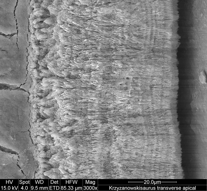
FIGURE 7.7. Original micrograph showing tooth enamel microstructure of (UCMP 165211), Krzyzanowskisaurus hunti in transverse section on the lingual side; EDJ oriented to left of picture.

 Poster Winners 2024
 The Princeton Field Guide to Mesozoic Sea Reptiles