

FIGURE 5. Scanning electron microscope images of NMMNH P-33798, Revueltosaurus callenderi maxillary tooth enamel microstructure in transverse section. 1, overview of tooth indicating approximate place where measurements and micrographs shown in this figure were taken; 2-8, close-up views showing enamel thickness variation, with EDJ oriented relative to overview in (1) and OES away from the same overview. Scale bars = 20 µm except 1 (2 mm), 3 (50 µm). Numbers in image are linked to further enlargements.
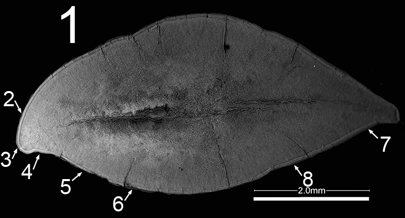
FIGURE 5.2. Original micrograph showing tooth enamel microstructure of maxillary tooth of Revueltosaurus callenderi (P-33798) in transverse section on labial side; EDJ to left.
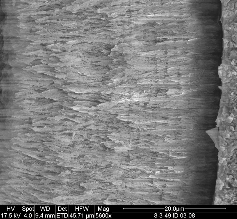
FIGURE 5.3. Original micrograph showing tooth enamel microstructure of maxillary tooth of Revueltosaurus callenderi (P-33798) in transverse section across one denticle; EDJ to right.
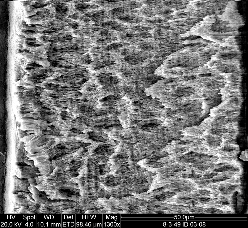
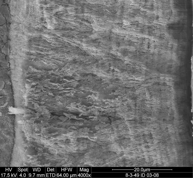
FIGURE 5.4. Original micrograph showing tooth enamel microstructure of maxillary tooth of Revueltosaurus callenderi (P-33798) in transverse section on lingual side; EDJ to left.
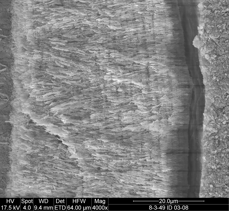
FIGURE 5.5. Original micrograph showing tooth enamel microstructure of maxillary tooth of Revueltosaurus callenderi (P-33798) in transverse section on lingual side; EDJ to left.
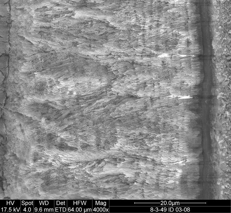
FIGURE 5.6. Original micrograph showing tooth enamel microstructure of maxillary tooth of Revueltosaurus callenderi (P-33798) in transverse section on lingual side; EDJ to left.
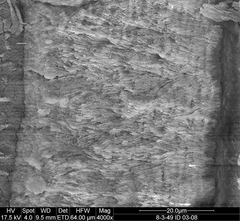
FIGURE 5.7. Original micrograph showing tooth enamel microstructure of maxillary tooth of Revueltosaurus callenderi (P-33798) in transverse section on lingual side; EDJ to left.
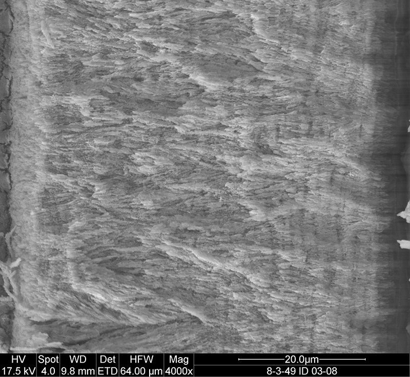
FIGURE 5.8. Original micrograph showing tooth enamel microstructure of maxillary tooth of Revueltosaurus callenderi (P-33798) in transverse section on lingual side; EDJ to left.

 Poster Winners 2024
 The Princeton Field Guide to Mesozoic Sea Reptiles