

Article Search
Volume 27.2
May–August 2024
Full table of contents
ISSN: 1094-8074, web version;
1935-3952, print version
Recent Research Articles
See all articles in 27.2 May-August 2024
See all articles in 27.1 January-April 2024
See all articles in 26.3 September-December 2023
 Antonino Briguglio. Faculty of Science, Universiti Brunei Darussalam, Jalan Tungku Link, Gadong BE1410, Brunei Darussalam, antonino.briguglio@ubd.edu.bn
Antonino Briguglio is currently a Senior Lecturer at the Faculty of Science at the University of Brunei Darussalam teaching sedimentology and micropalaeontology in a Petroleum Geology department. He was a lecturer at the Department of Palaeontology at the University of Vienna, Austria and collaborated as a research postdoctoral fellow at the Natural History Museum in the same city. He did his undergraduate degree at the university La Sapienza in Rome, Italy where he became interested in Eocene Larger Benthic Foraminifera. During his PhD he focused on the hydrodynamic behavior of nummulitids. His scientific interests cover mainly integrated biostratigraphy (Cenozoic), systematics, ecology and biology of Larger Benthic Foraminifera. During the last three years he focused his scientific attention on the functional morphology of Larger Foraminifera and he is routinely using computed tomography to investigate growth patterns and strategies of recent and fossil forms.

 Shunichi Kinoshita. Department of Palaeontology, University of Vienna, Geocenter, Althanstrasse 14, A-1090 Vienna, Austria. shunichi.kinoshita@univie.ac.at
Kinoshita-san is a Ph.D. students currently enrolled in a research project on Larger Benthic Foraminifera from Sesoko Island, Japan. He is very talented in using CT rendering programs to create animations and videos and is routinely using 3D software to study nummulitids.
 Johann Hohenegger. Department of Palaeontology, University of Vienna, Geocenter, Althanstrasse 14, A-1090 Vienna, Austria. johann.hohenegger@univie.ac.at
Johann Hohenegger is a retired professor of the Department of Palaeontology at Vienna University, Austria, where he got his undergraduate and postgraduate degrees. His main interests with more than 150 publications are focused on population dynamics, taphonomy and carbonate production of larger foraminifera in the present and past, recognition of species in the present and past based on population genetics, morphogenetic programs and their phylogenetic implication in comparison to molecular genetic trees, democoenoclines and morphocoenoclines along environmental gradients and their importance to decipher paleoenvironmental conditions, integrated stratigraphy in the Neogene of the Paratethys (Central Europe) and spatial distributions on a micro- and macroscale. He started his scientific work on Late Palaeozoic to Early Jurassic foraminifera. In 2012 he was awarded the Grzybowski Award for his life work on benthic foraminiferal ecology.
 Erik Wolfgring. Department of Geodynamics and Sedimentology, University of Vienna, Geocenter, Althanstrasse 14, A-1090 Vienna, Austria. erik.wolfgring@univie.ac.at
Erik is a micropalaeontologist currently employed at the University of Vienna. He is mainly focusing on the stratigraphy of Upper Cretaceous Tethyan pelagic sections and the expression of environmental changes on foraminifera assemblages. Furthermore he is focusing on the palaeobiology and biostratigraphy of larger benthic as well as planktonic foraminifera.
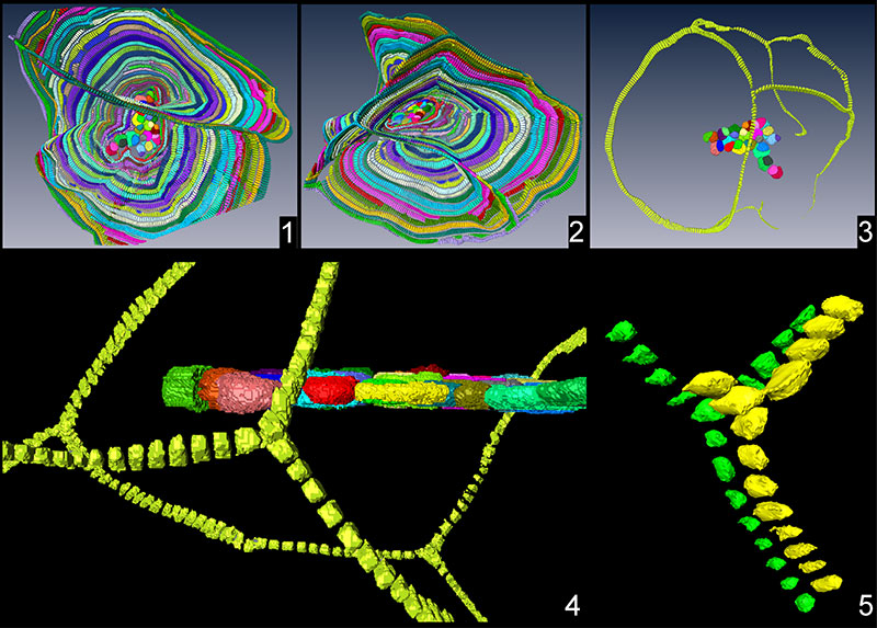
FIGURE 1. Specimen A1: 1) equatorial and axial sections of one specimen.; 2) equatorial view of the test; 3) lateral view of the test; 4) close-up to the nepiont and the first chambers in equatorial section; 5) segmentation of the entire nepiont. For more information, refer to text.
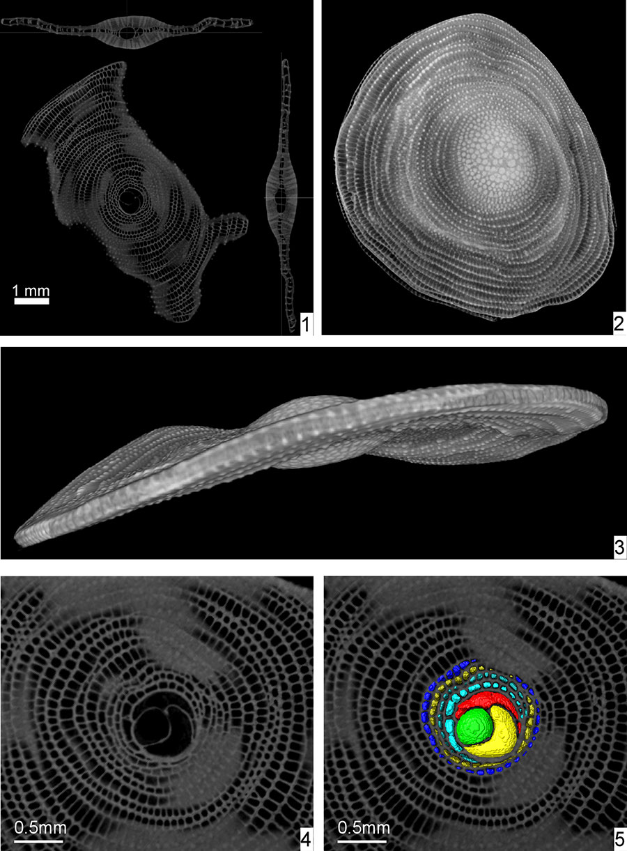
FIGURE 2. Segmentation and equatorial sections of specimens possessing multiple nepionts: 1) specimen A2; 2) specimen A3; 3) specimen A17; 4) specimen A10; 5) specimen A18; 6) specimen A5; 7) specimen A6. Scale bar equals 0.5 mm.
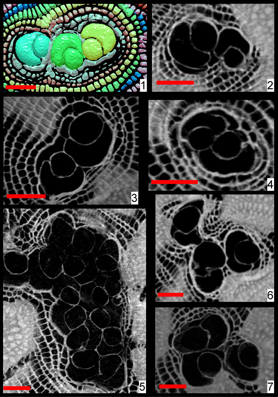
FIGURE 3. Proloculus diameters. 1) proloculus diameter for all individuals in the presented population, except for A18. Multiple bars indicate the presence of several proloculi. 2) diameters of all proloculi identified within the specimen A18.
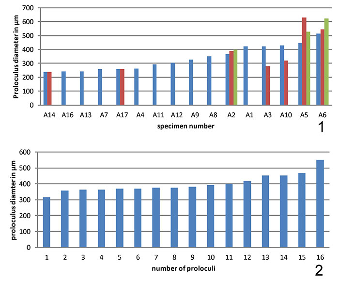
FIGURE 4. Relative position of embryos and secondary equatorial layers: 1) Specimen A2; 2) specimen A3; 3) specimen A6; 4) specimen A14.
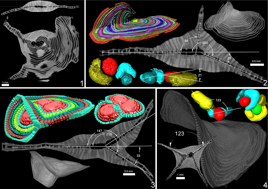
FIGURE 5. Multiple equatorial layers and T-connection in specimen A18.
APPENDIX 1.
External view of the specimen A1. Note the surface ornamentation and the gently wavy profile. This specimen has only one nepiont.
APPENDIX 2.
CT rendering of the three nepionts and the first chambers of specimen A6. Note that two nepionts are on the same plane whereas the third nepiont is tilted. Each of the three proloculus-deuteroloculus couplets builds its own ana-nepionic chamber; then the first two nepionts share some of their nepionic chambers, while those of the third nepionts are still clearly separated. The fusion of the third nepiont with the other two happens from the first neanic chamber (first in light blue). Note how each neanic chamber runs through both planes thus creating T-shape connections.
APPENDIX 3.
Portion of the external surface of specimen A18. Note the multiple equatorial planes all connected each other. Bryozoans, serpulids and other small shells fragments are visible on some surfaces.
APPENDIX 4.
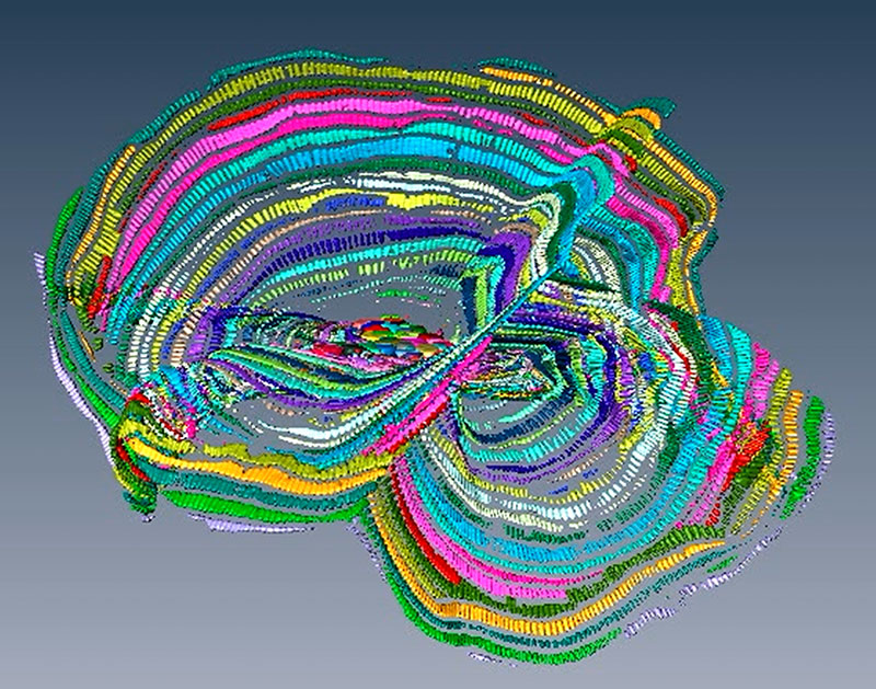
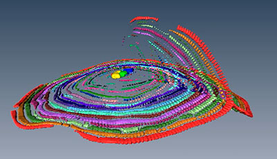
CT rendering of all nepionts and all equatorial planes in specimen A18. Note how each chamber runs through all equatorial planes with T-shape connections. Click on image to run.
APPENDIX 5.
CT rendering of all nepionts and all equatorial planes in specimen A18. Note how each chamber runs through all equatorial planes with T-shape connections. Click on image to run.
APPENDIX 6.
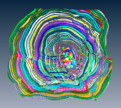
Close-up of all nepionts and one chamber running through all equatorial planes in specimen A18. Note the T-shape connections. Due to the resolution of the CT used and the size of the specimen, the smallest chamberlets could not be segmented, here only well visible chamberlets are presented.
APPENDIX 7.
Close-up of all nepionts and one chamber running through all equatorial planes in specimen A18. Note the T-shape connections. Due to the resolution of the CT used and the size of the specimen, the smallest chamberlets could not be segmented, here only well visible chamberlets are presented.
APPENDIX 8.
CT rendering of the nepionts and the first chambers of specimen A2. Note that the three nepionts share some nepionic and all neanic chambers. These three nepionts built one equatorial layer only.
APPENDIX 9.
External view of specimen A3. Note the two equatorial layer nicely connected by T-shape connections.
APPENDIX 10.
CT Rendering of the nepionts and of most of the neanic chambers in specimen A3. Note how each chamber runs through both equatorial planes. Click on image to run.
Morphological variations in Cycloclypeus carpenteri: Multiple embryos and multiple equatorial layers
Plain Language Abstract
In this work, 17 specimens of Cycloclypeus carpenteri have been imaged by computed tomography. C. carpenteri is a protist single celled organism that during its life calcifies a disc-like test which can reach a diameter of 13 cm. Computed tomography is a very advanced scientific technique which is commonly used to visualize the internal structures of objects. This technique also allows to quantify and measure a number of parameters which are impossible to obtain by cutting the object as it is normally done. It has been observed that in several cases, this species can produce abnormal shapes which in our explanation, can be related to environmental conditions and reproduction strategies. The specimens observed here keep a normal growth even though producing complex geometries. Furthermore, we have observed that shells of twins in this species can lead to very complex test shapes such as multiequatorial layers.
Resumen en Español
Variaciones morfológicas en Cycloclypeus carpenteri: embriones múltiples y múltiples capas ecuatoriales
En este trabajo, se han analizado 17 ejemplares de Cycloclypeus carpenteri mediante la exploración micro-CT. Se ha observado que muchos especímenes poseen múltiples embriones, múltiples nepiontes y más de una capa ecuatorial según muestran algunas pruebas. Se ha medido el diámetro de cada prolóculo, y parece que son muy variables incluso dentro del mismo espécimen, por tanto, ello cuestiona la teoría conocida desde hace mucho tiempo que indica que los esquizontes tienen prolóculos más pequeños que los gamontes y también cuestiona el hecho de que los prolóculos en la misma especie deben tener todos un tamaño comparable. Siempre que los nepiontes se colocan en planos diferentes, creando así un ángulo entre ellos, ese ángulo tiene una correlación significativa con el ángulo de la conexión de diferentes capas ecuatoriales. En la unión entre dos capas ecuatoriales se encuentran conexiones en forma de T; estas uniones se establecen por una camarilla (chamberlet), la cual posee un número inusualmente elevado de aberturas, por lo que se asemeja a la estructura de las camarillas del género Spiroclypeus.
Palabras clave: foraminíferos bentónicos de gran talla; Modelo 3D; micro-CT; tomografía; teratología; patrón de crecimiento
Traducción: Enrique Peñalver or Diana Elizabeth Fernández
Résumé en Français
Variations morphologiques chez Cycloclypeus carpenteri : multiples embryons et multiples couches équatoriales
Dans ce travail, 17 spécimens de Cycloclypeus carpenteri ont été analysés par microtomodensitométrie. Il a été observé que de nombreux spécimens possèdent de multiples embryons et de multiples népiontes, et que certains tests présentent plus d'une couche équatoriale. Le diamètre de chaque proloculus a été mesuré, et il semble qu'ils soient très variables même au sein d'un seul spécimen. Cela remet donc en question la théorie de longue date proposant que les schizontes ont des proloculi plus petits que les gamontes, et également que les proloculi de la même espèce devraient tous être de taille similaire. Quand les népiontes sont positionnés selon différents plans, créant ainsi un angle entre eux, cet angle est significativement corrélé avec l'angle connectant les différentes couches équatoriales. Les connections en forme de T sont situées à la jonction entre deux couches équatoriales. Ces jonctions sont formées par une loge qui possède un nombre inhabituellement élevé d'ouvertures, ressemblant à la structure de loge du genre Spiroclypeus.
Mots-clés : foraminifères benthiques de grande taille ; modèle 3D ; microtomodensitométrie (microTDM); tomographie ; tératologie ; schémas de croissance
Translator: Antoine Souron
Deutsche Zusammenfassung
Morpholgische Variationen bei Cycloclypeus carpenteri: multiple Embryonen und multiple Transversalebenen
In dieser Arbeit wurden 17 Stücke von Cycloclypeus carpenteri mit Mikro-CT untersucht. Es wurde beobachtet, dass viele der Stücke multiple Embryonen und multiple Nepionten besitzen und einige Tests zeigen sogar mehr als eine Transversalebene. Von jedem Proloculus wurde der Durchmesser gemessen und es scheint dass dieser sogar innerhalb desselben Stückes sehr variabel ist. Daher wird die seit langem bekannte Theorie infrage gestellt, nach der Schizonten kleinere Proloculi als Gamonten haben und ebenso stellen wir die Tatsache infrage, dass Proloculi derselben Art alle eine vergleichbare Größe haben. Wann immer die Nepionten auf verschiedenen Ebenen angeordnet werden und damit ein Winkel zwischen diesen erzeugt wird, hat dieser Winkel eine signifikante Korrelation zu dem Winkel der verschiedene Transversalebenen verbindet. T-förmige Verbindungen liegen am Knotenpunkt von zwei Transversalebenen. Diese Verbindungen entstehen durch ein Chamberlet, welches eine ungewöhnlich höhere Anzahl von Öffnungen besitzt und der Chamberlet-Struktur der Gattung Spiroclypeus ähnelt.
Schlüsselwörter: größere benthische Foraminifera; 3D Modell; Mikro-CT; Tomographie; Teratologie; Wachstumstruktur
Translator: Eva Gebauer
Arabic
Translator: Ashraf M.T. Elewa
-
-
-
Review: The Princeton Field Guide to Mesozoic Sea Reptiles
 The Princeton Field Guide to Mesozoic Sea Reptiles
Article number: 26.1.1R
April 2023

 Poster Winners 2024